Transcription
HindawiMediators of InflammationVolume 2021, Article ID 5582943, 14 pageshttps://doi.org/10.1155/2021/5582943Research ArticleProbiotic-Derived Polyphosphate Accelerates Intestinal EpitheliaWound Healing through Inducing Platelet-Derived MediatorsShotaro Isozaki,1 Hiroaki Konishi,1,2 Mikihiro Fujiya ,1,2 Hiroki Tanaka,3 Yuki Murakami,1Shin Kashima,1 Katsuyoshi Ando,1 Nobuhiro Ueno,1 Kentaro Moriichi,1and Toshikatsu Okumura11Division of Metabolism and Biosystemic Science, Gastroenterology, And Hematology/Oncology, Department of Medicine,Asahikawa Medical University, Asahikawa 078-8510, Japan2Department of Gastroenterology and Advanced Medical Sciences, Asahikawa Medical University, Asahikawa 078-8510, Japan3Division of Tumor Pathology, Department of Pathology, Asahikawa Medical University, Asahikawa 078-8510, JapanCorrespondence should be addressed to Mikihiro Fujiya; fjym@asahikawa-med.ac.jpReceived 20 January 2021; Revised 25 February 2021; Accepted 6 March 2021; Published 30 March 2021Academic Editor: Hirokazu FukuiCopyright 2021 Shotaro Isozaki et al. This is an open access article distributed under the Creative Commons Attribution License,which permits unrestricted use, distribution, and reproduction in any medium, provided the original work is properly cited.Inflammatory bowel disease (IBD), such as ulcerative colitis (UC) and Crohn’s disease (CD), is an intractable intestinalinflammation associated with the disruption of the intestinal mucosa. We previously demonstrated that Lactobacillus brevisderived long-chain polyphosphate (poly P) improved the intestinal barrier function by the upregulation of cell adhesion andrelieved intestinal inflammation, thereby exerting a curing effect on colitis in vitro, in vivo, and in an investigator-initiatedclinical study of UC. However, how poly P improves mucosal defects induced by intestinal inflammation has not beenelucidated. In this study, we detected the accumulation of platelets in inflamed tissues induced by poly P in a dextran sulfatesodium- (DSS-) induced colitis mouse model. A light transmission aggregometry analysis and scanning electron microscopyshowed that poly P promoted the platelet aggregation. An SRB assay and ki-67 staining showed that the supernatant of poly Ptreated platelet-rich plasma (PRP) increased intestinal epithelial cell growth. A wound healing assay showed that thesupernatant of poly P-treated PRP, but not poly P itself, accelerated wound healing. A Western blotting analysis indicated thatmitogen-activated protein kinase activation was induced by the supernatant of poly P-treated human PRP in the epithelial cellsand its wound healing effect was significantly decreased by the inhibition of ERK signaling. These data suggested that plateletderived mediators induced by poly P improved intestinal inflammation through the promotion of epithelial cell growth by theactivation of the ERK signaling pathway. The mechanism is a novel host-microbe interaction through mammalian plateletderived mediators induced by bacterial molecules.1. IntroductionVarious bacteria live in the host gastrointestinal tract andbuild a symbiotic relationship with their mammalian hosts.It has been shown that the disruption of the intestinal microflora results in harm to the host, such as the abnormal activation of immune systems and consequently the developmentof various diseases, including gastrointestinal inflammationsand tumors, liver dysfunction, atopic dermatitis, and brainand neurological disorders [1–3].Previous studies have suggested that the improvement ofthe intestinal condition by the administration of beneficialbacteria, such as Lactobacilli and Bifidobacterium, helpedresolve these diseases in in vitro and in vivo animal experimental models [4–6]. Other studies, including our own, haveshown that some probiotic bacteria released bioactive mediators that contributed to the maintenance of intestinalhomeostasis and the improvement of inflammation. Yanet al. identified p40 and p75 from the culture supernatantof Lactobacillus GG and showed how their antiinflammatory effects mediated the epithelial EGFR-Akt signaling pathway [7]. Likewise, we identified competence andsporulation factor (CSF) from Bacillus subtilis and showedthat CSF was incorporated into epithelial cells through
2OCTN2, thus activating the cell survival signaling, includingthe Akt and p38 mitogen-activated protein kinase (MAPK)pathways, resulting in an increase in the epithelial barrierfunction [8]. Kelly et al. found that microbiota-derivedshort-chain fatty acids stabilize the hypoxia-inducible factor(HIF) and strengthen the gut epithelial barrier function [9].We also identified long-chain polyphosphate (poly P) asa beneficial substance in the culture supernatant of L. brevisSBC8803 that supports the intestinal barrier function of themammalian host through the integrin β1-p38 MAPK signaling pathway [10]. Furthermore, poly P upregulated the intestinal barrier function and downregulated the macrophageactivation in a murine colitis model and dramaticallyimproved the symptoms and endoscopic findings of patientswith refractory ulcerative colitis (UC) in an investigatorinitiated clinical trial [11, 12]. Taken together, these previousfindings support the notion of host-microbe interactionmediated by probiotic-derived molecules through the modulation of epithelial and immune cells.Excess intestinal inflammation, such as inflammatorybowel disease, has been known to cause mucosal defects,including epithelial erosion and ulcers, which lead to thetranslocation of bacteria and/or harmful components,thereby exacerbating the intestinal inflammation [1, 13, 14].Because an investigator-initiated clinical trial showed thatpoly P frequently induces endoscopic mucosal healing inrefractory UC patients who exhibited multiple erosions andulcers, we speculated that poly P was effective for treatingmucosal defects as well as improving the intestinal barrierfunction [12]. However, the mechanism underlying the promotion of wound healing by poly P has not been elucidated.In the present study, we detected, for the first time, the aggregation of platelets, which is known to promote wound closure [15], at the surface of the mucosa in a DSS-inducedcolitis model after treatment with poly P. We thereforefocused on the wound healing effect of poly P through thecrossinteraction between poly P and host platelet-derivedsmall-molecule mediators in in vitro wound healing modelsand a mouse enteritis model.2. Materials and Methods2.1. Poly P. A mixture of 200 mM phosphoenolpyruvic acid(2 M Tris-HCl, pH 9.4), 30 mM phosphate buffer (pH 6.0),30 mM adenosine triphosphate (1 M Tris-HCl, pH 8.0),30 mM MgCl2, 600 mM acetic acid buffer (pH 6.0), 0.25 mLpoly P kinase, and 100 U/mL pyruvate kinase was incubatedat 37 C for 16–18 h. To purify the poly P, the mixture wasincubated with the same volume of 2 M acetic acid buffer at4 C for 2 h, and then, this was centrifuged at 3000 rpm for 5minutes. The precipitate was dissolved in distilled waterand incubated with the same volume of 2 M acetic acidbuffer at 4 C for 2 h. After another round of centrifugation, the precipitate was dissolved in distilled water andthe low-molecular weight components, including adenosinetriphosphate and short-chain poly P, were removed fromthe solution by dialysis using a tube equipped with a 3 kDamolecular weight cutoff (MWCO) membrane (ThermoFisher Scientific Ltd., Waltham, MA, USA). Poly P was dis-Mediators of Inflammationsolved in distilled water at 20 mg/mL and adjusted topH 10. CaCl2 was added to the poly P solution in a molecularratio (Ca : P 1 : 2) using a peristaltic pump. Insoluble polyP was collected using an Amicon Ultra 3K filter device(Merck KGaA, Darmstadt, Germany) at 3000 rpm, andethanol was added and centrifuged at 3000 rpm. The poly Pwas dried using a vacuum drying pump.2.2. Short-Chain Polyphosphate. Sodium hexametaphosphatewas purchased from FUJIFILM Wako Pure Chemical Corporation, Osaka, Japan. Sodium hexametaphosphate was dissolved in distilled water at 20 mg/mL and adjusted topH 10. CaCl2 was added to the poly P solution in a molecularratio (Ca : P 1 : 2) using a peristaltic pump. Insolubleshort-chain polyphosphate was collected using an AmiconUltra 3K filter device (Merck KGaA) at 3000 rpm, and ethanol was added and centrifuged at 3000 rpm. The shortchain polyphosphate was dried using a vacuum drying pump.2.3. Animals. The studies were approved by the InstitutionalAnimal Care and Use Committee of the Asahikawa MedicalUniversity (permit number 19027-2). BALB/c mice and Wistar Rattus were purchased from Charles River LaboratoriesJapan Inc. (Yokohama, Japan).2.4. The Induction and Assessment of Colitis. BALB/c micewere administered with 2.5% (wt/vol) dextran sulfate sodium(DSS) (molecular weight, 36–50 kDa) in the drinking waterfor 5 days and then switched to distilled water after administration. In the test group (n 8), 5 μg of L. brevis-derivedpoly P dissolved in 100 μL of phosphate-buffered saline(PBS) was perorally administered once a day throughout theexperimental period. In the control group (n 8), 100 μL ofPBS was perorally administered once a day throughout theexperimental period. The mice were sacrificed on the 7thday, and the entire colon was removed from the cecum tothe anus. The colon length was then measured as a markerof inflammation. A few pieces of colon mucosa were collectedto assess the mRNA expression using real-time reverse transcription polymerase chain reaction (RT-PCR), and the otherpieces were fixed in 10% buffered formalin, sectioned at4 μm, and used for hematoxylin and eosin staining followedby a light microscopy analysis.2.5. Histopathological Inflammation Score. The histologicalchanges were assessed according to Berg’s score [16]. Thegrade of intestinal inflammation was assessed at three representative parts of the colon in each mouse because the intestinal lesions were multifocal and the severity of the intestinallesions varied. The score was determined as follows: grade 0,no change from normal tissue; grade 1, one or a few multifocal mononuclear cells infiltrating the lamina propria accompanied by minimal epithelial hyperplasia and slight to nodepletion of mucus from goblet cells; grade 2, the lesions tendto involve more of the intestine than grade 1 lesions or aremore frequent, typical changes include mild inflammatorycell infiltrates in the lamina propria primarily composed ofmononuclear cells with a few neutrophils, small epithelialerosions are occasionally present, and inflammation rarelyinvolves the submucosa; grade 3, lesions involve a large area
Mediators of Inflammationof the mucosa or are more frequent than grade 2 lesions,inflammation is moderate and often involves the submucosabut is rarely transmural, inflammatory cells are a mixture ofmononuclear cells as well as neutrophils, and crypt abscessesand ulcers are occasionally observed; and grade 4, such lesionsusually involve most of the intestinal section and are moresevere than grade 3 lesions, with severe inflammation, including mononuclear cells and neutrophils, that is, sometimestransmural, while crypt abscesses and ulcers are present.2.6. Real-Time RT-PCR. Total RNA was extracted from thecolorectal tissue of mice using TRIzol (Invitrogen) and purified with an RNeasy Mini Kit (QIAGEN, Tokyo, Japan)according to the manufacturer’s instructions. The qualityand quantity of total RNA were verified by spectrophotometry. Single-stranded complementary DNA (cDNA) was synthesized using a High-Capacity cDNA Reverse TranscriptionKit (Applied Biosystems, Foster City, CA, USA). The geneexpression was measured with real-time PCR using specificprimers of TNF-α, IFN-γ, IL-6, and IL-1β (Thermo FisherScientific). The average mRNA expression was normalizedto the 18S rRNA expression.2.7. Immunohistochemistry. Formalin-fixed, paraffinembedded tissue samples were prepared in accordance witha standard protocol. CD61 was stained as a cell surfacemarker of platelets. For the immunohistochemical analysis,paraffin sections were sequentially treated before applicationof the primary antibody in the following way: deparaffinization, rehydration, quenching of endogenous peroxidase,and antigen retrieval. Sections were then incubated with primary antibody for anti-CD61 (Cell Signaling Technology,Danvers, MA, USA), followed by detection with horseradishperoxidase-conjugated secondary antibody for mice orrabbits (Vector Labs, Burlingame, CA, USA). The CD61positive area in the colon was quantified. Seven representative images of the colon from each mouse were taken under 40 magnification. After obtaining each CD61-positive areausing the ImageJ software program [17], the ratio of eacharea to the average area in control mice was calculated.2.8. Platelet-Rich Plasma (PRP) Preparation. Blood sampleswere collected from the postcaval vein in rats or peripheralvein in healthy human volunteers. The studies were approvedby the ethics committee of Asahikawa Medical University(number, 19015-2), and informed consent of each volunteerwas obtained. Blood samples were drawn into sample tubescontaining buffered 3.2% sodium citrate and centrifuged at800 g for 10 min, and PRP was collected from the supernatant.2.9. Preparation of the Supernatant of Poly P-Treated PRP.Poly P was diluted in Hanks’ Balanced Salt Solution (HBBS)in 10 mg/mL and added to the PRP (final concentration ofpoly P: 1 mg/mL), after which it was rotated for 20 min atroom temperature. The platelet aggregates were pelleted bycentrifugation for 10 min in a microcentrifuge at 2400 g, andpoly P-treated plasma was obtained for use in the enzymelinked immunosorbent assay (ELISA). To eliminate clottingfactors, plasma was separated by a 100 kDa molecular cutoff(MWCO) column (GE Healthcare, Chicago, IL, USA).32.10. Light Transmission Aggregometry. Aggregationresponses to HBSS, poly P (0.5 mg/mL), and ADP (40 μM)(FUJIFILM Wako Pure Chemical) were measured (n 5)using an EnSpire (PerkinElmer Japan, Kanagawa, Japan).2.11. Scanning Electron Microscopy. Scanning electronmicroscopy was used to study the morphology of the plateletaggregates formed in response to poly P (0.1 mg/mL) or ADP(40 μM). Following incubation for 20 min, platelet aggregateswere pelleted by centrifugation for 10 min in a microcentrifuge at 2400 g. Plasma was removed and fixed in 4% paraformaldehyde, followed by further fixation in 1% osmiumtetroxide with PBS. Specimens were examined using aHitachi S-4100 (Hitachi High-Tech Corporation, Tokyo,Japan) at the Center for Advanced Research and Education,Asahikawa Medical University, Asahikawa, Japan.2.12. Cell Culture. IEC-18 cells were grown in high-glucoseDulbecco’s Modified Eagle’s Medium (DMEM) (FUJIFILMWako Pure Chemical) supplemented with 10% (vol/vol) fetalbovine serum (FBS), 2 mM L-glutamine, 50 U/mL penicillin,and 50 mg/mL streptomycin (Thermo Fisher). HCEC-1CTcells were grown in ColoUp medium (Evercyte, Wien,Austria). Both cell lines were incubated in a humidifiedatmosphere of 5% CO2.2.13. A Wound Healing Assay. The cells were seeded on 12well microplates at 105 cells per well and cultured for 24 h.The scratches were created with a sterile 200 μL pipette tip,and then, the supernatant of HBSS or poly P or ADPtreated PRP diluted twofold by culture medium was addedto the cells. The wound areas were recorded using a digitalcamera system. The distance of scratches was measured usingthe ImageJ software program.2.14. Confocal Immunofluorescence Microscopy. HCEC-1CTcells were plated on chamber slides and allowed to culturein the supernatant of HBSS or poly P-treated PRP for 6 h.Slides were then fixed for 15 min in 4% paraformaldehyde,washed extensively with PBS, permeabilized with 0.2% Triton X-100 for 30 min, and blocked in SuperBlock BlockingBuffer (PBS) (Thermo Fisher) for 1 h at room temperature.Slides were then sequentially incubated with primary antibodies Ki-67 (Cell Signaling Technology) for 24 h at 4 C,washed with PBS, and then incubated with anti-rabbit antibody conjugated with Alexa Fluor 488 (Thermo Fisher) for1 h. The nuclei were counterstained with Hoechest33342(Thermo Fisher) for 3 min. The cells were mounted with anantifade mounting medium, and immunofluorescence wasvisualized using a confocal microscope. The Ki-67-positiverate (%) was calculated based on the proportion of the Ki67-positive area to the Hoechst-positive area, which wasmeasured using the ImageJ software program.2.15. SRB Assay. HCEC-1CT cells were seeded on 96-wellmicroplates at 1 104 cells/well and cultured for 24 h. Thecells were fixed in 5% trichloroacetic acid (TCA) for 1 h at4 C and washed 4 times in distilled water. The microplateswere then dehydrated at room temperature, stained in100 mL per well of 0.057% (wt/vol) SRB powder/distilled
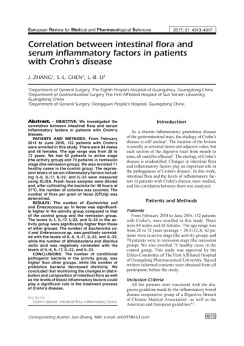
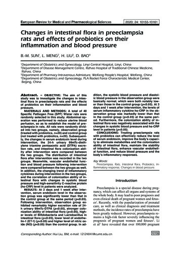
4water, washed 4 times in 0.1% acetic acid, and redehydratedat room temperature. The stained cells were lysed in 10 mMTris buffer, and the optical density (OD) was measured at510 nm [18].2.16. Western Blotting. IEC-18 cells were seeded on 12-wellmicroplates at 1 105 cells per well and cultured for 24 h.Cells were treated with the supernatant of HBSS or poly Ptreated PRP diluted twofold with medium. After incubationfor 30 min, the cells were lysed in NP-40 cell lysis buffer(Thermo Fisher) containing phosphatase inhibitor (ThermoFisher) and a complete protease inhibitor cocktail (RocheMolecular Biochemicals, Indianapolis, IN, USA). Protein(20 μg) in each sample was resolved using sodium dodecylsulfate-polyacrylamide gel electrophoresis (SDS-PAGE)(12%) and immediately transferred to a nitrocellulose membrane using transfer buffer (Bio-Rad, Hercules, CA, USA).The nitrocellulose membranes were incubated in SuperBlockBlocking Buffer (TBS) (Thermo Fisher) for 1 h at room temperature to block any nonspecific binding. The blots wereincubated overnight at 4 C with anti-phospho-AKT antibody(Cell Signaling), anti-phospho-ERK antibody (CellSignaling), antiphospho-p38 antibody (Cell Signaling), antiphospho-JNK antibody (Cell Signaling), anti-phospho-Rafantibody (Cell Signaling), and anti-phospho-MEK antibody(Cell Signaling) as the primary antibodies.The next day, the blots were quickly rinsed in distilledwater 5 times and then washed once for 5 min with T-TBSat room temperature, incubated for 60 min in speciesappropriate horseradish peroxidase-conjugated secondaryantibodies (R&D Systems, Minneapolis, MN,USA) in TTBS, quickly rinsed in distilled water 5 times, and thenwashed once for 5 min with T-TBS [19]. The blots weredeveloped using the SuperSignal West Pico EnhancedChemiluminescence System (Thermo Fisher) according tothe manufacturer’s protocols. The average protein expressionwas normalized to the actin expression (BD TransductionLaboratories, Lexington, KY, USA).2.17. An ELISA. The D-dimer levels in plasma were determined using the Mouse Fibrin Degradation Product DDimer (Competitive EIA) ELISA Kit (LifeSpan BioSciencesInc., Seattle, WA, USA) according to the manufacturer’sinstructions.The contents of growth factors in plasma were determined using the PDGF-BB, EGF, VEGF, and TGF-β Quantikine ELISA kits (Research and Diagnostic Systems Inc.,Minneapolis, MN, USA) according to the manufacturer’sinstructions.2.18. Inhibitors. The ERK1/2 inhibitor FR180204 (SigmaAldrich Co., LLC, St. Louis, MO, USA) was used at a terminalconcentration of 1 μM. The p38 inhibitor SB203580 (SigmaAldrich Co., LLC) was used at a terminal concentration of1 μM. The PI3K inhibitor LY294002 (Sigma-Aldrich Co.,LLC) was used at a terminal concentration of 20 μM. TheJNK inhibitor SP600125 (Sigma-Aldrich Co., LLC) was usedat a terminal concentration of 20 μM.Mediators of Inflammation2.19. Statistical Analyses. The assay data were analyzed usingStudent’s unpaired t-test for two-group comparisons, a oneway analysis of variance (ANOVA) followed by Fisher’s protected least post hoc test for three-group comparisons, and anANOVA followed by the Tukey-Kramer method post hoctest for four-group comparisons. The growth factor ELISAdata were analyzed using a paired t-test. The results of anSRB assay were analyzed using a two-factor repeated measureANOVA. Wound healing assays using MAPK inhibitorswere analyzed using a two-factor factorial ANOVA.P 0:05 was considered to indicate a statistically significantdifference.3. Results3.1. Poly P Improved the Intestinal Injuries of Mice Treatedwith DSS and Enhanced the Accumulation of Platelets inIntestinal Epithelia. BALB/c mice were allowed free accessto drinking water containing 2.5% DSS for 5 days. PBS or5 μg of poly P was orally administered to mice once per dayfrom day 0 to day 7, after which the mice were euthanized(Figure 1(a)). The colon length of PBS-treated mice was significantly shortened by 2.5% DSS treatment compared to thecontrol mice, while the length in 5 μg poly P-treated mice significantly recovered (Figure 1(b)). Hematoxylin-eosin (H&E)staining of the colonic sections showed that the exacerbationof the histopathological inflammation score induced by DSStreatment was significantly improved following treatmentwith poly P (Figure 1(c)). RT-PCR showed that the augmentation of the proinflammatory cytokines IL-1β, TNF, and IL6 was significantly lower in the DSS poly P group than inthe DSS PBS group (Figures 1(d)–1(g)). Interestingly, theaccumulation of platelets was detected at the surface of themucosa with the exfoliation of epithelia on H&E stainingand immunohistochemical staining of CD61, which is aknown surface antigen of platelets. The CD61-positive areaof the DSS poly P group was significantly larger than thatof the DSS PBS group (Figure 1(h)), suggesting that polyP induced the accumulation of platelets in the surface ofthe damaged epithelium due to intestinal inflammation.To assesses systemic thrombotic tendency, the plasma Ddimer levels in the DSS-induced model mice were measured.Although DSS treatment increased the D-dimer levels, therewas no significant difference between the DSS PBS groupand DSS poly P group (Figure 1(i)).3.2. Poly P Induced Platelet Aggregation. To assess whether ornot poly P directly induced the aggregation of platelets, polyP was added to the PRP obtained from the collected bloodsamples, and a platelet aggregation assay was performed.The absorption at 405 nm in suspended platelets was significantly decreased by the treatment with poly P (ratio of OD405nm at 1000 sec: 0:636 0:0072) as well as ADP (ratio ofOD405nm at 1000 sec: 0:817 0:0246), which stimulates theaggregation of platelets, suggesting that poly P directlyinduced platelet aggregation.It was previously reported that short-chain polyphosphate, whose chain length is under 60 mer, endogenouslyexists in platelets and is associated with platelet aggregation
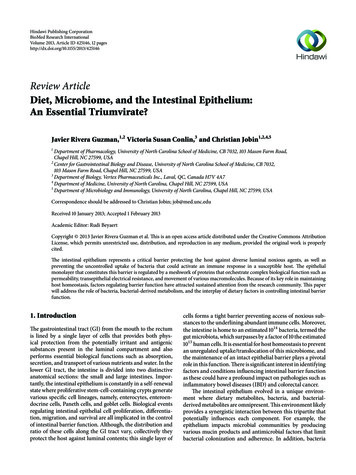
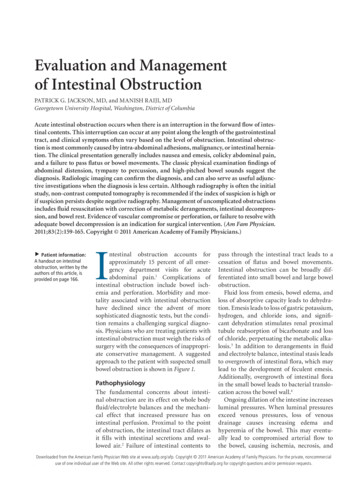
Mediators of Inflammation5Colon lengthColon length (mm)130Water2.5% DSSd1d8d5Poly P5 topathological score4IL1𝛽/18S (fold of normal) ⁎1200100080060040020000NCDSS-PBSDSS-poly PNC(c)DSS-PBSDSS-poly P(d)TNFIL-6⁎⁎900IL-6/18S (fold of 001000NCDSS-PBSDSS-poly PNC(e)DSS-PBS(f)IFN-𝛾450IFN-𝛾/18S (fold of normal)Grade 21⁎14003TNF/18S (fold of normal)DSS-poly Pn.s400350300250200150100500NCDSS-PBS(g)Figure 1: Continued.DSS-poly PDSS-poly P
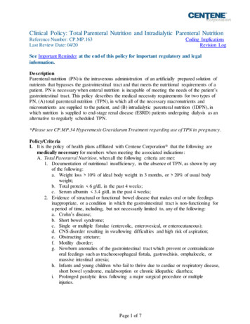
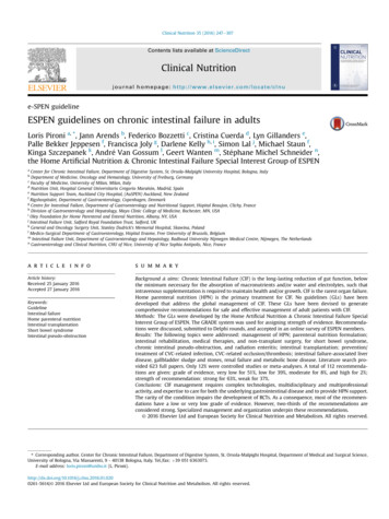
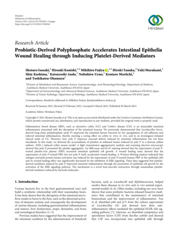
6Mediators of InflammationDSS-PBSNCDSS-poly P 400CD61 positive area(fold of normal) 100CD61-positive area⁎1086420NCDSS-PBS DSS-poly P(h)D-dimer1.6n.s.D-dimer (𝜇g/mL)1.41.210.80.60.40.20NCDSS-PBSDSS-poly P(i)Figure 1: Poly P improved intestinal inflammation and enhanced the accumulation of platelets at the intestinal epithelia in a DSS-inducedcolitis model. A schematic illustration of the curing schedule of the DSS-induced colitis study (a). The length of inflamed colon (b) andhistopathological changes (c) were significantly improved by poly P in DSS-induced colitis mice. RT-PCR showed that the expression ofmRNA of proinflammatory cytokines IL1β (d), TNF (e), and IL6 (f) was significantly decreased by poly P, while that of IFN-γ (g) wasnot. Immunohistochemical staining of CD61 showed the accumulation of platelets at the surface of the intestinal epithelia after treatmentwith poly P (h). An ELISA of plasma D-dimer levels showed no significant effect on the thrombotic condition by treatment with poly P (i).The error bars and numbers show the standard deviation. P 0:05 by a one-way ANOVA followed by Fisher’s protected least post hoc test.[20–22]. Short-chain polyphosphate also induced the aggregation of platelets (ratio of OD405nm at 1000 sec: 0:9445 0:021), but the degree of the aggregation in the short-chainpolyphosphate treatment group was lower than that in thepoly P treatment group (Figure 2(a)).To confirm the aggregation of platelets, an electronmicroscopic analysis was performed. The platelets aggregated following treatment with poly P as well as ADP andinsoluble particles, which appeared to correspond to poly Pnanoparticles, attached to the platelet surface (Figure 2(b)).These data suggest that poly P directly interacted with andactivated the naïve platelets, resulting in the aggregation ofplatelets.3.3. Poly P Improved the Wound Healing of IntestinalEpithelia Cells Mediating Platelet Activation. To assesswhether or not poly P exerts wound healing effects throughthe activation of platelets, the supernatant of poly Ptreated human PRP was collected and a cell scratch assaywas performed. The supernatant of poly P-treated humanPRP significantly improved the scratched wound compared to the supernatant of HBSS-treated human PRP inhuman normal epithelial HCEC-1CT cells (Figure 3(a)).Likewise, the wound healing effect was confirmed whenrat normal epithelial cells (IEC-18 cells) were treated withsupernatant of poly P-treated rat PRP (Figure 3(b)). Incontrast, poly P did not directly improve the epithelialwound of HCEC-1CT cells (Supplementary Figure 1A)nor did the supernatant of 40 μM ADP-treated PRP hasany such effect (Supplementary Figure 1B).To assess whether or not a poly P-treated platelet-derivedmolecule promotes cell growth, an SRB assay was performed.The cell growth of HCEC-1CT cells was significantlyincreased following treatment with the supernatant of polyP-treated human PRP compared to treatment with thesupernatant of HBSS-treated human PRP (Figure 4(a)).Immunocytochemistry showed that ki-67-positive cells weresignificantly more numerous following treatment with polyP-treated PRP (Figure 4(b)). These data suggest that poly Pinduced the secretion of growth-promoting molecules fromplatelets and thus promoted intestinal wound healing.3.4. Poly P Induced the Release of Small Mediators fromPlatelets and Accelerated Cell Growth Mediating ERKSignaling. Previous investigations have shown that MAPKassociated molecules are closely associated with wound
Mediators of Inflammation71.1OD 405 (fold of 0 sec)10.90.80.70.60 sec50 sec100 sec150 sec200 sec250 sec300 sec350 sec400 sec450 sec500 sec550 sec600 sec650 sec700 sec750 sec800 sec850 sec900 sec950 sec1000 sec1050 sec1100 sec1150 sec1200 sec1250 sec1300 sec1350 sec1400 sec1450 sec0.5HBSSPoly PADP1.1OD 405 (fold of 0 sec)10.90.80.70.60 sec50 sec100 sec150 sec200 sec250 sec300 sec350 sec400 sec450 sec500 sec550 sec600 sec650 sec700 sec750 sec800 sec850 sec900 sec950 sec1000 sec1050 sec1100 sec1150 sec1200 sec1250 sec1300 sec1350 sec1400 sec1450 sec0.5HBSSPoly P (short)ADP(a)HBSSPoly PADP(b)Figure 2: Poly P induced platelet aggregation. A platelet aggregation assay showed that long-chain poly P as well as short-chain poly Pinduced platelet aggregation and the effect of long-chain poly P on platelet aggregation was stronger than that of ADP (a). An SEManalysis indicated the morphological changes in naïve platelets by treatment with poly P or ADP (b). The arrow shows the insolubleparticles, suspected of being poly P nanoparticles. The error bars and numbers show the standard deviation.healing in numerous cell lines, including epithelial cells,keratinocyte, and cancer cells [23–28]. To clarify thegrowth-promoting mechanism of epithelial cells, the statusof MAPK signaling was assessed.Western blotting indicated that MAPK signaling-relatedmolecules (ERK, p38, JNK, and AKT), especially those concerning the ERK signaling pathway, were significantly activated by the treatment of the supernatant of poly P-treated
8Mediators of InflammationSupernatant of HBSStreated PRP24 hRecovery rate (%) at 24 hHCEC-1CTSupernatant of poly Ptreated PRPHCEC-1CT0h1009080706050403020100⁎HBSS treatedPoly P treatedRecovery rate (%) at 24 hSupernatant of poly Ptreated PRPIEC-18Supernatant of HBSStreated PRP(a)1009080706050403020100IEC-18⁎HBSS treatedPoly P treated(b)Figure 3: The supernatant of poly P-treated PRP increased the wound healing effect in epithelial cells. A cell scratch assay showed that thesupernatant of poly P-treated PRP accelerated the closure of wound in HCEC-1CT (a) and IEC-18 (b) cells. The error bars and numbers showthe standard deviation. P 0:05 by Student’s t-test.rat PRP in IEC-18 cells (Figure 5(a)). The epithelial healingeffect of the supernatant of poly P-treated rat PRP was attenuated by treatment with an ERK inhibitor (FR180204) butnot inhibitors of p38 (SB 203580), PI3K/Akt (LY294002),or JNK (SP600125) (Figures 5(b)–5(e)), suggesting thatERK signaling is responsible for promoting wound healing.Likewise, Western blotting also showed that the supernatantof poly P-treated rat PRP activated the upstream signaling ofERK, Raf-MEK signaling (Figure 5(f)), suggesting that polyP-induced molecules from platelets activated the epithelialRaf-MEK-ERK signaling pathway, resulting in the promotion of wound healing.Activated platelets are well known to release growthrelated molecules to support wound healing [15, 29]. Toidentify the molecules released from platelets by poly P treatment, the growth factors in the supernatant of poly P-treatedhuman PRP were assessed. An ELISA showed that theexpression of PDGF, EGF, VEGF, and TGF-β, which areknown to be components enclosing platelet granules [30–33], was not s
focused on the wound healing effect of poly P through the crossinteraction between poly P and host platelet-derived small-molecule mediators in in vitro wound healing models and a mouse enteritis model. 2. Materials and Methods 2.1. Poly P. A mixture of 200mM phosphoenolpyruvic acid (2M Tris-HCl, pH9.4), 30mM phosphate buffer (pH6.0),