Transcription
Introduction toElectroencephalography Eeg.jpg
Outline Introduction & History Neurophysiological Basis of EEG Recording Standards Applications Example ERP /2005027850 002/c imm 0003.jpg
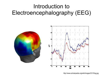
Introduction EEG: electrical activity recorded via electrodeson the scalpHigh temporal resolution (millisecond scale) andlow spatial resolutionRelatively cheap neuroimaging edia/2007/01/1-16-07-eeg.jpg
Brief History Vladimirovich (1912) Cybulski (1914) first animal EEG study (dog)first EEG recordings of induced seizuresBerger (1924) first human EEG recordings 'invented' the term electroencephalogram (EEG) American EEG Society formed in 1947 Aserinsky & Kleitman (1953) first EEG recordings of REM sleep(Swartz & Goldensohn, 1998)
Hans Berger (1924)
Neurophysiological Basis of EEG
Neurophysiological Basis of EEG Single neuron activity is too small to be pickedup by EEGEEG reflects the summation of the synchronousactivity of many neurons with similar spatialorientationsCortical pyramidal neurons produce most of theEEG signalDeep sources (subcortical areas) are muchmore difficult to detect than currents near theskull
Scalp EEG Recordingshttp://psyphz.psych.wisc.edu/ greischar/BIW12-11-02/neurons.jpg
Recording Standards
EEG le 10/Electroencephalogram figHead.jpg
Electrode Placementhttp://www.bbci.de/competition/ii/albany desc/image004.jpg
Electrode Placement(Blinowska & Durka, 2006)
Recording EEG Signalshttp://universe-review.ca/I10-63-EEG.jpg
2.htm
Perfect Reference?
Effect of theReferenceElectrode(Murray et al., 2008)
Applications
EEG Rhythms(Pinel, 2011)
Characteristic EEG rhythms, from the top: delta (0.5–4 Hz),theta (4–8 Hz), alpha (8–13 Hz), beta (13– 30 Hz).(Blinowska & Durka, 2006)
Brain-Computer dpress.com/2010/03/bci-eeg-surgery.jpg
Event Related Potentials .svg.png
ERPsAverage auditory ERP and visual ERP inlogarithmic time scale, showing thecommonly recognized components.Auditory components marked by romannumbers are the brainstem-evokedresponses (BAEP). They are followed bymid-latency exogenous components(MAEP).The first peak in exogenous visual ERPcomes from ERG (electroretinogram).Exogenous ERPs exhibit modality–specific featuresEndogenous ERP are similar in bothmodalities.(Blinowska & Durka, 2006)
Event-related potentials (ERP)(Pinel, 2011)
Comparing ERP ComponentsAcross 3/31/Eeg.jpg
Example ERP Study
Inhibition of Return (IOR)(Klein, 2000)
IOR andERPs(Klein, 2004)
Spatiotopic vs.Retinotopic IOR
ERPs (Blinowska & Durka, 2006) Average auditory ERP and visual ERP in logarithmic time scale, showing the commonly recognized components. Auditory components marked by roman