Transcription
ACLS 1310-654-8687ACLS Study GuidePlease STUDY FOR THE CLASS. This guide will assist your preparation for the class. The ACLS StudentManual is by far the best resource and can be accessed in the classroom, online, and from hospitals.2020 Edition1) CLASS INTRODUCTIONA) Instructor and student introduction-be prepared to actively participateB) Class philosophy-Don’t rush when providing care, but be aggressive (ex: Straight to the point, assertive, loud enough for people to hear, confident, specific)Note: when you rush you make mistakes and/or create safety issues. When you are not aggressive, communication is poor.C) Class format1. Lecture/Videos2. Practice3. Test; skills test, 50 question written exam2) BLS SURVEYA) C.A.B (Compressions, Airway, Breathing)B) Sequence: 1. Check the scene to make sure it is safe. Don’t get hurt trying to help.2. Check the patient for responsiveness and breathing3. Activate the Emergency Response System/ Get a Defibrillator4. Check for a Carotid pulse (no more than 10 secs, no less than 5 secs)5. Begin CPR, start with CompressionsC) C ompressions: 1. At a rate between 100-120 per minute and 2 inches deep2. 30;2 ratio of compressions to breaths3. Compress the center of the chest (lower half of the sternum)
ACLS 24. Switch compressors every 2 mins or when HR on monitor drops under100bpm during compressions5. Minimize ALL Interruptions to 10 secs or less during CPRD) A irway: 1. Open the airway with the Head/Tilt Chin Lift maneuver2. Use the Jaw Thrust Maneuver for Trauma PT.Note: do not apply Cricoid pressure any more to all patients.E) B reathing: 1. Give enough air for the chest to rise.2. Avoid excessive Ventilation to prevent vomiting, too much pressure around theheart, and 0xygen toxicity.F) Types of Breathing : 1. Normal CPR Breathing (2 breaths w/ 2 secs. in- between each breath)2. Rescue Breathing (1 breath every 6 secs)3. Breathing w/ an Advanced Airway (1 breathe every 6 secs)Cardiac ArrestIt may be reasonable for EMS providers to use a rate of 10 breaths/min (1 breath every 6seconds) to provide asynchronous ventilation during continuous chest compressions beforeplacement of an advanced airway.G) Defibrillation: 1. Assess and use the Defibrillator immediately after is arrives if necessary.2. Follow each shock immediately with CPR. Don’t reassess immediately.3. Defibrillation used to shock V-Fib and Pulseless V-Tach only in cardiac arrestShock Dosage: 200 J for first shock300 J for second shock360 joules for third shock and any shocks after4. Defibrillator pads can be used universally between defibrillators which resultsin faster defibrillation. It also all is faster to use than paddles because the padscan be left on the patient’s chest.NOTE: BLS takes priority over ACLS
ACLS 33) ACLS SURVEYA) Complete ACLS Survey after BLS SurveyB) Sequence: ABCD; A irway, B reathing, Ci rculation, D ifferential DiagnosisAIRWAY1. correct breathing by: Head/Tilt Chin Lift NPA (nasal pharyngeal airway) or OPA (oralpharyngeal airway) Advanced Airway PlacementQuestion: Is proper Placement of airway confirmed? Is the tube secured?2. Types of Advanced Airways : Laryngeal mask airway, Esophageal-tracheal tube, Endotrachealtube3. No more cricoids pressure. Not beneficial in all cardiac arrest.4. Measure NPA: From the tip of the PTs ear to the tip of the PTs noseMeasure OPA: From the tip of the PTs ear to the tip of the PTs mouth.CLASSIFICATION OF THE PATIENT: STABLE VS UNSTABLEA) STABLE-- TREAT REVERSABLE CAUSES-- MEDICATION THERAPY-CARDIOVERSION-- SPECIALISTPACING or S.B) UNSTABLE- PACING or S. CARDIOVERSION SPECIALIST1. Examples of signs and symptoms that can describe a patient that is UNSTABLE:SHORTNESS OF BREATH (different from respiratory distress)ALTERED MENTAL STATUSSBP 90,
ACLS 402 SATURATION 94 %.-If the patient has one of these signs or symptoms, they are unstable (in shock) and maydeteriorate very quickly.2. A patient who is unstable can deteriorate within minutes or seconds. They are critical andrequire EMS or Rapid Response/code Blue.3. The first question you should ask yourself (knowing the scene is safe) when you come incontact with the patient is: Are they stable or unstable?Note: the patients we are dealing with in this course are patients having cardiac and/orStroke signs and symptoms. Just because you may be having a cardiac emergency, it does notmean the patient is going to deteriorate quickly. In fact, some patients having heart attacksfor example die after 10 hours of onset, where others die in seconds.BREATHING1. For Cardiac Arrest PTs give 100% Oxygen.2. Monitor ventilations by using Quantitative Waveform Capnography &/or Oxygen Saturation.-O2 Saturation should be: equal to or greater than 90%-Capnography: PETCO normal range should be 35mm Hg – 40mm Hg or PACO range of35-40mm Hg. (Partial End Tidal CO2 measures Exhaled CO2 levels)CIRCULATION1. Monitor CPR quality (If PETCO is 10mm Hg , attempt to improve CPR quality)Questions: 1. What is the Cardiac Rhythm?2. Has IV/IO access been established?3. Are medications or fluids needed?4. Is Defibrillation or Cardioversion needed?DIFFERENTIAL DIAGNOSISQuestions: 1. Why did this PT develop symptoms of arrest?2. Is there a reversible cause that can be treated?
ACLS 53) TEAM DYNAMICSA) Understand your role AND the roles of other Team MembersB) Team Leader’s Role: 1. Organizes the group and assigns task2. Makes sure everything is done at the right time and the right way3. Trains and CoachesC) Closed-Loop Communications: The team leader gives an order, the team member repeatsthe order to confirm that they got it. When the task is done, the team member states itverbally.DO: 1. repeat medication orders2. Seek advice if necessary3. Question an order if the slightest doubt exists4. Talk professionally and avoid raising your voiceDON’T: 1. Forget to notify the team when a drug has been given2. Take on a task when you haven’t completed a task in progress3. Give unclear messages4. Take on assignments beyond your scopeD) Structured Team Debriefing1. Corrects Thought Process unlike Simple Feedback2. Summarize, analyze, create records, and set goals4) POST CARDIAC CARE (pg. 145-151)A) Therapeutic Hypothermia : 1. For PTs who are comatose after ROSC.2. Cool PT to 32*C to 36*C for at least 24hrs.3. Use Ice, surface cooling devices, and/or Ice cold isotonic fluidsB) PCI (Cath. Lab) and Hypothermia can be done concurrently.
ACLS 6C) EPI IV infusion for post care: 0.1-0.5mcg/kg per minute to assist in managing hypotension.D) Avoid O2 toxicity and hypotension. Get the PT to a ICU post-care specialty team. E) The main cause for patience to re-arrest relates to not optimizingventilations/oxygenation and not managing hypotension. The goal for Oxygensaturation is 92% to 98% 5) ACS: Acute Coronary Syndromes (pg. 93 & 94)A) Signs and symptoms: shortness of breath, chest pain, nausea, dizziness, sweating.B) Drugs used: Oxygen, aspirin ( 160 to 325mg ), nitro (none if SBP is 90, right ventricularinfarction , hypotension, bradycardia, tachycardia), 2-4mg. slow iv bolus of morphine(administer fluids if hypotension occurs. Morphine reduces the amount of oxygen that theheart needs for perfusion), fibrinolytic therapy ( clot busters . Can reduce mortality by 47% ifused in 1 st hr), and heparin. A newer drug that is being used: Brilinta 60-90mg.Don’t use morphine, vasodilators, nitro, or volume depleting drugs if inferior wall MI & RVInfarction.C) Focus for effective treatment: Time management and identification W/ 12 Lead ECGD) Out-of-Hospital Cardiac Arrest: Half of all patients who die of ACS do so before reaching thehospital. VF or Pulseless VT is the precipitating rhythm in most of these deaths. VF is most likelyto develop during the first 4 hrs after onset of symptoms.E) Reperfusion Therapy: opens an occluded coronary artery either w/ drugs or mechanicalmeans. (Fibrinolytics or PCI aka CATH LAB)F) EMS Treatment : Assessment, 0xygen, aspirin, nitro, morphine, obtain 12 Lead ECG, completea Fibrinolytic checklist, Inform receiving facility if ST elevation, Prep for CPR.G) Emergency Department Arrival: 1. Get 12 lead ECG w/ in 10 mins2. Use fibrinolytics w/ in 30 mins3. PCI w/ in 90 minsNOTE : HAVE STUDENTS REFER TO ACS ALGORITHM on pg. 77
ACLS 76) ACUTE STROKE CAREA) African American: highest risk, 795,000 strokes occur in US a yr, 1 of leading causes of death,B) Types: Ischemic - (87%) occlusion of artery, Hemorrhagic - (13%) vessel in brain ruptures.C) Rapid identification of Stroke: time of occurrence, assessment (Cinn. Test, glucose, history,12- lead ECG)D) Rapid transport to a facility that can provide stroke careE) Ischemic treatment: IV Fibrinolytic therapy (w/in 3-4 ½ hrs of recognition),Hemorrhage -SurgeryF) 8 D’s of stroke care: D etection, D ispatch (EMS), D elivery, D oor (triage), D ata (ED evaluation),D ecision (Expertise), D rug, and D isposition (rapid admission to stroke unit or critical care team).G) rTPA: can cause brain hemorrhage and other side effects, (contraindications: 80yrs or older,glucose 185, history of both stroke and diabetes, less than 18 yrs old, )7) Adult BradycardiaA) Definition: Heart rate less than 50. (signs: altered mental status, hypotension, signs of shock,chest discomfort, S.O.B.)B) Drugs : Atropine (0.5 mg every 3-5 mins with a max dose of 3mg) , Dopamine IV infusion(2-20 mcg/kg per min.) or Epinephrine (2-10 mcg per min). Adjust to PT’s response.C) Rhythms for Bradycardia: 1. Sinus Bradycardia2. First Degree AV Block3. Second Degree AV Block : TYPE 1TYPE 24. Third-Degree AV BlockD) When to look for AV BLOCKS : 1.Bradycardia(bad)(worse)(Complete Heart block)
ACLS 82. “P” waves QRS3. “P-R prolonged (0.20 secs)Question to ask: Is it regular or irregular?REAGULAR: 1ST degree (PR Interval is consistent and prolonged)3 rd degree (PR Intervals are really inconsistent & “P” waves are always indifferent positions from QRS)NOTE: 3 RD DEGREE BLOCK: Pace PT immediately or Asystoles will occur quickly)IRREGULAR: 2 ND degree : 1. TYPE 1 (PR inconsistent and missing some QRS)TYPE 2 ( PR waves consistent but missing QRS)Note that if a patient is unstable, pacing takes priority to medication therapy.Watch these videos before the class:https://www.youtube.com/watch?v U1zq4T7MEWwQUESTION: Was this code ran well? What was good and bad about the code?https://www.youtube.com/watch?v 90q19HTvm28QUESTION: Were good team dynamics present?https://www.youtube.com/watch?v AX7wKfTb2QkQUESTION: Why is capnography important?https://www.youtube.com/watch?v pliSsY23pwwhttps://www.youtube.com/watch?v Ts7gND-hyLYhttps://www.youtube.com/watch?v Iy1lV YzMIkhttps://www.youtube.com/watch?v d7ymmR8WuYE&t 392sQUESTION: When do you cardiovert? When do you pace?Optional:https://www.youtube.com/watch?v w32PUDL2lb8https://www.youtube.com/watch?v JypQxfW94a0
ACLS 9https://www.youtube.com/watch?v IEIaegVWnmABasic MEDICATIONS TO KNOW:Cardiac Arrest:EPINEPHRINE (used with all cardiac arrest patients)AMIODARONE/LIDOCAINE (used if a patient does not respond to defibrillation)Bradycardias:ATROPINE ( First drug used to treat bradycardias)EPINEPHRINE (Used if you can’t pace after atropine & also for hypotensionpost-cardiac arrest)DOPAMINE (also used to treat hypotension post-cardiac arrest)Stroke:TPA (Activase Stroke med used for clots)A.C.S:HEPARIN, MORPHINE, 02, NITRO, ASPIRIN/BRILINTA (chewable)Tachycardias:Adenosine (not used for all tachys.)KNOW THE FOLLOWING RHYTHMS:VENTRICULAR FIBRILLATION (shockable cardiac arrest rhythm)PULSELESS VENTRICULAR TACHYCARDIA (shockable rhythm)ASYSTOLES ( aka flatlined)
ACLS 10NORMAL SINUS RHYTHM ( will include normal HRbetween 60-100 bpm)SVT (very fast Tachycardia rhythm with a H.R. above180)TACHYCARDIAs (fast rhythm)BRADYCARDIAs (slow rhythms under 50 B.P.M.) including A.V. blocksATRIAL FIBRILLATIONATRIAL FLUTTERP.E.A. also known as pulseless electrical activity (normal sinusrhythm with no pulse)This website will allow easily prep you for rhythm interpretation and tor
ACLS 118) SYMPTOMATIC TACHYCARDIAA) The HR is greater than 100 and we get real concerned when it goes above 150.B) Signs and symptoms: signs of shock, hypotension, chest discomfort, acute heart failure, AMS,SOBC) Tachycardia rhythms: Atrial Fib, Atrial Flutter, SVT, VT, Uncertain Wide Complex Tachy.For the Stable Patient, we go with Vagal Maneuvers (works ¼ times) to Medications toCardioversion specialist.For Unstable Patients Cardioversion to SpecialistD) A wide complex tachycardia can be very severe and lead to hypotensive shock and cardiacarrest9) EMERGENCY TRIAGE- VERBAL ASSESSMENTWhat to ask the patient:S - SIGNS/SYMPTOMSA - ALLERGIESM- MEDICATIONSP- PAST PERTINENT HISTORYL- LAST ORAL INTAKEE- EVENTS LEADING UP TO THE EMERGENCYExample:1,” What’s wrong?” or “What brings you to the emergency department?”2. “Do you have any allergies?”3. “What medications should you be taking?” or “What medications are you on?”4. “What health issues do you have?”5. “What was the last thing you had to eat or drink?”
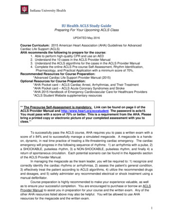
ACLS 126. “What were you doing when you started having chest pain?”ACS: Acute Coronary Syndromes Algorithm
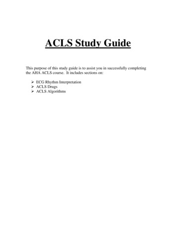
ACLS 13TACHYCARDIA ALGORITHM
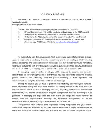
ACLS 14BRADYCARDIA ALGORITHM
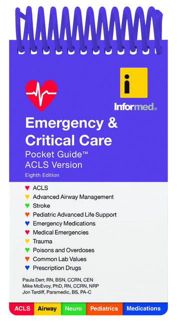
ACLS 15CARDIAC ARREST: SHOCKABLE & NON-SHOCKABLE ALGORITHM
ACLS 16
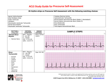
ACLS 17Cardiac Arrest in Pregnancy In-Hospital ACLS AlgorithmA revised algorithm is provided for cardiac arrest in pregnancy.Changes include: Layout is more streamlinedAdded step for administering 100% O 2 and avoiding excessive ventilationRemoved step to assess for hypovolemia/treatmentChanged “If no ROSC in 4 minutes” to “5 minutes”Maternal Cardiac Arrest box that highlights:o Team planningo Priorities of high-quality CPR and relief of aortocaval compressions with lateral uterinedisplacemento Goal of perimortem cesarean deliveryo Deliver in 5 minutes (depending on provider resources and skill sets)Cardiac Arrest in Pregnancy In-Hospital ACLS Algorithm
ACLS 18ROSC: Return of Spontaneous Circulation
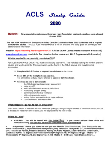
ACLS 19Waveform CapnographyNo Contraindications for Waveform Capnography
ACLS 20Physiologic Monitoring of CPR Quality2020 (Updated): It may be reasonable to use physiologic parameters such as arterial blood pressure orend-tidal CO 2 when feasible to monitor and optimize CPR quality.Why: Similar to the above, although the use of physiologic monitoring such as arterial blood pressureand/or end-tidal CO 2 to monitor CPR quality is an established concept, new data support its inclusion inthe Guidelines. Data from the AHA Get With the Guidelines-Resuscitation registry show higher likelihoodof ROSC when CPR quality is monitored by using either end-tidal CO 2 or diastolic blood pressure. Thismonitoring depends on the presence of an endotracheal tube or arterial line, respectively . Targetingcompressions to an end-tidal CO 2 value of at least 10 mmHg, and ideally 20 mm Hg orgreater, may be useful as a marker of CPR quality. An ideal target has not been identified.
ACLS 21
ACLS 1 310-654-8687 ACLS Study Guide P l e a se S T U D Y F O R T H E C L A S S . T h i s g u i d e w i l l a ssi st yo u r p re p a ra t i o n f o r t h e cl a ss. T h e A C L S S t u d e n t