Transcription
ORIGINAL ARTICLECLINICAL PRACTICE MANAGEMENTManagement of Incidental AdnexalFindings on CT and MRI: A White Paper ofthe ACR Incidental Findings CommitteeMaitray D. Patel, MD a , Susan M. Ascher, MD b, Mindy M. Horrow, MD c, Perry J. Pickhardt, MD d,Liina Poder, MD e, Mindy Goldman, MD f, Lincoln L. Berland, MD g,Pari V. Pandharipande, MD, MPH h, Katherine E. Maturen, MD, MS i, jCredits awarded for this enduring activity are designated “SA-CME” by the American Board of Radiology (ABR) andqualify toward fulfilling requirements for Maintenance of Certification (MOC) Part II: Lifelong Learning and Selfassessment. To access the SA-CME activity visit iew?CDId¼kDHaXjTjOzs%3d. SA-CME credit for this article expires December 27, 2022.AbstractThe ACR Incidental Findings Committee (IFC) presents recommendations for managing adnexal masses incidentally detected on CTand MRI. These recommendations represent an update of those provided in our previous JACR 2013 white paper. The AdnexalSubcommittee, which included six radiologists with subspecialty expertise in abdominal imaging or ultrasound and one gynecologist,developed this algorithm. The recommendations draw from published evidence and expert opinion and were finalized by iterativeconsensus. Algorithm branches successively categorize adnexal masses based on patient characteristics (eg, pre- versus postmenopausal)and imaging features. They terminate with a management recommendation. The algorithm addresses most, but not all, pathologies andclinical scenarios. Our goal is to improve quality of care by providing guidance on how to manage incidentally detected adnexal masses.Key Words: Adnexal cyst, CT, incidental findings, MRI, ovarian cystJ Am Coll Radiol 2020;17:248-254. Copyright 2019 American College of RadiologyOVERVIEW OF THE ACR INCIDENTALFINDINGS PROJECTThe core objectives of the Incidental Findings Project areto (1) develop consensus on patient characteristics andimaging features that are required to characterize anincidental finding; (2) provide guidance to manage suchfindings in ways that balance the risks and benefits topatients; (3) recommend reporting terms that reflect theahDepartment of Radiology, Mayo Clinic Alix School of Medicine, Phoenix,Arizona.bDepartment of Radiology, Georgetown University Medical Center,Washington, DC.cDepartment of Radiology, Albert Einstein Medical Center, Philadelphia,Pennsylvania.dDepartment of Radiology, University of Wisconsin School of Medicine,Madison, Wisconsin.eDepartment of Radiology, University of California San Francisco, SanFrancisco, California.fDepartment of Obstetrics, Gynecology & Reproductive Sciences, University of California San Francisco, San Francisco, California.gDepartment of Radiology, University of Alabama at Birmingham, Birmingham, Alabama.Department of Radiology, Institute for Technology Assessment, Massachusetts General Hospital, Boston, Massachusetts.iDepartment of Radiology, University of Michigan Medical School, AnnArbor, Michigan.jDepartment of Obstetrics and Gynecology, University of Michigan Medical School, Ann Arbor, Michigan.Corresponding author and reprints: Maitray D. Patel, MD, Mayo ClinicArizona, Department of Radiology, 5777 E Mayo Blvd, Phoenix,AZ 85054; e-mail: patel.maitray@mayo.edu.Dr Pandharipande reports grants from Medical Imaging and TechnologyAlliance, outside the submitted work. Dr Pickhardt reports personal feesfrom Bracco, other from SHINE, other from Elucent, outside the submitted work. The other authors state that they have no conflict of interestrelated to the material discussed in this article.ª 2019 American College of Radiology2481546-1440/19/ 36.00nhttps://doi.org/10.1016/j.jacr.2019.10.008
level of confidence regarding a finding; and (4) focusfuture research by proposing a generalizable managementframework across practice settings.THE CONSENSUS PROCESS: MANAGEMENTOF INCIDENTAL ADNEXAL MASSESThis article presents the ACR Incidental Findings Committee (IFC)’s updated recommendations for the management of incidental adnexal masses detected on CT orMRI. The material presented is updated from priorguidance from the ACR, published in 2013 [1]. Theprocess of developing this algorithm included naming asubcommittee chair, who appointed subcommitteemembers that included recognized experts in pelvicimaging, incidental findings, or the management ofpatients at risk for an adnexal mass. The subcommitteethen developed and gained consensus on preliminaryrecommendations. The subcommittee used publishedevidence as their primary source. When evidence wasnot available, they invoked the collective expertise oftheir team. The preliminary algorithm underwent reviewby additional members within the IFC, including theBody Commission chair and the IFC chair. The revisedalgorithm and corresponding white paper draft weresubmitted to additional ACR stakeholders to gain inputand feedback. Consensus was obtained iteratively aftersuccessive reviews and revisions. After completion of thisprocess, the algorithm and white paper were finalized.The IFC’s consensus processes meet policy standards ofthe ACR. However, they do not meet any specific,formal national standards. This algorithm and set ofrecommendations do not represent policy of the ACRPractice Guidelines or the ACR AppropriatenessCriteria. Our consensus may be termed “guidance” and“recommendations” rather than “guidelines,” which hasa more formal definition.ELEMENTS OF THE FLOWCHARTS: COLORCODINGThe algorithm is encapsulated in a flowchart. Within theflowchart, yellow boxes indicate using or acquiring clinical data (eg, lesion size), green boxes describe recommendations for action (eg, follow-up imaging), and redboxes indicate that work-up or follow-up may be terminated (eg, if the finding is presumed to be benign). Tominimize complexity, each algorithm addresses most—but not all—imaging appearances and clinical scenarios.Radiologists should feel comfortable deviating from thealgorithm in circumstances that are not represented in thealgorithm, based on the specific imaging appearance ofthe finding in question and patient characteristics—thealgorithm content must be viewed as recommendationsand should not be considered as “standard of care.”NATURE AND SCOPE OF THE PROBLEMIncidental adnexal findings are commonly identified inwomen on CT and MR studies that include the pelvis [24]. Normal physiologic changes in premenopausal womeninclude monthly development of a dominant follicle andsubsequent corpus luteum, resulting in a potentialincidental CT or MR finding in many premenopausalwomen [5]. Nonneoplastic cysts that may wax and wanein size are also common in postmenopausal women; in alarge series of postmenopausal women, 14% had cysts onan initial ultrasound examination, with 32% resolvingbut 8% developing a new cyst at 1-year follow-up [6]. Ina large CT colonography program, investigators reportedthat among 2,869 consecutive women aged 50 or olderwho underwent screening, none of the 118 women(4.1%) with an incidental adnexal mass on unenhancedCT colonography were found to have ovarian cancer [3].Incidental adnexal masses are detected even when pelviccoverage on an imaging examination is incomplete;Zidan et al found that 4 of 194 women (2%) had ovariancysts in their study of incidental extraspinal findings onlumbar spine MR examinations [4].An extensive body of ultrasound-based imagingliterature in surgical and clinically followed cohorts showsthat the risk of malignancy in simple cysts identifiedsonographically is negligible in both premenopausal andpostmenopausal women [7], a conclusion confirmed byrecent large studies showing no increased risk ofmalignancy in women with sonographically identifiedsimple adnexal cysts irrespective of cyst size [6-9]. Infact, invasive ovarian serous cystadenocarcinoma is nowknown to primarily arise from solid precursors in thefallopian tube, not the ovary [10]; this understandingsupports the conclusion that simple ovarian cysts arenot precursors to ovarian carcinoma.Based on this evidence, the Society of Radiologists inUltrasound (SRU) has revised consensus recommendationsfor the management of sonographically identified incidentaladnexal simple cysts [7]. Even with the understanding thatsimple adnexal cysts have negligible risk of malignancy,two primary concepts justify an approach in which someadnexal simple cysts on ultrasound merit a short period ofcontinued surveillance, even when asymptomatic. First, aswith any imaging observation, there is a small risk ofJournal of the American College of RadiologyClinical Practice Management n Patel et al n Incidental Adnexal Findings on CT and MRI249
mischaracterization, so the conclusion that an adnexal cyst issimple might be incorrect. Second, some simple adnexalcysts are benign ovarian neoplasms (cystadenomas) andmay grow at a rate that would eventually result in clinicalattention even if initially asymptomatic; patients andproviders may benefit from knowing that rate of growthso that appropriate clinical follow-up can be employed.These considerations must be balanced by the potentialharms introduced by imaging surveillance of likely benignfindings, including increased likelihood of unnecessarysurgical intervention as well as increased patient and provider anxiety and health care costs [11]. Size (maximum cystdiameter) plays an important role in determining whichsimple adnexal cysts might benefit most from sonographicsurveillance, targeting those more likely to be benignneoplasms rather than nonneoplastic cysts. The revisedSRU consensus recommendations have increased somesize thresholds for surveillance, now stating that simplecysts characterized with standard ultrasound quality donot require ultrasound follow-up when 5 cm in premenopausal women and 3 cm in postmenopausal women [7].When there is exceptional quality and documentation thatthe cyst is simple, the SRU panel opines that thesethresholds are justifiably increased to 7 cm inpremenopausal women and 5 cm in postmenopausalwomen, because the risk of mischaracterization is reduced[7]. The revised SRU consensus recommendation is animportant backdrop to this revision of the IFC’srecommendations related to the management of incidentaladnexal findings on CT and MR.REPORTING CONSIDERATIONSThe following four elements should be reported when anincidental adnexal mass is detected on CT or MRI:1. Mass characteristics (ie, simple-appearing cyst, featuresindicating a specific diagnosis, indeterminate features)2. Size (largest diameter)3. Technical considerations4. Known or presumed menopausal statusMass CharacteristicsIn broad terms, an adnexal mass identified on CT or MRIcan be placed into one of three categories: (1) a simpleappearing cyst; (2) a mass that is not a simpleappearing cyst but with characteristic features allowingpresumptive diagnosis; (3) a mass with features notallowing for confident diagnosis (uncertain diagnosis).The term “simple cyst” has been used for sonographically characterized cysts that are round or oval, withsmooth thin walls, no solid component or septation, and250no internal flow using color Doppler imaging [12]. In thefemale pelvis, these are called ovarian simple cysts whenshown to clearly arise from the ovary, and they arecalled adnexal simple cysts when the structure of originis uncertain because ovarian parenchyma is notvisualized (these are still usually presumed to havearisen from the ovary) [12]. To distinguish from thissonographically derived nomenclature, the term“simple-appearing cyst” is used to describe a round orovoid fluid-density mass on CT or fluid-signal cyst onMR without a solid component, with smooth, imperceptible, or thin walls; this terminology replaces the“benign-appearing cyst” term used in the prior IFC 2013recommendations regarding incidental adnexal findings[1,13]. On CT, multiple regions of interest should beexamined within different portions of the suspected cystto be assured that the mass is of fluid density;attenuation values between 10 and 20 Hounsfieldunits (HU) are considered fluid density [14]. On MR,fluid signal intensity is determined by comparison withother fluid containing structures such as the bladderand, broadly speaking, includes hyperintense signal onT2-weighted images, hypointense signal on T1weighted images, and absent postcontrast enhancement.As outlined in the prior IFC white paper on incidental adnexal findings [1], some adnexal masses on CT,and to a larger extent MRI, can show imaging featuresthat are characteristic for a particular diagnostic entity[1,15]. Benign examples include some hemorrhagiccorpus luteal cysts, para-ovarian or paratubal cysts,hydrosalpinges, peritoneal inclusion cysts, ovarian fibromas, subserosal or pedunculated uterine leiomyomas,endometriomas, and dermoids [1,16]. Some CT and MRfindings are also characteristic of ovarian or adnexalmalignancy [17]. It is important that radiologists whoreport CT or MR studies of the pelvis be familiar withthe features of adnexal masses that enable confidentbenign or malignant diagnosis, so that those featurescan be described in the reporting of these masses. Someof these features were described in Table 1 in the priorIFC Adnexal 2013 article [1] and have been the focusof other publications [18,19].When an incidental adnexal mass on CT or MRcannot be characterized as a simple-appearing cyst anddoes not have characteristic imaging features for specificadnexal pathology, the diagnosis is uncertain.SizeFor incidental adnexal masses on CT or MR, only thelargest single diameter needs to be reported when theJournal of the American College of RadiologyVolume 17 n Number 2 n February 2020
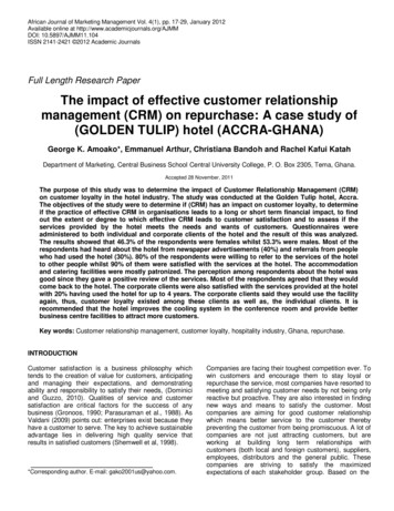
Table 1. Management recommendations for adnexal masses with characteristic features on CT or MRAdnexal MassHemorrhagic cyst 5 cm 5 cmPara-ovarian cystPeritoneal inclusion cystSimple hydrosalpinxOvarian fibromaUterine leiomyomaEndometriomaDermoidSuspected malignancyPremenopausal (or Age 50 If Unknown)Postmenopausal (or Age 50 If Unknown)US or MRI to characterize*No further imagingUS follow-up in 2-3 monthsUsually further imaging is unnecessary; clinical managementUsually managed by gynecologist, may require periodic imagingUS or MRI to characterize*US ¼ ultrasound.*The study should be performed promptly for further evaluation, rather than in follow-up to assess temporal changes.mass is round or ovoid in shape or the conclusion is thatthe mass does not need further imaging or follow-up.Reporting three orthogonal dimensions is helpful whenthe mass has a more irregular shape, especially whenimaging follow-up will be recommended.Technical ConsiderationsRelevant technical limitations in the assessment of theincidental mass should be reported. These may includelow signal-to-noise ratio, artifacts (eg, from hip prostheses), lack of intravenous contrast, and incompleteanatomic coverage; any mass imaged with these constraints should be described as having “limited assessment.” At the other end of the spectrum, MR hasexcellent diagnostic accuracy for adnexal mass characterization when it includes the following three elements: (1)T2-weighted images; (2) pre- and postcontrast T1weighted images; and (3) complete anatomic coverageof the mass in at least two imaging planes. In this scenario, reporting of the incidental adnexal mass shouldindicate that the mass has been “fully characterized” withMR, because this influences subsequent imagingrecommendations.Known or Presumed Menopausal StatusIf available, the menopausal status should be included aspart of the report when it impacts recommended management of incidental adnexal findings. When not available, as previously recommended in the IFC Adnexal2013 approach, 50 years of age can be used as a surrogatelandmark, with those findings in women less than 50years of age managed as if premenopausal, and thosefindings in women 50 years of age or greater managed asif postmenopausal [1]. Because the average age ofmenopause in the United States is 51 years, this willtend to apply more conservative size thresholds forfollow-up of simple-appearing cysts in patients who arejust at or slightly over 50 years of age with unknownmenopausal status.INCLUSION AND EXCLUSION CRITERIA FORUSE OF THE ALGORITHMThis algorithm should be applied to incidentally detectedadnexal masses 1 cm in maximum diameter in womenafter menarche; in keeping with the prior IFC 2013 algorithm, adnexal findings 1 cm are not includedbecause subcentimeter cysts (often follicles) are ubiquitous in premenopausal women, extremely common inpostmenopausal women, and often too small to characterize on CT or MR [1,5]. Because the average age ofmenarche in the United States is between 12 and 13years, age 16 may be used as a conservative estimate forassuming menarche. The algorithm does not apply toany CT or MR adnexal finding that is unchanged inappearance over 2 or more years, because malignancy iseffectively excluded by this stability [20]. The algorithmis not intended for use in women at high genetic riskfor ovarian cancer, in whom lower size thresholds forsonographic characterization of adnexal cysts may bejustified. The algorithm is aborted when a patientdevelops symptoms potentially related to a mass beingfollowed; an asymptomatic cyst may become painfulbecause of internal hemorrhage, rupture, or torsion,with symptoms justifying immediate imaging attention[21]. Adnexal masses 10 cm or larger in diameter areunlikely to be asymptomatic and are very unlikely to berelated to normal physiology [22]. For such masses, fullcharacterization with MR may be better thanultrasound, because ultrasound may have limitationsbecause of the size of the mass.Journal of the American College of RadiologyClinical Practice Management n Patel et al n Incidental Adnexal Findings on CT and MRI251
IMPLICATIONS OF IMAGING AND CLINICALFEATURESBasic Principles of the Algorithm1. Simple-appearing cysts on CT or MR have very lowrisk of malignancy. Sonographically identified simpleadnexal cysts have very low, if any, risk of malignancy,irrespective of cyst size [6-9]. There is good reason tobelieve that simple-appearing cysts on CT and MRhave similarly very low risk of malignancy [3]. MR haslong been known to have high specificity in thecharacterization of adnexal masses, with diagnosticperformance for benign cystic lesions comparable toultrasound with Doppler [23]. For CT, since priorpublication of the IFC 2013 recommendations,Baheti et al evaluated the agreement betweencontrast-enhanced CT and ultrasound in characterizing adnexal masses and showed that simpleappearing cysts on CT correspond to simple cysts onultrasound [24]. A subsequent investigation alsoshowed that simple-appearing cysts on CT had norisk of malignancy [25]. Thus, although there is farmore evidence confirming the absence of increasedmalignancy risk in sonographically characterizedsimple adnexal cysts, based on the available currentevidence, it is reasonable to assume that simpleappearing cysts on CT or MR are similarly benign.2. Small, incidental, simple-appearing adnexal cysts onCT or MR do not justify sonographic characterizationeven when assessment is limited. Paralleling the revisedSRU consensus recommendations for incidental simplecysts on ultrasound, our committee consensus uses 3cm (postmenopausal) and 5 cm (premenopausal) asthe default threshold for not pursuing follow-up ofincidental simple-appearing cysts on CT or MR [7].Because many adnexal cysts are 5 cm inpremenopausalwomenand 3cminpostmenopausal women, the mere existence of a small(ie, below size threshold) simple-appearing cyst withlimited assessment is not enough to justify sonographicrecharacterization or follow-up when it is an incidentalfinding. On the other hand, sonographic characterization of incidental simple-appearing cysts on CT orMR 3 cm (postmenopausal) or 5 cm (premenopausal) is justified when assessment is limited, becausecysts of this size are less common and the likelihood ofCT or MR mischaracterization may be higher.3. The optimal timing of sonographic follow-up of largersimple-appearing cysts balances the small risk of CTor MR mischaracterization against the desire to gain252information about cyst growth with as few imagingstudies as possible. The distinction between recommending ultrasound to characterize an adnexal massand recommending ultrasound follow-up of anadnexal mass identified on CT or MR is important[5]. The former is intended to better demonstrate cystcontents and the latter to provide surveillance overtime. When an incidental simple-appearing cyst isadequately characterized by CT or MR but justifiessonographic follow-up because of its size, the sonographic evaluation is reasonably delayed by 6 to 12months to provide evidence regarding the cyst growthrate. By delaying the sonographic follow-up by 6 to 12months instead of immediately recharacterizing thecyst with ultrasound, the cyst has a chance to resolveor involute, allowing for diagnosis as a nonneoplasticcyst that requires no further follow-up, or to grow,favoring a benign cystic neoplasm. However, the desireto wait at least 6 to 12 months to more confidentlyassess cyst resolution, involution, or growth nevertakes precedence over new symptoms, which mayreflect cyst hemorrhage, rupture, or torsion; luckily,these developments are uncommon, occurring in lessthan 1% of cases [21].4. Fully characterized simple-appearing cysts on MR havevery low risk of mischaracterization, justifying highersize thresholds for pursuing follow-up. Similar to theapproach taken by the revised SRU consensus recommendations, when an incidental simple-appearingcyst has already been fully characterized (as definedin the “Technical Considerations” subsection), thecommittee reasoned that the risk of mischaracterization is substantially reduced, justifying a higherthreshold for sonographic follow-up: 5 cm forpostmenopausal women and 7 cm for premenopausal women. For such masses, imaging follow-upserves only as a measure of the rate of cyst enlargement to inform subsequent clinical management.5. CT or MR may show characteristic features of anadnexal mass that enable confident diagnosis of a specificentity. Table 1 outlines typical management strategiesemployed when an adnexal mass has CT or MRIfeatures characteristic of specific adnexal pathology.Overview of the AlgorithmFigure 1 addresses the gamut of incidental adnexal masseson CT or MR over 1 cm and without documentedstability for 2 or more years. Because follicles and othercysts 1 cm are pervasive in normal premenopausalJournal of the American College of RadiologyVolume 17 n Number 2 n February 2020
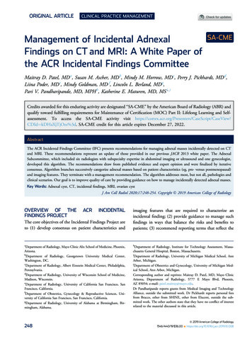
Fig 1. Management of incidental adnexal masses detected on CT or MR. aExclusions: (1) normal findings, including crenulatedenhancing wall of corpus luteum, asymmetric ovary without mass, with normal shape; (2) calcifications without associatednoncalcified mass; (3) previous characterization with ultrasound (US) or MRI; or (4) documented stability in size and appearancefor 2 years. bLimited assessment on CT or MR: As defined in the article, this means the cyst is consistent with a simpleappearing cyst, but characterization is limited by low signal-to-noise ratio, artifact, lack of contrast assessment, or incompleteanatomic coverage. cUS or MRI to characterize means that the study should be performed promptly for further evaluation, ratherthan in follow-up to assess temporal changes. dFully characterized by MR: As defined in the article, this means the cyst has beencharacterized with (1) T2-weighted images; (2) pre- and postcontrast T1-weighted images; and (3) complete anatomic coveragein at least two imaging planes. eAssumes mass has not already been fully characterized by MR. Yellow boxes indicate using oracquiring clinical data (eg, lesion size), green boxes describe recommendations for action (eg, follow-up imaging), and red boxesindicate that work-up or follow-up may be terminated (eg, if the finding is presumed to be benign).ovaries (some with hemorrhage), common inpostmenopausal women, and potentially too small tocharacterize, these incidental findings are not included.Furthermore, adnexal findings that have demonstratedstability in appearance and size over 2 or more years arenot ovarian malignancies; the ultrasound literatureshows that these malignancies demonstrate change in 7months or less [20]. Radiologists should be familiarwith CT and MR features of normal ovarian physiologyand inconsequential findings, including crenulatedenhancement of the wall of a corpus luteum, minorasymmetry of ovarian size when no mass is present,and adnexal calcifications without mass [5,26,27].When an incidental adnexal mass has an uncertaindiagnosis, ultrasound is usually the first imaging test forfurther characterization, but there may be instances inwhich contrast-enhanced MRI is preferred [28]. Forexample, a mass 10 cm in diameter may be toolarge for optimal sonographic characterization, justifyingcontrast-enhanced MR to further evaluate.Journal of the American College of RadiologyClinical Practice Management n Patel et al n Incidental Adnexal Findings on CT and MRI253
TAKE-HOME POINTS---Incidental adnexal findings on CT and MR examinations of the female pelvis are common; weprovide an algorithm to guide management of theincidental adnexal mass based on whether the massis (1) a simple-appearing cyst; (2) has reasonablydiagnostic imaging features; or (3) has an uncertaindiagnosis.Simple-appearing cysts on CT or MR have very lowrisk of malignancy. Imaging follow-up is justifiedonly when the cyst is relatively large for the patient’smenopausal status. The primary goals of imagingfollow-up are to limit the risk of cyst mischaracterization and to understand the rate of cyst growth,which may inform subsequent clinical decisionmaking.Recommendations regarding the optimal timing ofsonographic follow-up for a large simple-appearingcyst balances the small potential risk of CT orMR mischaracterization against the desire to gaininformation about cyst growth using as few imagingstudies as possible.REFERENCES1. Patel MD, Ascher SM, Paspulati RM, et al. Managing incidentalfindings on abdominal and pelvic CT and MRI, part 1: white paper ofthe ACR Incidental Findings Committee II on adnexal findings. J AmColl Radiol 2013;10:675-81.2. Pooler BD, Kim DH, Pickhardt PJ. Indeterminate but likely unimportant extracolonic findings at screening CT colonography (C-RADScategory E3): incidence and outcomes data from a clinical screeningprogram. AJR Am J Roentgenol 2016;207:996-1001.3. Pickhardt PJ, Hanson ME. Incidental adnexal masses detected at lowdose unenhanced CT in asymptomatic women age 50 and older:implications for clinical management and ovarian cancer screening.Radiology 2010;257:144-50.4. Zidan MMA, Hassan IA, Elnour AM, et al. Incidental extraspinalfindings in the lumbar spine during magnetic resonance imaging ofintervertebral discs. Heliyon 2018;4:e00803.5. Patel MD, Dubinsky TJ. Reimaging the female pelvis with ultrasoundafter CT: general principles. Ultrasound Q 2007;23:177-87.6. Greenlee RT, Kessel B, Williams CR, et al. Prevalence, incidence, andnatural history of simple ovarian cysts among women 55 years old ina large cancer screening trial. Am J Obstet Gynecol 2010;202. 373.e371-9.7. Levine D, Patel MD, Suh-Burgmann EJ, et al. Simple adnexal cysts:SRU Consensus Conference update on follow-up and reporting.Radiology 2019;293(2):359-71.8. Sharma A, Gentry-Maharaj A, Burnell M, et al. Assessing themalignant potential of ovarian inclusion cysts in postmenopausalwomen within the UK Collaborative Trial of Ovarian CancerScreening (UKCTOCS): a prospective cohort study. BJOG 2012;119:207-19.2549. Smith-Bindman R, Poder L, Johnson E, Miglioretti DL. Risk ofmalignant ovarian cancer based on ultrasonography findings in a largeunselected population. JAMA Intern Med 2019;179:71-7.10. Erickson BK, Conner MG, Landen CN Jr. The role of the fallopiantube in the origin of ovarian cancer. Am J Obstet Gynecol 2013;209:409-14.11. Grossman DC, Curry SJ, Owens DK, et al. Screening for OvarianCancer: US Preventive Services Task Force Recommendation Statement. JAMA 2018;319:588-94.12. Levine D, Brown DL, Andreotti RF, et al. Management of asymptomatic ovarian and other adnexal cysts imaged at US: Society ofRadiologists in Ultrasound Consensus Conference Statement. Radiology 2010;256:943-54.13. Boos J, Brook OR, Fang J, Brook A, Levine D. Ovarian cancer: prevalence in incidental simple adnexal cysts initially identified in CT examinations of the abdomen and pelvis. Radiology 2018;286:196-204.14. O’Connor SD, Silverman SG, Ip IK, Maehara CK, Khorasani R.Simple cyst-appearing renal masses at unenhanced CT: can they bepresumed to be benign? Radiology 2013;269:793-800.15. Kalish GM, Patel MD, Gunn ML, Dubinsky TJ. Computed tomographic and magnetic resonance features of gynecologic abnormalitiesin women presenting with acute or chronic abdominal pain. Ultrasound Q 2007;23:167-75.16. Griffin N, Grant LA, Sala E. Adnexal masses: characterizationand imaging strategies. Semin Ultrasound CT MR 2010;31:330-46.17. Iyer VR, Lee SI. MRI, CT, and PET/CT for ovarian cancer detectionand adnexal lesion characterization. AJR Am J Roentgenol 2010;194:311-21.18. Thomassin-Naggara I, Aubert E, Rockall A, et al. Adnexal masses:development and preliminary validation of an MR imaging scoringsystem. Radiology 2013;267:432-43.19. Forstner R, Thomassin-Naggara I, Cunha TM, et al. ESUR recommendations for MR imaging of the sonographically indeterminateadnexal mass: an update. Eur Radiol 2017;27:2248-57.20. Suh-Burgmann E, Hung YY, Kinney W. Outcomes from ultrasoundfollow-up of small complex adnexal masses in women over 50. Am JObstet Gynecol 2014;211 623.e621-7.21. Froyman W, Landolfo C, De Cock B, et al. Risk of complications inpatients with conservatively managed ovarian tumours (IOTA5): a2-year interim analysis of a multicentre, prospective, cohort study.Lancet Oncol 2019;20:448-58.22. Schallert EK, A
Corresponding author and reprints: Maitray D. Patel, MD, Mayo Clinic Arizona, Department of Radiology, 5777 E Mayo Blvd, Phoenix, AZ 85054; e-mail: patel.maitray@mayo.edu. Dr Pandharipande reports grants from Medical Imaging and Technology Alliance, outside the submitted work. Dr Pickhardt reports personal fees
![Clinical significance of incidental [18 F]FDG uptake in the .](/img/32/s12876-016-0545-x.jpg)