Transcription
University of EdinburghSchool of Informatics - DTC NeuroinformaticsSpike dynamics in Purkinje cells: invivo recordings and computationalmodellingMSc Thesis in NeuroinformaticsYear 2010Student: Paolo PuggioniSupervisor: Dr. Ian Duguid
ii
AbstractHCN1 channels and Purkinje cell voltage-clamp in vivo. In collaborationwith the laboratory of Dr. M. Nolan, we are currently trying to understand howthe inferior olivary-cerebellar circuit is affected by selective HCN1 deletion and whatbehavioural outcomes this produces. Preliminary data suggests that HCN1 channels have a large influence on the initiation of action potentials in inferior olive (IO)neurons, upstream of the cerebellum. In vitro recordings show that the deletion ofHCN1 subunits have two main effects: (i) increasing the spontaneous firing rate and(ii) preventing the emergence of spikelets during the long afterdepolarising potential.In order to test these predictions in vivo, it was proposed to perform voltage-clamprecordings from Purkinje cells (PCs) of HCN1 receptor deficient mice using urethaneanaesthesia. Because each large excitatory post synaptic current (EPSC) correspondsto a spike originated upstream in the IO, the voltage clamp recordings from PCs allow an indirect measurement of the IO activity. This work is to be considered a proofof concept to determine the feasibility of the study.Purkinje cell dynamics in vivo: analysis of the spike train temporal structure. Purkinje cells (PCs), the only output of the cerebellar cortex, show twodistinct types of spiking activity: complex (CSs) and simple (SSs) spikes. Recentfunctional cerebellar theories, such as the adaptive-filter model, consider the SSs asthe carrier of the information, while the CSs as a teaching signal, responsible of thelearning process. At the same time, the most recent coding theories propose thatthe PC information is stored in the rate and pauses of the spike train. These twoperspectives are somehow incompatible, as it was shown that CSs induce pauses andalter the SS temporal structure and therefore necessarily carry information. Therefore it will be of interest to examine the reciprocal influence that SSs and CSs haveon each other on a dynamical basis. This study investigates the dynamical temporalstructure of SS and CS trains in PC recordings from urethane-anaesthetised mice.The findings suggest the conception of two simple models describing (i) the pausesas a possible intrinsic feature of PC dynamics and (ii) the effect of a CS on the nextSS interval. The huge variability of responses to the same inputs implies the presence of different ’states’ in which the PC can operate. This sort of state-dependentcomputation, in which the state of the network is determined by the previous inputs,could potentially be described in a liquid-state machine framework.iii
iv
DeclarationAll the surgeries and in vivo recordings were performed by the author of this reportunder the patient supervision of Dr. Ian Duguid, except where specified in the text.All the MATLAB codes used for data analysis, modelling and simulations wereconceived and written by the author of this report.v
vi
AcknowledgementsThis work would have not been possible without the outstanding human and technical qualities of my supervisor Dr. Ian Duguid. His enthusiasm and patience havebeen my reference point in the days and in the nights of my short journey throughthe marsh of the wet-lab. Ian, it is a pleasure to work with you. Thank you.A special thank is for Marti Jelitai, who shared with me endless days, nights andweekends of catastrophic mistakes and long-awaited recordings in the Lab. Marti,also in the worse moments you were able to find a nice, appropriate word.Thanks also to Derek Garden, who shared with me his fundamental in vitro data.I would like also to thank all team-mates of the Doctoral Training Centre, studying with you guys has been a continuous source of fun and motivation.Grazie Luigi, for all the 5 minute tea-breaks that naturally turned into all-nightlong dense discussions about our work and hopes. In te non solo ho trovato uninaspettato amico, ma anche l’ideale interlocutore ogni volta che urge il bisogno diun διάλoγoς.D’akujem Lukas, and not only for showing us the path with your pessimisticgospel. I know I can count on you.Thank you Colin, every night I came to the Forum at 5 am, I was sure to find afriend in the office.vii
viii
ContentsIIIPrologue1Original work51 Purkinje cell voltage-clamp in vivo1.1 Introduction . . . . . . . . . . . . . . . . . . . . . . . . . . . . . . . .1.2 Materials and methods . . . . . . . . . . . . . . . . . . . . . . . . . .1.2.1 Surgery . . . . . . . . . . . . . . . . . . . . . . . . . . . . . .1.2.2 In vivo voltage clamp . . . . . . . . . . . . . . . . . . . . . .1.3 Results . . . . . . . . . . . . . . . . . . . . . . . . . . . . . . . . . . .1.3.1 Feasibility of obtaining an adequate space-clamp of Purkinjecells in vivo . . . . . . . . . . . . . . . . . . . . . . . . . . . .1.3.2 The effect of urethane on the inferior olive activity in vitro .1.4 Discussion . . . . . . . . . . . . . . . . . . . . . . . . . . . . . . . . .2 Purkinje cell dynamics in vivo2.1 Introduction . . . . . . . . . . . . . . . .2.2 Materials and methods . . . . . . . . . .2.2.1 Electrophysiology . . . . . . . . .Surgery . . . . . . . . . . . . . .In vivo cell attached recordings .2.2.2 Data analysis . . . . . . . . . . .Spike detection and sorting . . .ISI dynamics . . . . . . . . . . .Entropy and mutual information2.2.3 Modelling and simulations . . . .CS generator . . . . . . . . . . .SS generator . . . . . . . . . . .2.3 Results . . . . . . . . . . . . . . . . . . .2.3.1 Basic statistical proprieties . . .Continuous firing Purkinje cells .Pausing Purkinje cells . . . . . .2.3.2 Dynamics analysis . . . . . . . .ix.7. 8. 9. 9. 9. 10. 10. 12. 12.151618181819191920212222222527272731
xCONTENTS2.42.A2.B2.C2.DContinuous firing Purkinje cells . . . . . . . .Pausing Purkinje cells . . . . . . . . . . . . .2.3.3 Intrinsic pausing dynamics in Purkinje cells .2.3.4 The role of the CS . . . . . . . . . . . . . . .2.3.5 Analysis of Awake data . . . . . . . . . . . .Discussion . . . . . . . . . . . . . . . . . . . . . . . .2.4.1 SS spike train dynamics . . . . . . . . . . . .2.4.2 Interaction between CS and SS . . . . . . . .2.4.3 A possible functional role for the bi-stabilityAppendix A: return maps and probability tables . .Appendix B: SS generator . . . . . . . . . . . . . . .Appendix C: Effect of CS on the SS train . . . . . .Appendix D: Supplemental dynamic analysis . . . .31313538404444464749505152
List of SScontinuous firing Purkinje cellaction potentialclimbing fibercomplex spikedeep cerebellar nucleusexcitatory post-synaptic currentgranule cellinferior oliveinhibitory post-synaptic currentinter-spike intervallong term depressionlong term potentiationmossy fiberparallel fiberpausing Purkinje cellPurkinje cellsingle spikexi
xiiCONTENTS
Part IPrologue1
2
One of the research goals of Nolan Lab is to study the role of ion channels in learnedbehaviours. By combining in vitro electrophysiology and computational modelling,M. Nolan et al. are investigating the role of HCN1 channels in coordinated motorbehaviour. On the other hand, Ian Duguid is one of the pioneers of in vivo awakewhole-cell recordings from the mice cerebellar cortex. Duguid Lab is currently oneof the few places in the world practising this ground-breaking technique.Last year Dr. Ian Duguid and Dr. M. Nolan set-up a collaboration to investigatethe role of HCN1 channels, regulating the olivary cerebellar activity, by voltageclamping single Purkinje cells (PCs) of awake mice. In vivo recordings are the onlyway to prove that the phenomena extensively studied in vitro are relevant also inreal physiological conditions.Originally, under the supervision of Dr. Ian Duguid, my task was to produce asimple proof of concept verifying feasibility of in vivo voltage-clamp recordings ofsingle Purkinje cells (PCs) in urethane-anaesthetised mice. It was chosen to startwith anaesthetised preparations because the technical requirements are much lessdemanding than recording from awake animals. However, the possible drawback isthat the anaesthetic might have side-effects on the micro-circuit under study. Thepreliminary results suggested that the experiment could not be performed accordingto the original plans, as described in Chapter 1.Fortunately experimental data are always an endless source of ideas for any curious investigator. During the first cell-attached PC recordings, that were supposed tobe just a short prelude before the space-clamp phase, I found a cell having a strangespiking dynamics. While normal 1 PCs have a rather constant high firing rate, this1It must be read as the most common type of Purkinje cell.3
4cell showed time-varying interspike intervals and even pauses.Seven years ago I was reading about a bizarre mathematician that studied indetail the dynamics of a dripping faucets (in Chaos: making a new science [11],original work [28]). He plotted the successive inter-drop-intervals on a (x,y) graphand found regular patterns that were not reproducible with a general stochasticprocess.A PC is not so different from a dripping faucet. An intrinsic dynamic process,modulated by external excitations and inhibitions, produces a spike every time thatthe cell membrane potential reaches a certain threshold. The spikes of the cell arelike the drops of the faucet. The information transmitted by the cell is coded in theinter-spike intervals (ISIs). I immediately thought about applying to the PC spikesthe same kind of analysis used to study the dynamics of the dripping faucet. I soonrealised that my idea was not absolutely original and I could rapidly find dozens ofpapers on the topic [9].However, these methods had an enormous utility for the present work. The nonlinear time-series analysis of the experimental data suggests that the pauses observedin the simple spike firing could be generated by an intrinsic mechanism of the celland do not need an external triggering signal. The recordings, methods and findingsof this work are described in Chapter 2.
Part IIOriginal work5
6
Chapter 1HCN1 channels and Purkinjecell voltage-clamp in vivoAbstractIn collaboration with the laboratory of Dr. M. Nolan, we are currently trying tounderstand how the inferior olivary-cerebellar circuit is affected by selective HCN1deletion and what behavioural outcomes this produces. Preliminary data suggeststhat HCN1 channels have a large influence on the initiation of action potentials ininferior olive (IO) neurons, upstream of the cerebellum. In vitro recordings show thatthe deletion of HCN1 subunits have two main effects: (i) increasing the spontaneousfiring rate and (ii) preventing the emergence of spikelets during the long afterdepolarising potential. In order to test these predictions in vivo, it was proposed toperform voltage-clamp recordings from Purkinje cells (PCs) of HCN1 receptor deficient mice using urethane anaesthesia. Because each large excitatory post synapticcurrent (EPSC) corresponds to a spike originated upstream in the IO, the voltageclamp recordings from PCs allow an indirect measurement of the IO activity. Thiswork is to be considered a proof of concept to determine the feasibility of the study.7
8CHAPTER 1. PURKINJE CELL VOLTAGE-CLAMP IN VIVO1.1IntroductionMuch attention has been given recently to HCN1 channels, mediating the hyperpolarization-activated current (Ih ) and mainly responsible for the resetting of the restingmembrane potential after spike-induced hyperpolarization. The HCN1 subunit wasshown to be involved in motor-learning and motor-coordination tasks controlled bythe cerebellar cortex. Previous studies in HCN1 global knockout mice proved thatthe deletion of the channels causes major changes in the firing rates of Purkinje cells(PC) leading to profound deficits in motor learning [23].Current interest in the Nolan Lab is the investigation of how the inferior olivarycerebellar circuit is affected by selective HCN1 deletion and what behavioural outcomes this produces [22]. Preliminary data suggests that HCN1 channels have alarge influence on the initiation of action potentials in IO neurons, upstream of thecerebellum. In vitro recordings show that deletion of HCN1 subunits have two maineffects:- increasing the spontaneous firing rate (this is paradoxical for depolarising channels);- preventing the emergence of spikelets during the long afterdepolarising potential (ADP).The molecular mechanisms underlying these phenomena are currently under study.Recent evidences have demonstrated that IO bursts are faithfully transmittedto the PCs via the climbing fibers (CF) pathway [19]. A successful space-clamprecording of a PC is necessary to distinguish the EPSCs of CF inputs. As eachlarge EPSC in the PC corresponds to a spike originated upstream in the IO [19], byanalysing the statistics of the EPSCs we can infer the statistics of the IO bursts.The goal of the present work is to verify feasibility of an in vivo investigationof the role of HCN1 channels regulating the olivary cerebellar activity by voltageclamping single PCs in anaesthetised mice. The choice of the correct anaestheticis always critical for obtaining unbiased results but, unfortunately, the offer is verylimited. The basic requirement is that the drug does not act directly on the channel
1.2. MATERIALS AND METHODS9under study. In the present case, urethane was selected the most suitable anaestheticbecause other drugs were recently proved to act on HCN channels (e.g. ketamineand isofluorane [3] [4]).1.21.2.1Materials and methodsSurgeryC57BL6 KOPSI mice (age 6-7 weeks) were anaesthetised with isofluorane and putin a stereotaxic frame. A first surgery was performed to attach a head post, neededto fix the head during the recordings. Mice were given post-operative analgesia andwere returned to their cage for at least 24 hours. On the recording day mice wereanaesthetised again with isofluorane, a 500 µm 300 µm craniotomy was made overlobules V of the cerebellar vermis and the dura mater was removed. Mice were thaninjected with urethane (1.6 mg Kg 1 ) and carried to the recording rig.1.2.2In vivo voltage clampWhole cell recordings were made with Multiclamp 700A amplifier (Axon instrument),filtered at 4 kHz and digitised at 10-20 kHz. The pipette contained a Cs basedinternal solution with QX314 (Cs MethSo4 140 mM, NaCl 8 mM, HEPES 10 mM,MgATP 2 mM, Na ATP 2 mM, Na GTP 0.5 mM, EGTA 0.3 mM, QX314 Cl 5mM). The Cs blocks K channels while QX314 blocks Na channel in order to avoiddendritic spikes. Artificial cerebro-spinal fluid (aCSF: NaCl 125 mM, NaHCO3 25mM, NaH2 PO4 1.25 mM, KCl 2.5 mM, CaCl2 1.5 mM, MgCl2 1 mM, Glucose 25mM) was used as external solution. The electrodes (resistance range 5-6 MΩ) werepositioned by the means of a 4-axis manipulator. PCs were univocally identified fortheir typical high capacitance ( 100 pF) and depth from brain surface (190-300µm).
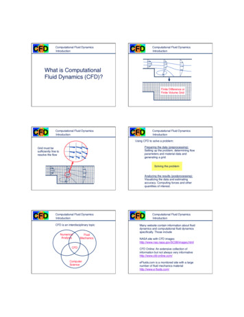
10CHAPTER 1. PURKINJE CELL VOLTAGE-CLAMP IN VIVO1.31.3.1ResultsFeasibility of obtaining an adequate space-clamp of Purkinjecells in vivoAn optimal space-clamp of the PC is required in order to distinguish the number ofEPSCs in the burst of CF inputs. If the cell is not space clamped correctly, spurious currents and potentially also unclamped dendritic spikes appear on the traceand could be misinterpreted as a genuine EPSC. Unfortunately, the PC has a hugedendritic tree that makes the space clamp a demanding task [26]. Moreover, in vivoaccess resistance in whole-cell configuration is typically higher than in vitro and theclamping quality is worse [17].The plots of Fig. 1.1 show two typical CF evoked EPSC of a PC [this voltageclamp recording was made by Ian Duguid]. The holding potential was set at -100mV because a strong hyperpolarisation enhances the probability that proximal anddistal dendrites were at least clamped at -70 mV. This technique reduces the impactof a relative inadequate space-clamp. Nevertheless, the result is suboptimal: theevidence for unclamped currents is the long duration of the EPSC, whose decay timeis ruled by at least two different decay constants. This observation makes unsure theinterpretation of the second peak: it is not possible to determine with certainty if itis a second EPSC or an unclamped dendritic conductance.For comparison, an in vivo voltage clamp recording from a granule cell is shownin Fig. 1.2 [this voltage-clamp recording was made by Paolo Puggioni]. Granule cellsare very small, diameter 10 µm, and therefore easy to space-clamp correctly. Theleft picture shows a typical EPSC recorded with holding potential -70 mV, while theright one represents a typical IPSC with holding potential 20 mV. The signal isclean and the cell is space clamped correctly, as the kinetic of each event indicate anadequate space-clamp.
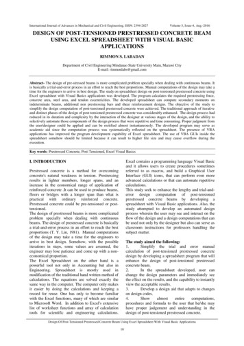
1.3. RESULTS11Figure 1.1: PC voltage-clamp recording. Two typical CF inputs recorded with holding potential -100 mV. Despite the Cs internal solution and the strong hyperpolarised membranepotential, the space-clamp is not optimal. The evidence for unclamped currents is the longduration of the EPSC, which display a bi-exponential decay reflecting unclamped dendriticconductances. [This recording was made by Ian Duguid]Figure 1.2: Granule cell voltage clamp recording. (Left) Typical EPSC with holding potential-70 mV and (Right) typical IPSC with holding potential 20 mV. The signal is clean andthe cell is space clamped correctly, as proved by the exponential decay.
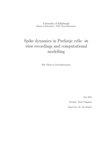
12CHAPTER 1. PURKINJE CELL VOLTAGE-CLAMP IN VIVO1.3.2The effect of urethane on the inferior olive activity in vitroAt the same time, the effect of the urethane on the activity of IO was tested in vitro[in collaboration with Nolan Lab, this study was conducted by Dr. D. Garden andis reported here due to its relevance for this work].As spontaneous action potential (AP) frequency is low in vitro the IO have beenstimulated by 2 ms depolarising steps. Each plot of Fig. 1.3 (Top) shows 10 consecutive APs before and after application of 20 mM urethane. In control conditions800 pA was sufficient to generate an AP while in 20 mM urethane the amplitudehad to be increased to 1100 pA. Interestingly, the plateau potential and spikelets arecompletely abolished in urethane. Finally, the resting membrane potential in controlconditions is about 5 mV more depolarised.1.4DiscussionAs stated in Sec. 1.1, the goal of the present work is to verify feasibility of an in vivoinvestigation of the role of HCN1 channels regulating the olivary cerebellar activityby voltage-clamping single PCs in anaesthetised mice. The quality of the PC spaceclamp is fundamental to distinguish the number of CF evoked EPSC in each IOburst and test the two predictions based on in vitro data (increased CF activity butsuppression of the bursts).This study showed that the in vivo measurements cannot be made according tothe original plans for two reasons:- the poor quality of the PC space clamp does not allow determination of individual EPSCs in a CF burst;- the urethane anaesthesia reduces the occurrence of CF inputs and abolishesthe bursts.The only possibility left for an in vivo investigation is to switch to awake animals,because it is now proved that both of the two available anaesthetic drugs (ketamineand urethane) affect directly and strongly the CF inputs.
1.4. DISCUSSION13Figure 1.3: Effect of urethane on the IO activity. (Top) 10 consecutive APs before (left) andafter (right) application of 20 mM urethane. As spontaneous action potential (AP) frequencyis low in vitro the IO have been stimulated by 2 ms depolarising steps. In control conditions800 pA was sufficient to generate an AP while in 20 mM urethane the amplitude had to beincreased to 1100 pA. (Middle) The plateau potential and spikelets are completely abolishedin urethane. (Bottom) The resting membrane potential in control conditions is about 5 mVmore depolarised.
14CHAPTER 1. PURKINJE CELL VOLTAGE-CLAMP IN VIVO
Chapter 2Purkinje cell dynamics in vivo:analysis of the spike traintemporal structureAbstractPurkinje cells (PCs), the only output of the cerebellar cortex, show two distincttypes of spiking activity: complex (CSs) and simple (SSs) spikes. Recent functionalcerebellar theories, such as the adaptive-filter model, consider the SSs as the carrierof the information, while the CSs as a teaching signal, responsible of the learningprocess. At the same time, the most recent coding theories propose that the PCinformation is stored in the rate and pauses of the spike train. These two perspectivesare somehow incompatible, as it was shown that CSs induce pauses and alter the SStemporal structure and therefore necessarily carry information. Therefore it will beof interest to examine the reciprocal influence that SSs and CSs have on each otheron a dynamical basis. This study investigates the dynamical temporal structure ofSS and CS trains in PC recordings from urethane-anaesthetised mice. The findingssuggest the conception of two simple models describing (i) the pauses as a possibleintrinsic feature of PC dynamics and (ii) the effect of a CS on the next SS interval.The huge variability of responses to the same inputs implies the presence of different15
16CHAPTER 2. PURKINJE CELL DYNAMICS IN VIVO’states’ in which the PC can operate. This sort of state-dependent computation, inwhich the state of the network is determined by the previous inputs, could potentiallybe described in a liquid-state machine framework.2.1IntroductionPurkinje cells (PCs) are the only output of the cerebellar cortex, known to play afundamental role in motor coordination and learning. Two distinct types of spikingactivity characterise PC recordings. Complex spikes (CSs), generally occurring at lowfrequencies (0.05-2 Hz), are massive depolarising events driven by excitatory inputsfrom a single climbing fiber (CF) coming from the inferior olive (IO) [5]. Single spikes(SSs) generally occur at higher frequency (30-100 Hz) and are intrinsically generatedwithout the need of any synaptic input [2]. Two different mechanisms modulate SSdynamics: excitation from the mossy fiber (MF) - granule cell (GC) - parallel fibers(PF) pathway and inhibition from basket cell interneurons (BC). PCs inhibit deepcerebellar nuclear (DCN) cells which, in turn, inhibit the IO. The IO completes theloop by exciting back the DCN and, as already described, by inducing CSs in PCs.The picture, already very complicated, is not complete yet. Classical theories onthe function of cerebellum (Albus-Ito-Marr [1] [13] [18]) consider the SS as the carrierof the information, while the CS as the responsible of the learning process. In recentstudies, the cerebellum is modelled as a filter whose inputs (coming from the brainstem to the PC through the MF-GC-PF path) are weighted by synapse strengths inthe PCs and then recombined to form the SS output. Moreover, the filter is adaptive,because synapses activated synchronously with a CS (an error teaching signal) aredown-regulated by long term depression mechanisms (LTD), while synapses carryinginputs negatively correlated with CSs are up-regulated by long term potentiation(LTP) [7]. The adaptive-filter model, although mathematically elegant and consistent with many experimental findings, is still not fully convincing.Present theoretical works are investigating the nature of the code adopted by PCsto transfer information. The most up-to-dated theories supported by experimentalevidences suggest that the DCN (target of PCs) is sensible to two parameters: (i)
2.1. INTRODUCTION17the rate of the spikes and (ii) the length of the pauses of PC spike trains. PCsinhibit DCN through GABA synapses: their regular firing controls DCN membranepotential and acts as a rate code. The pauses, when sufficiently synchronised acrossafferent PCs, generate bursts of activity in DCN and are interpreted to be a temporalcoding signal [6].A recent hot and rather controversial topic is related to the observed bi-stabilityin the firing rate of a relevant fraction of PCs. This phenomenon was shown with invitro [25] and in vivo anaesthetised [16] preparations, but its importance was harshlycriticised by objecting that it is rarely observed in awake animals [27]. Recently,awake recordings from cats not engaged in any behavioural task revealed that about50% of the cells show frequent long pauses suggesting the presence of up- and downstates [32]. However, the functional relevance of bistability is not clear.In this view, CSs cannot be considered as a pure teaching signal as in the adaptivefilter cerebellar theory, but necessarily they carry information. As a matter of fact,experimental evidence collected in the last years showed that CSs could affect therate and the pauses of PCs in many ways:- CSs introduce a typical 8-10 ms refractory period eventually followed by apause whose length depends on the number of Ca2 dendritic spikes triggeredby the full blown somatic Na action potential [5];- CSs can help the synchronisation of spike trains between paired PCs (definedas PCs with significant correlation in CF inputs, usually located in the samemicrozone [31]);- CSs can act as a switch between up- and down- states of the PC and thereforeproducing directly a pause or a regular firing period [16] [25].The last case is subtle and tricky from a coding point of view, as the result of theCS action depends on the state of the PC. Therefore, if we assume that the IO codesin a deterministic way, we should admit that it somehow knows the state of the PCand changes it deliberately.
18CHAPTER 2. PURKINJE CELL DYNAMICS IN VIVOThe study of the interaction of CSs with SS trains, the emergence of pauses andthe possible coding consequences requires the application of statistical techniquesthat go beyond the characterisation of overall averaged quantities. Processes havingcompletely different dynamic behaviours could produce the same mean result [9].The present work should be considered as an effort to propose the study of neuraldynamics by applying tools originally developed for the analysis of general dynamicalsystems. The main challenge is that the stochastic component is often dominant andtherefore the detection of regularities and recurrences is quite difficult.A total of 15 cell-attached recordings were made from PCs of urethane anaesthetised mice. The results presented in Sec. 2.3.1 and 2.3.2 give the flavour of thehuge variability characterising both average and dynamical properties of the PCs.Continuous firing, pausing and bistable PC are described in detail and the data areconsistent with published studies conducted on anaesthetised and awake preparations.The second part (Sec. 2.3.3 and 2.3.4) is devoted to the description of two simplemodels explaining (i) a possible intrinsic mechanism of pause generation, (ii) thesynchronisation effect that CSs have on the subsequent SSs.Section 2.3.5 contains the analysis of 2 awake extracellular recordings that areconsistent with the previous findings, although the range of the firing frequencyshown in one of the cells is massive.The discussion (Sec. 2.4) is focused on outlining a possible robust method to cooperate with and possibly explain in a probabilistic framework the striking biologicalvariability of the dynamical properties of the PCs.2.22.2.1Materials and methodsElectrophysiologySurgeryC56BLJ6 mice (age 6-7 weeks) were anaesthetised with isofluorane and put in astereotaxic frame. A first surgery was performed to attach a head post, needed tofix the head during the recordings. Mice were given a post-operative analgesia and
2.2. MATERIALS AND METHODS19were returned to their cage for at least 24 hours. On the recording day mice wereanaesthetised again with isofluorane, a 500 µm 300 µm craniotomy was made overlobules V of the cerebellar vermis and the dura mater was be removed. Mice werethan injected with urethane (1.6 mg Kg 1 ) and carried to the recording rig.In vivo cell attached recordingsCell-attached recordings were made with Multiclamp 700A amplifier (Axon instrument), band-pass filtered at 300 Hz-4 kHz and digitised at 10-20 kHz. The pipettecontained a HEPES based in vivo external solution (NaCl 150 mM, KCl 2.5 mM,HEPES 10 mM, CaCl2 2 mM, CaCl2 1 mM). Artificial cerebrospinal fluid (NaCl 125mM, NaHCO3 25 mM, NaH2 PO4 1.25 mM, KCl 2.5 mM, CaCl2 1.5 mM, MgCl2 1mM, Glucose 25 mM) was used as external solution. The electrodes (resistance range2.5-3.5 MΩ) were positioned by the means of a 4-axis manipulator. PCs were identified for their high frequency rate ( 30 Hz) and depth from brain surface (190-300µm). Although the presence of a CS is a final proof for PC identification, not allPCs show CSs (see Sec. 2.3.1). Therefore the detection of CSs cannot be used as theunique PC identification strategy.2.2.2Data analysisAll the data analysis was performed off-line by using a custom-made code implemented in MATLAB (MathWorks, Natick, MA, USA).Spike detection and sortingThe spikes are detected by setting thresholds at a proper value chosen carefully foreach cell after a visual inspection of peak amplitude and noise level. Spikes are thensorted as CS of SS by checking the signal between 1.5 and 3.5 ms after a spike. If themembrane current is higher than a given threshold, the spike is s
University of Edinburgh School of Informatics - DTC Neuroinformatics Spike dynamics in Purkinje cells: in vivo recordings and computational modelling