Transcription
Dissection of the Rabbit
Dissection of the RabbitContentsIntroduction . . . . . . . . . . . . . . . . . . . . . . . . . . . . . . . . . . . . . . . . . . . . 1Unit 1: External Anatomy . . . . . . . . . . . . . . . . . . . . . . . . . . . . . . . . . 3Unit 2: The Skeletal System . . . . . . . . . . . . . . . . . . . . . . . . . . . . . . . 5Unit 3: The Muscular System . . . . . . . . . . . . . . . . . . . . . . . . . . . . . 6Unit 4: The Digestive System . . . . . . . . . . . . . . . . . . . . . . . . . . . . . 9Unit 5: The Respiratory System. . . . . . . . . . . . . . . . . . . . . . . . . . . 15Unit 6: The Circulatory System . . . . . . . . . . . . . . . . . . . . . . . . . . . 17Unit 7: The Urogenital System . . . . . . . . . . . . . . . . . . . . . . . . . . . 20Unit 8: The Nervous System . . . . . . . . . . . . . . . . . . . . . . . . . . . . . 23Rabbit illustration on pg. 1 DILEEP/Shutterstock.com 2012 Carolina Biological Supply Company Printed in USA.
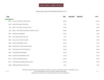
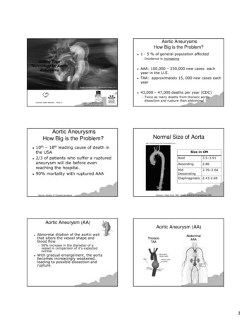
IntroductionAs you progress through this investigation ofrabbit anatomy, consider how the form of eachparticular structure is appropriate for itsfunction. For example, the rabbit’s sharpincisors enable it to cut the herbaceous plantsthat it eats and to gnaw through roots whenburrowing. Correlating form with function is apart of any in-depth anatomical study. Beforeyou begin the dissection, read the entireprocedure carefully and familiarize yourselfwith the following list of commonly used terms.Important terms are boldfaced if they appear ina corresponding figure.Until the early 20th century, rabbits wereclassified as rodents; however, they differ fromrodents in several ways and are now classed inthe order Lagomorpha, with hares and pikas.The majority of domestic rabbits, includingyour dissection specimen, are derived from theEuropean rabbit, Oryctolagus cuniculus. In thewild, this species lives in groups that formelaborate systems of burrows referred to aswarrens. Domestic rabbits have been eitheraccidentally or purposely introduced to manyhabitats in which they then over-proliferated,disrupting the native ecosystem.Midsagittal ximalDistalLateralMedialTransverseplaneFigure 1. Body planes and directions.1
SafetyWear safety glasses or goggles, gloves, and alab apron when dissecting the specimen.Perform the dissection only on the dissectiontray. Follow proper hygiene practices before,during, and after the lab.Direction or PlaneDefinitionLateralToward the right or left sideMedialToward the midlineProximalNear or toward the point of referenceDistalAway from the point of referenceDorsalToward the backVentralToward the bellyAnterior (cranial)Toward the headPosterior (caudal)Toward the tailSuperficialToward or along the surfaceDeepA significant distance below the surface2
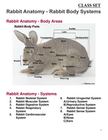
Unit 1External AnatomyLay your specimen on its ventral surface.Examine its exterior carefully. Note that therabbit can be divided into three sections, thehead, trunk, and tail. The head is separated fromthe trunk by the neck.Note the hair. The presence of hair, at least atsome point in their development, is adistinguishing feature of mammals. Hair serves avital role in thermoregulation in most species ofmammals and also protects the skin. Specializedhairs, vibrissae, or whiskers, are typically foundon the face. They are used as part of the rabbit’ssensory system and help the animal gatherinformation about its surroundings. Examine thehead to view the vibrissae.While examining the head, note the othersensory structures. The rabbit’s eyes are largeand set laterally on the head. Lateral positioningincreases field of vision but reduces depthperception. Also due to the placement of theeyes, a rabbit has a small blind spot directly infront of its face. This eye positioning is commonTailTrunkHeadFigure 2. The three sections of the rabbit3
much as 10 percent of the overall surface areaof a rabbit.Open the mouth of the rabbit and examine itsdentition. Like a rodent, a rabbit has incisorsthat are open-rooted and grow continuouslythroughout the animal’s life. Unlike a rodent,a rabbit has four incisors in its upper jaw. Thesmaller pair behind the front incisors are referredto as peg teeth. As you examine the dentition,consider the rabbit’s diet. Observe the sensoryorgans located in the mouth, the taste buds.Rabbits have a good sense of taste. Like humans,rabbits can taste the four basic categories offlavors: sweet, sour, bitter, and salt. It is not yetknown if they are able to taste the umami flavor.The sense of taste helps a rabbit determinepotential toxicity and nutritional value of theplants that it nibbles.It is in the mouth that the digestion of foodbegins. Food is mechanically broken down bythe teeth, while salivary enzymes begin thefood’s chemical breakdown.After examining the rabbit’s head, examine itstrunk. Note the paired feet and legs of the rabbit.The feet and legs of the rabbit are modified forhopping, also known as saltatorial locomotion.Mammals with saltatorial locomotion tend tohave enlarged muscular hind limbs and reducedforelimbs and to live in open spaces.in animals that have many predators. Comparedwith humans, rabbits tend to be farsighted—theirfocus is worse on things that are nearby butbetter on things at a distance (such asapproaching predators). Rabbits tend to becrepuscular (most active near twilight and dawn)and have good vision in low light conditions.Protecting the eyes are upper and lower lids anda partial nictitating membrane located in themedial corner of the eye. Because it istransparent, the nictitating membrane allows forsome protection and moistening without as greatof a loss of vision as blinking of the eyelids. Inmany animals, including some mammals, acomplete nictitating membrane can cover theentire front of the eye. Humans retain only avestige of the nictitating membrane.The nares, or nostrils, are not only theopenings to the respiratory system but are alsocrucial to the sensory system, allowing rabbitsto gather olfactory information from theirenvironment. The nares are enclosed by lateralfolds which radiate from the median cleft.The external part of the ear, the pinna (pl.,pinnae), directs sound waves into the auditorymeatus, or ear canal. A rabbit can hear agreater range of frequencies than a human. Thepinnae function also in thermoregulation; infact, the surface area of the ears may occupy cisorsFigure 3. The external anatomy of the rabbit’s head4
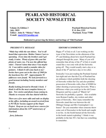
Unit 2The Skeletal SystemThe skeletal system of the rabbit is comprisedof both cartilage and bone. The system servesto support the body mass and to protect internalorgans. The skeletal system consists of twomain parts, the axial skeleton and theappendicular skeleton. The axial skeleton is theskull, vertebral column, ribs, and sternum. Theappendicular skeleton includes the pectoral and8 910pelvic girdles and their appendages. Thepectoral appendages are the forelimbs, and thepelvic appendages are the hind limbs. Duringyour dissection of the rabbit, refer to theskeleton diagram to identify the bones thatyou encounter. Observe the structure andarrangement of the bones as you consider theirspecific functions.1176155171612131443 NasalPremaxillaMaxillaFrontalOptic .20.21.22.23.24.25.26.3938Cervical vertebra (7)Thoracic vertebra (12)Ribs (12)Lumbar vertebra (7)Sacral vertebra (4)Caudal vertebra (16)ScapulaClavicleHumerusSternumXiphoid processOlecranon processRadiusFigure 4. The skeletal structure of the rFibulaTibiaPatellaTarsalsMetatarsals
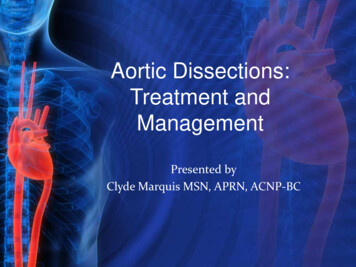
Unit 3The Muscular SystemThe muscular system consists of skeletalmuscle, which contracts to move bones. Atypical skeletal muscle is attached to two bonesby tendons. The attachment point to the bonethat is moved during contraction is called themuscle’s insertion. The muscle’s origin is thestationary point of attachment. To observe themusculature, you must first skin the rabbit asdescribed below. After removing the skin, referto the muscle diagrams to identify the superficialmuscles of the rabbit. Consider the function ofeach muscle as you examine it. Try to find theorigin and insertion of some of the muscles thatyou examine.skin away from the body and then cut throughthis muscle’s attachment points to the trunkalong the midline and in the axillary (armpit)region of the rabbit. Be careful not to damageany of the other muscles. Finally, remove theskin by pulling it down from the ventral cranialregion toward the caudal region. Skin shouldremain only on the feet, head, tail, and genitaliaof the rabbit.Skinning the rabbitLay the rabbit on its ventral surface. Massagethe skin in the dorsal neck area to loosen aportion of the skin from the underlying muscle.With a scalpel, make a small incision in the centerof the area of loosened skin. Using blunt-tippedscissors, cut a shallow incision down the dorsalmidline toward the tail. As you cut, pull the skinaway from the underlying muscles. From thiscut, make additional cuts down the lateral surfaceof each leg and around the wrists and ankles.Additionally cut around the neck, tail, anus, andexternal genitalia. Ensure that you do notdamage the genitalia while making the cuts. Afterall cuts are made, separate the skin from theunderlying muscles by using a blunt probe totear through the superficial fascia while pullingthe skin away from the body. As you separate theskin near the neck you will see the platysma, fineparallel muscle fibers that are attached to theskin. Remove this muscle as you remove theskin. As you separate the skin from the muscleson the rabbit’s ventral surface, you will see thatthe skin is attached to a thin sheet of muscle, thecutaneous maximus, which originates at thelinea alba (midline). To remove the skin, pull theFigure 5. Incision lines for skinning6
Dorsal Superficial Pectoral MusclesVentral Superficial Pectoral sLevator scapulae dExtensor carpi radialis longusExtensor brevis pollicisRadiusTransverse ligamentUlnaExtensor carpi ulnaris12.13.14.15.16.17.18.19.20.21.Extensor communis digitoriumExtensor digiti propriusFlexor carpi ulnarisOlecranon processTriceps brachii, lateral headAcromiotrapeziusLatissimus dorsiSpinotrapeziusExternal obliqueSternohyoidFigure 6. Superficial pectoral dJugular veinBasioclavicularisCleidomastoidPectoralis tenuisCleidohumeralisPectoralis majorCutaneous maximusLatissimus dorsiExternal oblique
Dorsal Superficial Pelvic MusclesVentral Superficial Pelvic 5.6.7.8.9.10.11.Dorsal aponeurosisGluteus mediusGluteus maximusRectus femorisTensor fascia lataBiceps femoris anteriorVastus intermediusVastus lateralisBiceps femoris posteriorExtensor digitorum longusGastrocnemius anosisAdductor magnusGracilisExternal obliqueRectus femorisVastus medialisSartoriusAbductor caudaeSemimembranosisGracilisAdductor magnusFigure 7. Superficial pelvic muscles8
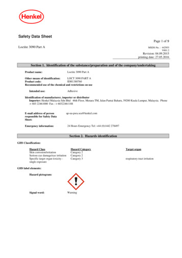
Unit 4The Digestive SystemSecure the skinned rabbit on its dorsal side inthe dissection tray. Following the incision linesdepicted in the diagram, cut through themuscular wall of the abdomen from the pubicarch to the most posterior rib. Cut two additionalincisions from the linea alba to the dorsal side ofthe rabbit, almost to the vertebral column.Carefully fold the flaps of skin and musclelaterally. Do not disturb the underlying organs.Pin the flaps to the tray to expose the abdominalcavity. There are two body cavities separated bya thin sheet of muscle called the diaphragm. Theexposed cavity is the abdominal cavity. Anteriorto that cavity and separated by the diaphragm isthe thoracic cavity.A prominent organ in the anterior of theabdominal cavity is the liver. This reddishbrown organ is divided into right and left lobesby the falciform ligament. Each of these lobes isfurther divided into median and lateral lobes,for a total of four lobes of the liver. The liverproduces some amino acids, filters the blood,converts harmful substances into harmlessones, and helps to regulate blood sugar.Additionally, in the digestive system, the liverproduces bile, which emulsifies fat duringdigestion. The bile produced by the liver isstored in the gallbladder. Raise the right lobe ofthe liver and observe the gallbladder, a dark,greenish-yellow sac.LiverLarge intestineDiaphragmSmall intestineFigure 8. Abdominal cavity9
LiverGallbladderLiverFigure 9. Abdominal cavity showing the gallbladderEsophagusAs you continue to examine the abdominalcavity, trace the path of food through thedigestive system. Each swallow of food forms abolus, which travels from the mouth down theesophagus. The majority of the esophagus,located in the thoracic cavity, is best examinedduring observation of the circulatory system.Some small portion may be visible at the mostanterior portion of the stomach. After leaving theesophagus, the food reaches the stomach,located on the left side of the rabbit, beneath thediaphragm. Lift the liver to view the entirestomach. The stomach consists of four portions:the cardia, the fundus, the body, and the pylorus.The cardiac portion is the area where thestomach joins the esophagus. In this section, thecardiac sphincter separates the esophagus fromthe stomach. (Malfunction of this sphincter is acause of acid reflux in humans.) The fundus isthe upper curve of the stomach that lies tothe left side of the esophagus. The body isthe main portion of the stomach. Thepyloric section lies to the right of theesophagus and terminates in the pyloricsphincter, which separates the stomach from theduodenum, the first portion of the smallCardiac regionFundusGreater cuLesrvaserturecurvatureCardiacsphincterPyloric regionPyloricsphincterFigure 10. Digestive system diagram10
LiverStomachFigure 11. Abdominal cavity showing the stomachStomachSpleenFigure 12. Abdominal cavity showing the spleen11
sections of the small intestine, which aredifficult to distinguish. The caudal end of theileum is modified into a round muscularenlargement called the sacculus rotundus. Thesacculus rotundus is a structure found only inrabbits. It has a large amount of lymphoid tissueand is involved in the immune system. Carefullycut longitudinally along a portion of the smallintestine and examine the lining with a handlens. The small intestine is lined with thousandsof circular folds, the plicae circulares. There arealso small projections called villi. Each villus hasits own projections, the microscopic, hair-likemicrovilli. The folds and projections increasethe surface area of the small intestine andmaximize the absorption of nutrients. The smallintestine ends at the ileocecal valve. Within themesentery of the small intestine is the pancreas,a pinkish-brown, somewhat granular mass. Thepancreas opens to the small intestine throughthe pancreatic duct at the caudal end of theduodenum. It is part of both the digestive andendocrine systems and secretes both enzymesand hormones.intestine. In the stomach, chemical andmechanical digestion continue as muscularcontractions mix the bolus with digestivesecretions, forming a mixture called chyme.Open the stomach and remove any food from theinner surface. Examine the internal surface of thestomach. Note the deep folds, or rugae, thatallow the stomach to expand without pressure.(A hands lens may help you see greater detail.)Though not part of the digestive system, thespleen is best viewed at this point. The spleen islocated on the greater curvature of the stomach,the larger convex curve. It is a small, dark,reddish-brown organ. The spleen is part of thelymphatic system and is designed to filter blood.From the stomach, the chyme travels throughthe pyloric sphincter into the duodenum, thefirst section of the small intestine. It is here thatthe common bile duct from the liver andgallbladder and the pancreatic duct from thepancreas meet the small intestine to releaseadditional enzymes to continue the process ofdigestion. From the duodenum, the mixturetravels to the jejunum and ileum, the otherStomachPancreasDuodenumFigure 13. Abdominal cavity showing the pancreas12
digestive system. The rabbit extracts additionalnutrients from the food this time, due to thefermentative changes that occurred in the cecum.Rabbits’ digestion exemplifies one type ofhindgut fermentation. Cattle and other ruminantsare called “foregut fermenters” becausemicrobial fermentation of their food occursbefore the food reaches the intestine.Although the rabbit colon can be divided intothe ascending, transverse, and descending colon,it is usually discussed in terms of the proximaland distal colon. A short, muscular portion calledthe fusus coli marks the division between thetwo. This section helps regulate the complexevents that move materials either forward orbackward and that determine whethercecotropes or dry feces are being produced. Thebeginning section of the proximal colon has alumpy appearance because it includes manypouches called haustra between longitudinalbands called taenia. The distal colon is a long,twisted tube that terminates with the rectum.Both dry feces and cecotropes exit the anus.Materials exit the small intestine through thesacculus rotundus to the large intestine—first toa short compartment called the ampulla cecaliscoli (a connection between the cecum and thecolon) and then to the colon. In the first section ofthe colon, the proximal colon, specialcontractions separate the more-digestible fromthe less-digestible materials and force thenutritious components backward into the cecum.The rougher materials continue along the distalcolon. The cecum is a large, coiled blind sac thatextends from the sacculus rotundus. The cecummakes up a larger proportion of the digestivetract in rabbits than in any other group ofanimals, and it is critical to the digestion of theirfood. Within the cecum, bacteria break down thematerial through fermentation. At certain times,material from the cecum is released to the colonwhere it is coated in mucus and excreted throughthe anus in the form of soft balls calledcecotropes, also known as night feces or softfeces. Rabbits immediately eat their cecotropes,and the materials pass again through theDistal colonHaustraColonTaeniaProximal colonCecumSacculus rotundusSmall intestineFigure 14. Abdominal cavity showing intestinal tract13Appendix
Dorsal Superficial Pelvic MusclesVentral Superficial Pelvic iform ligamentLiver, right central lobeOmentumTaeniaColonCecumRectumLiver, left central lobe10.11.12.13.14.15.16.17.18.Liver, left lateral lobeStomachSmall intestineAppendixEsophagusCystic ductGallbladderBile ductLiver, caudate lobeFigure 15. Stomach Pyloric stomachCardiac stomachSpleenPancreasIleumSacculus rotundusMesentery
Unit 5The Respiratory SystemTo view the respiratory system, open thethoracic cavity. Cut along the midline, from thediaphragm to the base of the neck. As you makeyour incisions, you will be cutting through somemusculature as well as the ribs. Fold back themusculature and ribs on both sides to expose thecavity. Pin the musculature down to expose thethoracic cavity.You have already examined the nares, theexternal portion of the rabbit’s nose. Rabbits areobligate nasal breathers; a rabbit cannot breathethrough its mouth due to the position of theglottis and epiglottis. These structures serve asthe opening of the respiratory system from thepharynx, the area just posterior to the mouthand common to both the respiratory anddigestive systems.As you examine the features of the respiratorysystem, follow the path of an inhaled breath.From the nares, air travels to the pharynx,past the glottis and epiglottis, to the larynx, andthen the trachea. To view the larynx and trachea,extend the midline incision toward the chin,making a shallow cut to avoid damaging theunderlying structures. Then, using a bluntinstrument, tease away any surrounding tissue.EsophagusTracheasLungHeart enclosed inthe pericardiumLungsAortic archFigure 16. Thoracic cavity, with the heart15
The larynx, a cartilaginous structure, protectsthe entrance to the trachea and is important invocalization. Posterior to the larynx is thetrachea. The trachea is a long tube ringed withbands of cartilage that provide support andprevent collapse. The trachea continuesposteriorly until it divides into left and rightbronchi dorsal to the heart. These bronchi lead tothe left and right lungs. In the lungs, the bronchibranch further into smaller bronchioles, whichterminate in vestibules containing alveoli, wheremost gas exchange occurs. The lungs of therabbit are divided into three portions: theanterior, middle, and posterior lobes. Due to theposition of the heart on the left side of the cavity,the left anterior lobe is much smaller than itscounterpart on the right.TracheasLungBronchusLungsFigure 17. Thoracic cavity, without the heart16
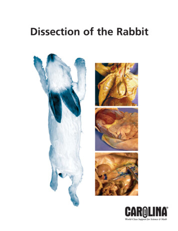
Unit 6The Circulatory SystemThe circulatory system transports varioussubstances through the body, includingdissolved gases, nutrients, hormones, defensivecells, and cellular wastes. Like other mammals(and like birds), rabbits possess a highlybranched network of vessels and a fourchambered heart.There are two circulatory pathways: pulmonaryand systemic. In pulmonary circulation,deoxygenated blood is pumped from the rightside of the heart to the lungs, and oxygenatedblood flows back to the left side of the heart. Insystemic circulation, this oxygenated blood ispumped from the left side of the heartthroughout the body, and deoxygenated bloodtravels back to the right side of the heart. To fullyview the heart, carefully remove the thymusgland, a component of the endocrine system.The heart is covered by a membrane calledthe pericardium, or pericardial sac.Carefully lift it away from the heartand make an incision in it. In a livingrabbit, the pericardial sac is full offluid, which protects and cushionsRight anteriorthe heart. Once you have removedvena cavathe pericardial sac, examine thefour chambers of the heart. Twodark, thinner-walled atria areanterior to the two thicker-walledventricles. The organization of theRight atriumheart keeps oxygenated anddeoxygenated blood separated.To trace the flow of bloodthrough the heart, slice a ventralopening by making a smooth,continuous partial incision into theheart’s left side. Carefully fold thePosteriorventral portion away from thevena cavadorsal portion, as if opening abook. Deoxygenated blood fromRight ventriclethe body enters the right atrium viathe right anterior vena cava, the leftanterior vena cava, and theposterior vena cava. From the rightatrium, blood flows through thetricuspid valve into the right17ventricle. The blood is then pumped through thepulmonary valve to the lungs by way of thepulmonary artery. Arteries are vessels that carryblood away from the heart. Valves prevent theblood from flowing back into the chamber fromwhich it came. In the lungs, the blood becomesoxygenated through gas exchange in the alveoli.The blood then returns to the heart through thepulmonary veins. Veins are vessels that carryblood toward the heart. The blood enters theheart again in the left atrium and travels throughthe bicuspid valve to the left ventricle. From theleft ventricle, the blood is pumped through theaortic valve and out of the heart through theaorta, the largest artery in the body.AortaLeft anteriorvena cavaLeft atriumLeft ventricleFigure 18. Heart, ventral view
brachiocephalic artery supplies blood to the rightsubclavian artery (which leads into the rightshoulder) and the right common carotid artery(which leads to the head). Likewise, the leftbrachiocephalic artery supplies the left subclavianartery and the left common carotid artery.Veins have less-muscular walls than arteriesand tend to be more superficial (i.e., majorarteries tend to lie deeper in the body than themajor veins). The veins merge until they reachthe venae cavae, which return blood to the rightatrium of the heart. The venous system is insome ways the inverse of the branching arterialsystem. The paired common iliac veins flow intothe inferior vena cava. As you trace the venacava cranially, you can locate the pairediliolumbar veins, the paired gonadal veins, andthe paired renal veins. The hepatic portal veindrains the liver and merges with the inferiorvena cava near the diaphragm before flowinginto the right atrium. Blood drains from thehead via the transverse jugular vein and theexternal jugular veins, and from there into thesubclavian veins, which merge to form thesuperior venae cavae.Following the path of the aorta, you will seethat it immediately makes a posterior turn, theaortic arch. Continuing caudally from the arch isthe dorsal aorta, which supplies blood to theposterior portion of the body (and may be calledthe abdominal aorta below the diaphragm). Thefirst branch is the celiac artery (or celiac trunk). Itdiverges to form the hepatic artery, whichsupplies the liver, and the gastrosplenic artery,which further subdivides to serve the stomachand spleen. Continuing caudally, locate thesuperior mesenteric artery, which supplies bloodto the small intestine. Next, find the paired renalarteries that carry blood to the kidneys and thegonadal arteries, which deliver blood to thetestes in males or the ovaries in females. Fartheralong the dorsal aorta you will see the pairediliolumbar arteries that carry blood to themuscles of the back. The last major branch formsthe right common and the left common iliacarteries, which supply blood to the legs.Blood travels through the aortic arch and to theanterior regions of the body through severalimportant vessels. The coronary arteries supplyblood to the heart muscle itself. The rightAnterior vena cavaPosterior vena cavaFigure 19. Heart, dorsal view18
Thoracic cavity, superficialHeart, 5Thoracic cavity, deep17Heart, 25121.2.3.4.5.6.7.8.9.10.11.12.Thymus glandHeartMediastinumRight lung, superior lobeRight lung, middle lobeRight lung, medial inferior lobeRight lung, lateral inferior lobeLeft lung, middle lobeLeft lung, inferior lobeRibXiphoid processDiaphragm, , centrum tendineumPhrenic v.EsophagusTransverse jugular v.External jugular v.Vertebral v.Left ventricleRight ventricleLeft atriumRight atriumLeft anterior vena cavaFigure 20. Diagram of the thoracic cavity with heart1924.25.26.27.28.29.30.31.32.33.34.Right anterior vena cavaPosterior vena cavaPulmonary v.Pulmonary a.Ligamentum arteriosumAortic archBrachiocephalic a.Right subclavian a.Left subclavian a.Left common carotid a.Pericardium
Unit 7The Urogenital SystemThe urogenital system consists of the urinaryand the reproductive systems, which share somecomponents. The urinary system removesnitrogenous wastes from the body as urine, andit also regulates osmotic pressure. Thereproductive system produces the gametes (themale’s sperm and female’s ova) necessary for theformation of offspring. It also includes thestructures involved in the sexual transfer ofsperm to ova and in the protection, development,and birth of young.into and out of the kidney. Also notice the adrenalglands, part of the endocrine system, one ofwhich is medial to each kidney.The kidneys filter waste from the bloodstreamand produce urine. The urine moves from thekidneys through the ureter to the urinary bladder,where it accumulates. As the bladder fills, it isstimulated to contract, expelling the urine outthrough the urethra. In the female rabbit, theurethra empties into the urogenital sinus,whereas in the male it continues to the penis,joining the reproductive system.Urinary SystemTo view the urinary system, remove therabbit’s liver and push the visceral organsto the right side of the specimen. Partiallyembedded on the dorsal surface of theabdominal cavity are two bean-shapedorgans, the kidneys. The kidneys filterwaste from the bloodstream, regulate theamount of water in the rabbit, andmaintain appropriate concentrations ofcertain ions. Using a sharp scalpel, cutone of the kidneys in half longitudinally,along its flattened plane. As you examinethe interior of the kidney, note the outerportion, the renal cortex, and the innerportion, the renal medulla. As youexamine the kidneys, notice the renalartery and renal vein, which carry blood11011928763451.2.3.4.5.6.KidneyHilusAbdominal aortaInferior vena cavaUreterRenal pelvis7.8.9.10.11.Renal medullaRenel cortexRenal a. and v.Dorsolumbar a. and v.Adrenal glandFigure 21. Kidneys and associated structuresUninary bladderKidneyUreterFigure 22. Upper portion of urinary system20
Reproductive Systemare released from the epididymis to the ductusdeferens, which transfers sperm from a testis tothe urethra. At the base of the bladder lies theseminal vesicle, which secretes a fluid into theductus deferens. This fluid combines with thesperm and with fluid from the prostate toproduce semen. The prostate is seen as athickening of the dorsal wall of the seminalvesicle. The semen passes through the urethraand out through the tip of the penis. The free endof the penis, the glans penis, typically lies withina pouch of skin called the prepuce. Inside theglans penis is a small bone called the baculum,which stabilizes the penis during copulation.MaleIn males the hormone testosterone and thesperm (the male ga
organs located in the mouth, the taste buds. Rabbits have a good sense of taste. Like humans, rabbits can taste the four basic categories of flavors: sweet, sour, bitter, and salt. It is not yet known if they are able to taste the umami flavor. The sense of taste helps a rabbit determine potential toxicity and nutritional value of the