Transcription
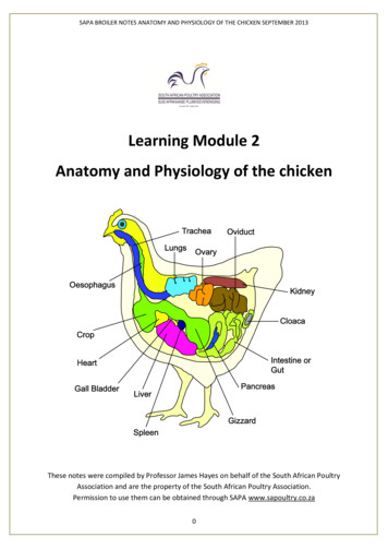
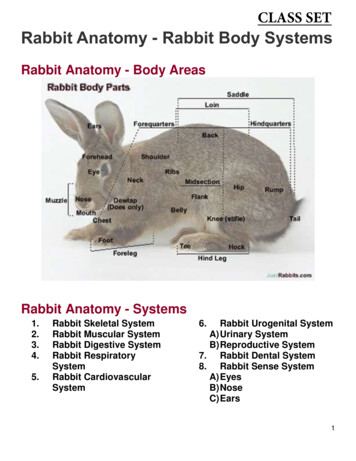
CLASS SETRabbit Anatomy - Rabbit Body SystemsRabbit Anatomy - Body AreasRabbit Anatomy - Systems1.2.3.4.5.Rabbit Skeletal SystemRabbit Muscular SystemRabbit Digestive SystemRabbit RespiratorySystemRabbit CardiovascularSystem6.Rabbit Urogenital SystemA) Urinary SystemB) Reproductive System7. Rabbit Dental System8. Rabbit Sense SystemA) EyesB) NoseC) Ears1
Rabbit Anatomy - Skeletal System The rabbit has a delicate skeleton compared with other mammals. It makes only 8% of the body weight, as compared to12 to 13% in cats.Here are some other important points to note about the rabbit skeletal system:There are 46 bones that make up the spinal column alone, 7 cervical (the neck), 12 thoracic (the chest), 7 lumbar (thelower back), 4 sacral (the pelvis) and 16 coccigeal (the tail).A rabbit's bones have extremely thin cortices and are easily shattered.The lumbar vertebrae are elongated to allow for considerable flexion and extension during hopping, but this makes themsusceptible to fracture.The powerful hind limb musculature and light skeleton enable powerful jumping over long distances.The hopping movement is made possible due to the hind legs being longer than the fore legs. Most of the elongation isbelow the stifle (the knee) in two bones, the tibia and fibula. The tibia is also prone to fracture.Rabbit's hind legs can kick out with extreme force and if they struggle when they are picked up, or even when they stamptheir feet violently on the ground, they are prone to fracture of their backbones (usually the 7th lumbar vertebra) anddamage their spinal cord.Rabbits have 7 tarsal bones (the ankle) and 4 digits on both hind legs, and 9 carpal bones (the wrist) and 5 digits on bothfore legs. Each digit has an associated toenail.Weakened bones and bones affected by osteoporosis are easily injured or broken.Various types of spinal deformations have been observed in a rabbit's anatomy. The degree of curvature is variable andmay range from mild and barely visible to severe and causing gait problems. The origin of these congenital deformationsis not well understood, they include:hemivertebrae - abnormal birth defect in which the vertebra fails to develop completely. As a result of the growth defectof the spine, a wedge-shaped vertebra develops, and neighboring vertebrae expand or tilt to fit the deformityspondylosis - a condition of the spine marked by stiffness of a vertebral jointkyphosis - humplike curvature of the spinelordosis - abnormal, increased degree of forward curvature of any part of the spine)These conditions may relate to a number of factors, a lack of calcium in the food, improper absorption of calcium in theintestine, lack of exercise, wrong posture due to being kept in a small cage, or defective genes.Female rabbits appear more frequently affected by these spinal abnormalities than males. It is linked to their higher needsin calcium, especially during pregnancy and lactation.Rabbits suffering from weak muscles and poorly mineralized bones and/or bone degeneration are at increased risk ofspine fracture when there is an inadequate support of the heavily muscled hindquarters, walking on a slippery floor, ortwisting of the lumbosacral junction when frightened or restrained.2
Rabbit Anatomy - Muscular System Wild rabbits are very athletic animals that are built to move rapidly in order to find food, water, find or fight mates, orflee predators over greater distances to find a hiding place.This daily exercise strengthens the locomotive muscles, fortifies the muscles in their heart and lungs and increasestheir resistance against stress.Just like us, exercise is wonderful for fortifying muscle and bones, it will stimulate blood circulation and the activityand functioning of other organs including the digestive system.The vertebrae of the spine provide support for the back. If this is accompanied by poorly developed transversospinalisspine muscles and trunk muscles, the normal balance of the spinal structure and the biomechanics can be altered,which can lead to degenerative processes.Deformations appear that will prevent the development of a good locomotric activity.Intrinsic muscle imbalance leads to degenerative changes of the lumbar vertebrae, even changes to femoral headhave been observed in rabbits that lack exercise.The rabbit muscular system, like in any vertebrate, is controlled by the nervous system, it's the basic concept of anymuscular system. Muscles are controlled through electrical signals between the body's parts and the brain.3
Rabbit Anatomy - Digestive System As total herbivores, rabbits have an extremely long digestive tract in order to process their food in the most efficientway.The whole of a rabbit anatomy has evolved to survive on a very poor diet, the digestive tract especially. A specialfeature of the process, known as caecotrophy, is a remarkable way the rabbit 'recycles' waste fecal matter in order toextract any nutrients that may have been missed on the first, second or even third time round in the digestion system.The whole digestive system of the rabbit is huge and may account for between 10-20 per cent of its total body weight.Let's follow the process through the rabbit's anatomy.Stomach In an adult rabbit the total length of the alimentary canal is 4.5 to 5 m. After a short esophagus there is a simplestomach which stores about 60-80 g of a rather pasty mixture of feedstuffs. Food eaten by the rabbit quickly reaches the stomach where it remains for a few hours, and although in an acidenvironment it has little chemical change.4
Liver & PancreasTwo major glands secrete into the small intestine: the liver and the pancreas. Bile from the liver contains bile salts and many organic substances but no enzymes. Bile aids digestion catalytically. The reverse is true of pancreatic juice which contains a sizable quantity of digestive enzymes allowing the breakdownof proteins (trypsin, chymotrypsin), starch (amylase) and fats (lipase).Small Intestine If the small intestine of a rabbit were laid out it would be more than 10 times the length of the rabbit. The contents of the stomach are gradually 'injected' into the small intestine in short bursts, by strong stomachcontractions. The small intestine is about 3 m long and nearly 1 cm in diameter. The contents are liquid, especially in the upperpart. Normally there are small tracts, about 12 cm long, which are empty. The small intestine ends at the base of thececum. This second storage area is about 40-45 cm long with an average diameter of 3-4 cm. It contains 100-120 g ofa uniform pasty mix with a dry matter content of about 20 percent. As the contents enter the small intestine they are diluted by the flow of bile, the first intestinal secretions and finally thepancreatic juice. After enzymatic action from these last two secretions the elements that can easily be broken downare freed and pass through the intestinal wall to be carried by the blood to the cells.Large Intestine The large intestine is made up of the cecum and colon. The cecum is very large, (about 10 times the volume of thestomach, and about 40 per cent of the total volume of the gastrointestinal tract). The colon separates the large and small fiber particles. The large particles of indigestible fiber are moved straightthrough the colon to form the hard droppings. The smaller fiber particles and other small incomplete digested foodparticles are moved backwards (by special muscles in the colon called haustrae). This 'slurry' enters the cecum where it is broken down and fermented.Cecum The particles that are not broken down in the small intestine enter the cecum after less than 2 hours. There they haveto stay for about 2 to 12 hours, while they are attacked by bacterial enzymes. Elements which can be broken down by this new attack (mainly volatile fatty acids) are freed and in turn pass throughthe wall of the digestive tract and into the bloodstream. Very near the end of the small intestine, at the entrance to the cecum, begins the exit to the colon. The cecum has ablind pouch branching off from the small intestine-colon axis. Physiological studies show that this blind pouchreservoir forms part of the digestive tract: the contents circulate from the base to the tip passing through the center ofthe cecum, then return towards the base, along the wall. The content of the cecum is then evacuated into the colon. Approximately half consists of both large and small foodparticles not already broken down, while the other half consists of bacteria that have developed in the cecum, fed onmatter from the small intestine.Colon The colon is about 1.5 m and follows on from the cecum, it is creased and dented for about 50 cm (proximal colon)and smooth in the terminal section (distal colon). The rabbit's digestive tract is virtually the same as that of other monogastric animals, however in the rabbit anatomy,the digestive tract has a uniqueness that lies in the dual function of the proximal colon. If the cecum content enters the colon in the early part of the morning it undergoes few biochemical changes. If the caecal content enters the colon at another time of day the reaction of the proximal colon is entirely different. Colon Dual Action Successive waves of contractions in alternating directions begin to act; the first to evacuate thecontent normally and the second to push it back into the cecum. The dual action of the colon produces two types of excrement: soft and hard.Hard Pellets (fecal droppings)These are round, relatively dry and composed mostly of undigested fiber. Normal fecal droppings are round balls, dark tolight brown in color, and fairly inform in size. They are slightly moist when fresh but dry out quickly. If you examine themclosely, or break one apart (they should crumble relatively easily), you will see the tiny rectangles of undigested plantfiber that they are formed from.5
Droppings that are small, very dark in color, or irregularly shaped are asignal that your rabbit is not processing enough fiber through his digestivetract. This may be because the diet is too low in fiber or because anotherproblem has slowed down the travel of food through the digestive system.If your rabbit stops producing any droppings, the gut may havecompletely stopped processing food; this should be treated as apotentially life threatening emergency and you should seek urgentveterinary attention.Some rabbits eat their fecal pellets, this is normal as long as they are alsoeating the caecotrophes. Soft Pellets (caecotrophes)The colon wall secretes a mucus which gradually envelops pellets formedby the wall contractions. Under the varying pressure and rhythm ofcontractions the content is squeezed like a sponge.Most of the liquid part, containing soluble products and small particles ofless than 0.1 mm, is forced back into the cecum. These pellets gather in elongated clusters and are called soft or nightpellets (caecotrophes).The soft pellets are recovered by the rabbit directly upon being expelled from the anus. To do this the rabbit twists itselfround, sucks in the soft feces as they emerge from the anus, then swallows without chewing them.By the end of the morning there are large numbers of these pellets inside the stomach, where they may comprise threequarters of the total content.From then on the soft pellets follow the same digestive process as normal feed. Considering the fact that some parts ofthe intake may be recycled once, twice and even three or four times, and depending on the type of feed, the rabbit'sdigestive process lasts from 18 to 30 hours in all, averaging 20 hours. Half the soft pellets consist of imperfectly broken-down food residues and what is left of the gastric secretions, and half ofbacteria. The latter contain an appreciable amount of high-value proteins and water-soluble vitamins. The practice ofcaecotrophy therefore has a certain nutritional value.The composition of the soft pellets and the quantity expelled daily are relatively independent of the type of feed ingested,since the bacteria remain constant.In particular, the amount of dry matter recycled daily through caecotrophy is independent of the fiber content of the feed.The higher the crude content of the feed and/or the coarser the particles, the sooner it passes through the digestive tract.Digestible & Indigestible FiberThe dual action of the colon requires fiber but if the feed contains few large particles and/or it is highly digestible, most ofthe caecal content is pushed back to the cecum and loses elements which nourish the 'normal' bacteria living in thececum.This would appear to increase the risk of undesirable bacteria developing in this impoverished environment, some ofwhich might be harmful.Therefore it is advisable to minimize roughage in the feed, enabling the rabbit's digestive process to be completed fairlyrapidly.In theory, roughage is provided by the crude fiber content of the feed, as this is normally rather hard to digest. However,certain fiber sources (beetroot pulp, fruit pulp in general) are highly digestible (digestibility of crude fiber varies from 60 to80 percent).For this reason recommendations are now made on quantities of indigestible crude fiber to be fedAdrenaline EffectsThe digestive process of the rabbit appears to be highly dependent on adrenaline secretions.Hypersecretion associated with stress slows down digestive activity, and entails a high risk of digestive ailments.Stress can be brought about by a number of factors, some that you may not even have considered. For example, a simplechange in brand of food pellets can stress your rabbit, also children chasing them to try and pick them up can also causean upset to their eating habits.6
Rabbit Anatomy - Respiratory SystemRabbits are nasal breathers. Blocking their nasal passages for any reason, including oral examinations, can lead torespiratory compromise due to the ineffectiveness of mouth breathing. If a rabbit is breathing through its mouth, it is in severe respiratory distress. The rabbit has a small chest cavity compared with the size and weight of its digestive system. Rapid breathing may sometimes be a symptom of pain elsewhere in the body, and is seen in rabbits with bladderstones or womb cancer. The nasal passages are in close proximity with the maxillary dental arcade, and changes in either the nasal passagesor molar tooth roots may affect each other adversely. Diseases invading the nasal passages may alter bone structure, and may ultimately lead to molar tooth movementand malocclusion; conversely, molar abnormalities and root elongation may impinge on nasal passages andcompromise respiration. The respiratory channel is separated from the food channel by the presence of the palate. The respiratory system of arabbit comprises nasal chambers, larynx, trachea, bronchi and lungs.Respiratory Organ & Related Parts:Nasal chamber: The air passes through the external nares into two large respiratory passages, which are hollow cavities presentabove the plate. The respiratory passage is divided in to right and left halves by the mesethm bone. Each side, therespiratory passages is divided in to two regions: An anterior nasal chamber & A posterior respiratory tube. The nasal chamber is bounded dorsally by nasal bone and ventrally by hard palate. The turbinals, very much foldedscroll kike bones, are present within the nasal chambers. The turbinals are covered by richly vascular, glandular andciliated epithelium. One respiratory tube opens behind the soft palate through the internal nares into the pharynxdirectly and that too very close to the glottis. The nasal chambers serve as very efficient filters, i.e. it removes fine and coarse dust particles, germs etc. throughmucous covering the turbinals. It helps to warm up the air before it is to be inhaled and enters the lungs. Due to thecovering of sensory epithelium, it also helps in the detection of smell.Larynx The pharynx opens into a larynx or sound blood through the glottis. The larynx lends in to a trachea. This is theanterior end of the windpipe or trachea. The larynx is a cylindrical box like structure. Inside the larynx, there is a cavity known as laryngeal chamber, whichcontains vocal cords. These vocal cords can be set into vibration by the passage of air over them to produce the‘voice’ – in rabbits mainly a squeak.7
Trachea After larynx, the trachea is a long respiratory tube extending through the neck into the thoracic cavity. It is linedinternally by ciliated cartilaginous rings. These ‘c’ shaped cartilaginous rings help to prevent the collapse of tracheaand to keep it expanded allowing free passage of air to and from the lungs. The mucous glands present in the wall of trachea help keep its inner surface moist and hold dust particles andbacteria. Thus only the clear air is allowed to pass into the lungs. The dust particles are swept towards the pharynx bycilia. The rabbit trachea is deeply recessed within the oral cavity behind the torus of the tongue. The trachea itself is narrowrelative to body size.Bronchi The thoracic cavity is small in comparison with the large abdominal cavity. Because of the small thoracic cavity,rabbits have more referred upper airway and bronchial sounds and may sound somewhat harsh. In the thorax, the trachea divides into left and right bronchi which enter the lungs. The bronchi possess the similarstructure as the trachea. After entering in to the lungs, each bronchus divides into many thinner branches, calledbronchioles. Which again divide into finer branches of less diameter known as respiratory bronchioles. Each respiratory bronchioledivides again into many finer branches, called alveolar ducts or infundibulum. The alveolar ducts end in small hollowair sacs, known as alveolar sacs. Each alveolar sac is formed of many small thin walled hollow alveoli or air cells. The respiratory bronchiole alveolar duct, alveolar sac and alveoli are devoid of cartilaginous rings but their wallspossess cilia. There is a network of capillaries of pulmonary artery and pulmonary vein around each alveolus. This complicatedbranching increases the total respiratory surface and allows the air to penetrate into every portion of the lungs.Lungs The lungs are soft compact and spongy mass of tissues lying in the pleural cavity within the thorax. Each lung iscovered by a fold of coelomic epithelium, which is in contact with the organ (visceral pleura). Each lung is divided intolobes. The right lung has four lobes. But left lung has got two lobes.8
Rabbit Anatomy - Cardiovascular System Function of the Cardiovascular SystemBy circulating blood throughout the body, the cardiovascular system functions to supply the tissues with oxygen andnutrients, while removing carbon dioxide and other metabolic wastes.As oxygen-rich blood from the heart flows to the tissues of the body, oxygen and other chemicals move out of the bloodand into the fluid surrounding the cells of the body's tissues.Waste products and carbon dioxide move into the blood to be carried away. As blood circulates through organs such asthe liver and kidneys some of these waste products are removed.Blood then returns to the lungs, receives a fresh dose of oxygen and gives off carbon dioxide. Then the cycle repeatsitself.This process of circulation is necessary for continued life of the cells, tissues, and ultimately the whole organism. Up anddown the evolutionary ladder, there are different forms of cardiovascular systems with different levels of efficiency, butthey all perform this same basic function.However the cardiovascular system of rabbits is slightly unique.Both the right and left atrioventricular valves are bicuspid in rabbits.The heart is small relative to total body size, comprising only 0.3% of the body weight.Rabbits have the most muscular pulmonary artery of anyspecies, which contributes to their predisposition for pulmonaryhypertension.Other vessels in rabbits are thin-walled, and prone to collapseand hematoma formation with venipuncture. The external jugularvein provides the main route for venous drainage from the head,as compared to the internal jugular vein in most mammals.There is a lack of anastomoses between the external andinternal jugular veins. This is clinically significant because ligationor thrombosis of the external jugular vein can lead to temporaryexophthalmos. Ligation of the external carotid artery will causeocular necrosis on that side.1K 9
1.2.Let's take a closer look at the different areas:The Heart:ChambersThe rabbit heart is divided into two sides; the left side pumps the blood to the body, and the right side pumps blood to thelungs.The left side is more muscular than the right as it needs to generate enough pressure to pump blood round the entirebody of the rabbit.The right side receives blood carrying carbon dioxide (waste product) and pumps it to the lungs. This carbon dioxide isthen expelled from the lungs as the rabbit breathes out.In return, oxygen is transferred to the bloodstream from the lungs. The oxygenated blood passes to the left side of theheart where it is pumped to the body, leaving the heart through a large vessel called the aorta.ValvesBetween each chamber of the heart there are special valves which ensure that the blood does not flow backwardsbetween beats. The characteristic "lub-dub,lub-dub" heart sounds heard through a stethoscope are the result of vibrationscaused by the closing of the respective valves.Electrical NodesThere are two different electrical nodes, or groups of specialized cells, located in the cardiac tissue.The first is the sinoatrial node (SA), commonly called the pacemaker. The pacemaker is embedded in the wall of the rightatrium. This small patch of tissue experiences rhythmic excitation and the impulse rapidly spreads throughout the atria,causing a muscular contraction and the pumping of blood from the atria to the ventricles.The other node, the atrioventricular (AV) node, relays the impulse of the SA node to the ventricles. It delays the impulse toprevent the ventricles from contracting at the same time as the atria, thus giving them time to fill with blood. The cycle of contraction of the heart muscle is called a heartbeat, the rate of which varies greatly between organisms.The average heart rate of a rabbit is about 205 beats per minute. The normal range can vary from 123 to 304 beats perminute. Which, if you compare to a human, at an average heart rate of 70 beats per minute, you can see why a rabbit'sheart can be easily affected by change or stress. Heart failure can occur in a similar way to those of man. Their arteries can become 'furred up', affecting the function ofthe heart. Rabbits that are fed a high calcium diet can develop calcification of the aorta. This results in a loss of elasticity of theblood vessel wall and leads, ultimately, to heart failure.Vessels:A blood vessel is a hollow tube for transporting blood. There are three main types of blood vessels:1. Arteries2. Capillaries3. VeinsThese main blood vessels function to transport blood through the entire body and exchange oxygen and nutrients forcarbon dioxide and wastes.Arteries The arteries carry blood away from the heart, and are under high pressure from the pumping of the heart. To maintaintheir structure under this pressure, they have thick, elastic walls to allow stretch and recoil. The large pulmonary artery carries deoxygenated blood from the right ventricles to the lung, where it gives off carbondioxide and receives oxygen. The aorta is the largest artery. It carries oxygenated blood from the left ventricle to the body. The arteries branch and eventually lead to capillary beds.Capillaries The capillaries make up a network of tiny vessels with extremely thin, highly permeable walls. They are present in all of the major tissues of the body and function in the exchange of gases, nutrients, and fluidsbetween the blood, body tissues, and alveoli of the lungs.Veins At the opposite side of the capillary beds, the capillaries merge to form veins, which return the blood back to the heart. The veins are under much less pressure than the arteries and therefore have much thinner walls. The veins also contain one-way valves in order to prevent the blood from flowing the wrong direction in the absence ofpressure. The pulmonary vein returns oxygenated blood from the lungs to the left atria. The vena cava returns blood from the body to the right atria. The blood that is returned to the heart is then recycled through the cardiovascular system.10
Rabbit Anatomy - Urogenital SystemThe Female Rabbit Urogenital SystemIn a female rabbit the urogenital system consists mainly of the following: ovary uteri urogenital sinus infundibulum cervix mammary gland oviduct vagina11
The Male Rabbit Urogenital SystemIn a male rabbit the urogenital system consists mainly of the following: scrotum vas deferens bulbourethral glands testes urethra penis epididymis prostate1K 12
Reproductive SystemFemale Reproductive System The reproductive organ of the female rabbit is considered a little primitive. Indeed, the split two-horned system is onlyobserved in monotreme egg lying mammals and in lagomorphs (pika, hare and rabbit). The organ is held in place by a broad ligament that is anchored at 4 points under the vertebral column. Sex differentiation occurs during the embryonic phase, on the 16th day post fertilization. The ovaries grow from anaggregate of cells that is lying near the original testes. The development of the ovaries is accompanied by thedegeneration of the testes. The development of the ovules (female reproductive cell) starts around the 21st day and continues till birth, aroundthe 30th day. The first ova and follicles start to develop only 13 days after birth. The reproductive organ of the female rabbit is duplex: the uterus is formed by two independent horns, split over theirwhole length. Each horn possess its own cervix. The ovaries, ellipsoid bodies that have a maximal length of 1-1.5 cm, are located at the end of the uterus, right underthe kidneys. They are hidden by the mesometrium (portion of the broad ligament that separates and encloses theuterus) and fat. The vagina does not present any particularities. This part of the reproductive tract is large, with the urethra joininghalfway, at the level of the vaginal vestibule. At the end of the vagina, the glands of Bartholin and prepucial glands can be recognized. Female rabbits do not have an estrus (heat) cycle with regular periods of heat (estrus), as do other small animals likedogs or cats. In fact, adult female rabbits,(does) are considered to be more or less always in estrus and are inducedovulators, (or 'reflex ovulators'), which means that intercourse stimulates ovulation. Then after about 40 seconds ofmating , the egg is emitted for fertilization.A certain cycle does nevertheless exist. The presence of the estrogenhormone will influence the size and the color of the vulva. Most female rabbits are receptive to a male and prone to mate when their vulva is colored reddish/purple, and willrefuse to mate when their vulva is pale and small. However, this is no clear indication though, as some female rabbits will mate when their vulva is pale and small. To avoid problems related to mating, health (ovarian adenocarcinoma, endometritis) or/and unwanted litter,ovariohysterectomy is recommended, starting at the age of 6 months. The breed of the rabbit must be taken into account, before performing the operation.Sexual Maturity The age at which sexual maturity is reached depends on the size and the breed: while small and middle sized rabbitsbecome adult between 4 and 6 months, it may take between 5 to 8 months for giant breeds. As a rule, it is considered that a rabbit is adult and able to reproduce when it has reached 75 to 80% of its adult size.Male Reproductive System The reproductive tract of a male rabbit is similar to most mammals. The testes of male rabbits are located within hairless scrotal sacs which are located cranial to the penis. Paired testesproduce sperm, which become fertile as they are transported through the epididymis. Passage through the entire epididymis takes approximately 4-7 days, and the resulting spermatozoa are stored in thetail of the epididymis, or cauda region. Upon sexual excitement, this sperm is transported through the ductus deferens to the pelvic urethra. It is then mixed with fluids from the accessory sex glands, and ejaculated through the penis. Most rabbits ejaculate avolume between 0.5-1.5 ml of semen. Differentiating males from females can be difficult, as the anogenital distance is the same in males and females,however eversion of the cranial orifice will reveal either a circular entrance to the penis or a slit entrance to the vagina.13
Urinary System Urinary TractThe urinary tract of the rabbit is similar to that of other mammals.The upper urinary tract is comprised of:the kidneys (one each on the left and right side of the body)andthe ureters (tubes that connect the kidneys to the urinary bladder)The lower urinary tract is comprised of:the urinary bladder (simple collection bag for urine)andthe urethra (the tube connecting the bladder to the outside of the body and is the pathway for urine to travel duringurination)Urine is formed as blood goes into the kidneys and is filtered of waste products.Depending on the hydration status of the rabbit, urine is diluted or concentrated as the kidneys allow more or less waterinto the urine.Once formed, urine travels through collecting ducts in the kidney to the ureters. Via the ureters, urine flows to the urinarybladder.When the rabbit is ready to urinate, the muscles of the bladder contract and the sphincter at the exit hole relaxes andurine is expressed out of the bladder and through the urethra and to the outside of the b
Various types of spinal deformations have been observed in a rabbit's anatomy. The degree of curvature is variable and . is controlled by the nervous system, it's the basic concept of any muscular system. Muscles are controlled through electrical signals between the body's parts and the brain. 4 Rabbit Anatomy - Digestive System .