Transcription
Fetal PigDissection Manual2019Turner College & Career High School
IntroductionFetal pigs are readily available, since farmers find it profitable to breed female pigswhich they plan to sell. Thus, pig fetuses are byproducts of the slaughter houses. Theperiod of gestation is 112 to 115 days, and there are, on the average, about seven to eightoffspring in a litter. At birth the pigs vary from 12 to 14 inches in length. Theapproximate age of the fetus can be determined by measuring the length of the bodyfrom the tip of the snout to the rump (not including the tail). The following areapproximate body length to age relationships:Gestation TableLength of SpecimenApproximate Age in Days from Fertilization4 cm56 days20 cm75 days25 cm100 days30 cm105 days35 cm111 days40 cm115 days (full term)As a laboratory animal the fetal pig has several advantages. It is relatively inexpensiveso that usually a maximum of two students can be assigned to an animal. Since they aresmall, they do not require much storage space. The animals are mammals and, therefore,their structures are like those of humans. In addition to relatively mature organs, thereare also fetal structures present that are directly comparable to those of human beings.These include the umbilical cord and the circulatory structures which are specialized forfetal circulation.As the fetal pig is dissected and studied, the structures identified should becompared with those of the human. Dissection is not merely ‘‘cutting” the animal, but asystematic technique of bringing into view structures which, in their normal position,cannot readily be seen. Follow instructions exactly. Do not cut or remove any structureunless directed to do so. Always separate structures carefully, especially blood vessels,by moving connective tissues out of the way. It is best to use the dull probe for this task.You may find that the substances used to preserve the specimens are irritating toyour skin. If so, wear thin vinyl or plastic gloves. Remove as much of the preservativefrom your specimen as possible by frequently washing it with tap water. Keep yourfingers away from your eyes during dissection.At the conclusion of each laboratory period, clean up the working area thoroughly.Put the pig in the container provided by your teacher. To identify your pig, you shouldattach an earring that is unique, making it easy for you to find your pig each time adissection is made. Do not leave any solid material in the sink. Clean and dry thelaboratory table and the dissection tools that were assigned to you.The terms right and left always refer to the pig’s right and left. In a quadruped,anterior or cranial refers to the head end; posterior or caudal to the tail end; dorsal orsuperior to the back; ventral or inferior to the belly. Lateral refers to the side, medial tothe position of a structure nearer the midline of the body.
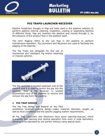
Day 1: External Anatomy Examination1. Each group should obtain a dissecting tray, fetal pig, dissecting equipment (scalpel,scissors, probe, forceps), and personal equipment (goggles and gloves). Use theautopsy report for recording of all observations.2. Cut the fetal pig bag near the top and carefully drain all the preservative fluid intothe sink. If any splashes on the counter, rinse off thoroughly. To make the roomsmell as best as possible, run the water for a few seconds to wash all the fluid downthe drain, making sure to rinse the whole bottom of the sink off.Image 1: Fetal Pig3. Examine the pig for body hair, although this is usually not conspicuous currently. Isbody hair present?a. Look under the chin for some longer hairs.4. Note the epitrichium, the layer of embryonic skin that is visibly peeling. This is lostas the hair develops. It may be removed by rinsing the pig in tap water. Use a sinkwith a disposal, if possible. The fetal skin will easily plug a sink; and care should betaken to prevent this.5. On the head locate the following structures:a. The mouth, bounded by upper and lower jaws and soft lips, is sometimespartially open, revealing a soft tongue. The front end of the head is prolongedinto a snout. The snout is used for rooting around in the soil for roots, insects,and other materials used by the pig for food. Do you have a snout?b. Observe the two nostrils (external nares) at the end of the snout.
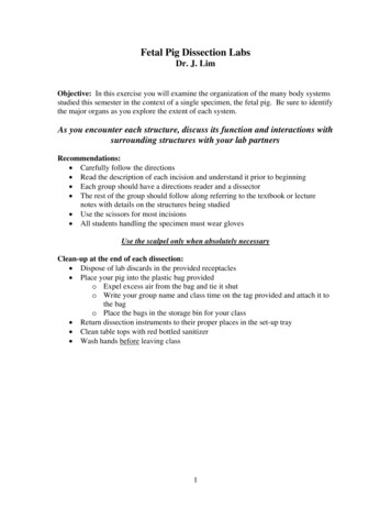
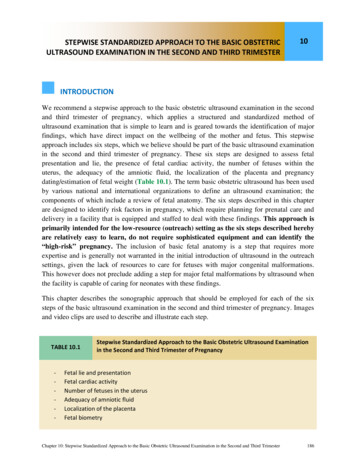
c. The eyes (usually closed) are covered by upper and lower eyelids fringed witheyelashes. Use a probe and pull the upper eyelids apart. The nictitatingmembrane should be visible in the medial corner of the eye. This transparentmembrane, which is referred to as a third eyelid, can move across the eyeballwith the eye open, thus providing protection. Check your partner’s eye for thisstructure. Is it present?d. Observe the eyes of the pig, carefully remove the eyelid so that you can viewthe eye underneath. Does it seem well developed? Do you think pigs are bornwith their eyes open or shut?e. The opening into the ear is called the external acoustic (auditory) meatus andthe flattened flap of skin is called the pinna, or auricle. The pinna and theexternal acoustic meatus make up the external ear in the pig as well as in thehuman.6. Image 2: Lateral view of pig’s head with salivary glands exposed: Label duringdissection. Carefully lay the pig on one side in your dissecting pan and cut away theskin from the side of the face and upper neck to expose the masseter muscle thatworks the jaw, lymph nodes, and salivary glands. The salivary glands kind of looklike chewing gum and are often lost if you cut too deeply.Image 2: Lateral View of Fetal Pig Head7. Open mouth: Label during dissection. Open the pig's mouth and locate the hard andsoft palate on the roof of the mouth. Can you feel your own hard and soft palateswith your tongue?8. Note the taste buds (also known as sensory papillae) on the side of the tongue.Locate the esophagus at the back of the mouth. Feel the edge of the mouth for teeth.Does the fetal pig have teeth? Are humans born with teeth?
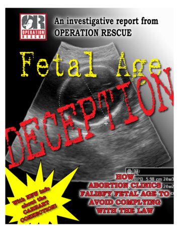
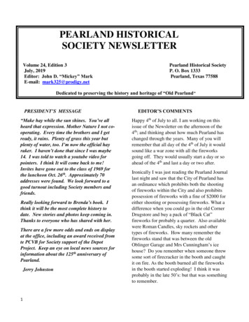
9. Locate the epiglottis, a cone-shaped structure at the back of the mouth, a flap of skinhelps to close this opening when pigs swallow. The pharynx is the cavity in the backof the mouth - it is the junction for food (esophagus) and air (trachea).10. Using your scissors, cut the corners of the jaw so that the mouth will remain open.Locate the following structures and label them on the diagram below: tongue, teeth,nasopharynx, glottis, epiglottis, hard palate, soft palate, salivary glands, and the opening ofthe esophagus.1Identify the structures on the diagram.1.22.3.4.345675.6.7.88.99.Image 3: Open Mouth of Fetal Pig11. Carefully examine the external features of your pig beginning with the head. Payattention to the amount and color of hair, birth marks, and other unique markings.Record your findings as you examine the mouth, nostrils, tongue, ears, and eyes.12. Note that the short neck joins the thorax in front of the first pair of legs. There isusually an incision in the right lateral part of the neck where the blood waswithdrawn, and colored latex was injected. The arteries should be represented with ared latex and while the veins were filled with a blue latex rubber.13. The trunk can be divided approximately into two general regions, consisting of ananterior thorax and a posterior portion, the abdomen.a. Note that the front limbs are attached to the thorax. The ribs making up thethorax are soft at this stage of development because they are made of cartilage.b. Locate the mammae which are present in both sexes. These form a double rowof small teats or mammary papillae on the ventral surface of the abdomen. Thenumber and location of the mammary glands vary in different species, but theglands are one of the distinguishing characteristics of all mammals.c. Observe the umbilical cord near the center of the ventral surface of theabdomen. If the cord is long enough, make a fresh cut across the end of it.
Three large blood vessels should now be visible. The largest of these is theumbilical vein which carries blood from the placenta to the fetal pig. Thisvessel may contain blue latex. The other two, smaller and with thicker walls,are the umbilical arteries, which may contain red latex. These vessels carryblood from the fetus to the placenta. Between or near the umbilical arteries; is asmall, hard core of tissue called the allantoic stalk. All the structures present inthe cord are embedded in a gelatinous connective tissue. Look up the functionof the placenta and record its purpose below. Do all mammals develop from aplacenta?Image 4: Umbilical Cord of Fetal Pigd. Locate the anus just ventral to the tail. This is the posterior opening of thedigestive tract. The anus is a sphincter muscle. Describe a sphincter muscle.14. Examine the ventral side and note any unusual markings. Note the number andlocation of mammary papillae (teats). Observe the end of the umbilical cord andidentify the umbilical cord vein, artery, and allantoic duct. The allantoic duct isconnected to the fetus’ bladder and is used for removing metabolic wastes. Thesestructures may be easier to view if you cut off a small portion of the chord.Image 5: Urogenital View of Fetal Pig
15. Determine the sex of your pig by looking for the urogenital opening. On females,this opening is located near the anus. On males, the opening is located near theumbilical cord. Record this in your Notebook.a. If your pig is female, you should also note that urogenital papilla is presentnear the genital opening. Males do not have urogenital papilla.b. Both males and females have rows of nipples, and the umbilical cord will bepresent in both.16. Determine the gender of your pig by using the figures above. If your pig is male,you will see the urogenital opening posterior to the umbilical cord. It serves as apassageway for urine and semen. Depending on the age you may or may not seescrotal sacs. The penis is not visible, but you can feel it by pressing the skin betweenthe urogenital opening and the scrotal sacs. The males’ mammary papillae willnever develop but in females they will develop into teats during pregnancy. If youpig is female, you will see the genital papillae under the tail. This releases metabolicwastes and is the opening to the reproductive system. Be sure to observe anothergroup’s pig of the opposite gender.17. Note that there are only four toes or digits on each limb as compared to five inhumans.18. Examine the legs and note that they have the same general structure as that ofhumans and other animals, although they are somewhat modified.a. Examine the posterior surface of one of the hind legs and note the largeprotuberance about two inches above the toes. This is comparable to thehuman heel, and the region from it to the toes corresponds to the human foot.Since the pig walks on the tips of the toes, the ankle and most of the foot isabove the ground.b. Locate the wrist and elbow of the forelimb and the knee and ankle of the hindlimb.19. Use a piece of string to measure the length of your pig. Stretch the string along itsback from the base of its tail to the tip of its nose. Make your measurement incentimeters. How many centimeters is it? Use the data from the Gestation Table todetermine the age of your pig in terms of gestation. How many days was the pig ingestation?20. Place a distinctive earring on your pig so that you will be able to find it easilyamongst the others. Most of the fetal pigs will look similar and it will be hard toidentify yours. Once pinned, you may now return the pig to its designated container.Wash your hands and clean up the area. Dissecting tools should be cleaned, driedoff, and returned to their proper spot.21. Lateral view of fetal pig: Label during lecture. Make sure you are familiar with termsof reference: anterior, posterior, dorsal, ventral. In addition, you'll need to know thefollowing terms: Medial: toward the midline or middle of the body; Lateral: towardthe outside of the body; Proximal: close to a point of reference; Distal: farther from apoint of reference.
Image 6: Lateral View of Fetal PigDay 2: Muscular SystemIntroductionIn today’s lab you are to locate and draw the gastrocnemius muscle of the pig. In manthis is the large calf muscle which terminates in the large Achilles tendon. This tendonthen inserts on the heel bone. In the pig the same thing occurs, however, you must becareful when locating the femur, the tibia, the fibula, and the calf muscle area on a pig.The gastrocnemius is covered by a thin sheet of muscle, the biceps femoris.Image 7: Lateral View of Fetal Pig Muscles
Directions for Today’s Dissection1. Work on the left side of the animal. Preserve the right side for circulatory andnervous system dissection. Skin the left leg. Cut carefully through the skin. Make acircle cut around the upper leg where it blends into the trunk of the body. The cutshould be about a one to two mm in depth. Cut from the top of the circle downwardtowards the foot. Now you should be able to remove the skin from the leg.2. Clear away any fascia which covers the muscles of the shank. Fascia will appearsolid sheet-like (no fibers) and will be from clear to white in color.3. Refer to Figure 7. Notice the biceps femoris covers the origin of the gastrocnemius.The biceps femoris should be removed carefully. The key to success is that muscletissue is fibrous and the direction the fibers run helps in identifying the muscle.Biceps femoris run obliquely; gastrocnemius fibers run parallel to the bone.4. The gastrocnemius originates on the lower end of the femur. It inserts by the Achillestendon on the calcaneus, the heel bone. This muscle extends the foot.5. Isolate the muscle at the origin and insertion, include the tendon. Have yourdissection okayed at this point. The only muscle fiber that can be cut is the bicepsfemoris. A good dissection will reveal a major nerve trunk running through themuscle in its upper one-third.6. Make a drawing of the lower leg below, showing the foot, knee, and upper leg. Labelthe following parts in your drawing: body of the muscle, origin and insertion pointsof the gastrocnemius, Achilles tendon, femur, calcaneus, and nerve.Analysis and Conclusion1. Make a drawing of your dissection and label the parts.
2. Describe the difference between muscle and tendon as to function and structure.Include a visual description of the differences between size, color, and texture ofeach.3. Is the gastrocnemius muscle considered a flexor or extensor? Explain your answer.4. What can a person who has just broken his or her Achilles tendon unable to do?Why?Day 3: Thoracic Cavity1. Put paper towels under the dissecting tray. Place the pig in the dissecting tray on itsback. Remember: when observing structures from the ventral side, left and right will bereversed. Slide a “body block” (sponge) under the back until is rests under itsshoulders (front legs). The block causes the neck and arms (pig’s front legs) to fallback will elevating the chest so that the 1st incision of the trunk is easier to make.2. For a better view of the abdomen, you can pull the legs farther apart by tying stringaround each leg and twisting the string around the spools in each corner of the tray(Image 8). You can also pull the string underneath the dissecting pan and tie it to theother leg. Don’t tie this too tight as you may want to adjust the ties as you open thechest cavity.Image 8: Tying the Fetal Pig
3. With a scalpel, the diener makes a Y-shaped incision (Image 9). Use the figure belowto guide you as to how to make the cuts and where to cut according to the gender ofyour pig. Do not cut off pieces of skin! You will need to sew the chest cavity backtogether once the autopsy is complete. The arms of the Y start from the top of eachshoulder anterior to the front legs and come down to the sternum which is directlyover the heart between the front legs. The incision should be just deep enough to cutthrough the muscular chest wall. Cut away the tissue from the underside of the flapof skin formed by the arms of the Y. Continue to cut the tissue as you pull the flapback toward the nose until the protruding larynx is exposed.Image 9: Incision GuidelinesThe organ systems that we will be exploring in depth during this dissection will be: The Respiratory SystemThe Circulatory SystemThe Digestive SystemThe Excretory SystemThe Reproductive SystemPrior to exploring each system in the fetal pig, you may virtually dissect at the followingwebsite: ation Procedures1. Make a median longitudinal incision through the muscles in the neck in order toexpose the larynx and trachea. Do not sever the blood vessels or nerves located oneither side of the trachea. Use Image 3, 11 and 12 as a guide for identification of thesestructures.a. The trachea contains rings of cartilage in its walls. Determine whether theserings are complete on the dorsal surface of the trachea.2. Remove muscular tissue from the larynx. Make a longitudinal incisionthrough the ventral wall of the larynx and locate the vocal folds, which aretwo small, shelf-like membranes. These are poorly developed in the fetal pig.
Image 10: Fetal Pig Internal Organs3.4.5.6.b. Locate the hyoid bone anterior to the larynx.c. The sublingual and submandibular glands are now visible adjacent to thelarynx.Identify the thymus gland. the large gland ventral to the heart. This gland consists oftwo major lobes which extend anteriorly into the neck region on either side of thetrachea. The thymus is relatively large in the fetus.The ventral neck muscles and the cervical part of the thymus gland cover the thyroidgland, the small, dark gland which lies on the upper trachea. Part the muscles andthymus gland to expose this gland.Observe the large right and left common carotid arteries and the internal jugularveins on each side of the trachea.The vagus nerve is the conspicuous white band that is bound to the dorsal surface ofthe common carotid artery. This nerve connects many of the thoracic and abdominalorgans as part of the autonomic nervous system.
7. Free the trachea, laterally, from the preceding blood vessels and nerves. Lying alongthe trachea, and attached to it, are the two slender inferior laryngeal nerves. Thesenerves which are essential for speech in humans originate from the vagus nerve and,although they are small and delicate, are easily seen against the trachea on eitherside.8. Locate the esophagus, the muscular tube dorsal to the trachea.9. Examine the interior of the thoracic cavity.a. Note that the thoracic cavity is divided into two lateral pleural cavities. whichcontain the lungs. The pericardial sac, which contains the heart. is in the space(mediastinum) between the lungs.b. The pleura is a double layered membrane which lines the thorax. That portionof the pleura lining the thoracic wall is called the parietal pleura; that whichcovers and adheres to the lungs is called the visceral pleura.c. The pericardium, the membrane surrounding the heart, is also composed oftwo layers: the outer parietal layer and the inner visceral attached to the heart.Much of the parietal pleura forming the medial walls of the pleural cavities istightly bound to the parietal pericardium.9. Remove thymus tissue in the thoracic cavity in order to study the lungs.a. Note that the lung is attached to other structures in the thorax only by the root.The root of the lung is formed by the bronchus, pulmonary artery and vein,bronchial arteries and veins, nerves, lymphatic vessels, and bronchial lymphnodes, all encircled by pleura.Image 11. Superficial View of the Thoracic Cavity with the Neck Dissected
10.11.12.13.b. Determine the number of lobes in each lung. Each lung is divided into threemajor lobes: apical, cardiac, and diaphragmatic. The right lung has anintermediate lobe beneath the apex of the heart.c. Cut off a small section of the left lung and note the density of the lung. Thelungs have not yet filled with air, since they are nonfunctional before birth.The trachea branches into a right and left bronchus dorsal to the heart. In order tolocate the right bronchus, push the heart to the left side of the thoracic cavity; thenlocate the inferior end of the trachea dorsal to the heart and right pulmonary bloodvessels. Try not to sever the pulmonary blood vessels.Locate the apical bronchus which leaves the trachea anterior to its termination andsupplies the right apical lobe. Note the right main (primary) bronchus whichsupplies the right cardiac and diaphragmatic lobes, and the small branch of thebronchus which supplies the intermediate lobe. Then scrape away the right cardiaclobe of the lung, bit by bit, noting the organization of the bronchial tree and bloodvessels and locate a primary bronchus. Leave the vessels intact. The branches of thebronchi can be identified by the cartilage in the walls.Locate the phrenic nerve. It is the conspicuous white line that passes along thepericardium to the diaphragm on either the right or left side of the heart.Lift the left lung and remove some of the parietal pleura dorsal to the lung to locatethe esophagus. Follow the esophagus to the diaphragm.14. At this point, make sure your teacher has seen your work. She will providedirections for cleanup.Image 12. Dissection of the Organs of the Thoracic Cavity of the Fetal Pig
Day 4: Abdominal CavityExamination Procedures1. Expose the organs in the abdominal cavity by making the incisions through the bodywall as shown in Figure3. Arrows indicate the directions in which they should bemade. First trace each incision by making a shallow cut with a scalpel through theskin, then continue the cut through the rest of the body wall with a pair of scissors.Lift the body wall toward you as you do this to avoid cutting internal organs.2. Make a pair of cuts from just in front of the umbilical cord and extend themposteriorly to the mammary papillae (nipples). The midventral strip of tissue lyingbetween this pair of incisions contains the umbilical arteries (injected with red latex),urinary bladder (the large sac situated between the two umbilical arteries), and, inthe male, the penis. This strip of tissue can be turned back by cutting the umbilicalvein that extends cranially from the umbilical cord to the liver. Cut the vein near theumbilical cord, leaving a long stump attached to the liver. You will need to find thisvein again later.3. Make a short cut that extends cranially from the umbilical cord to the posterior endof the sternum (breastbone), which you can feel. Look into the abdominal cavity andnotice the muscular diaphragm that forms a border between the abdominal andthoracic cavities.4. Make lateral incisions through the body wall (incision 3) just posterior to theattachment of the diaphragm, which you can feel with your fingers. These cutsshould follow the attachment of the diaphragm all the way to the back muscles. Youshould now have the abdominal cavity exposed. If the body cavity is filled with adark fluid, flush it out with water; be careful not to damage any organs.Image 13. Superficial View of the Digestive Organs of the Fetal Pig
5. Use Images 13 and 14 to assist you in locating the following organs:a. Locate the large, reddish-brown colored liver, posterior to the diaphragm. Notethat the superior surface of the liver is convex to match the concavity of thediaphragm. The liver produces bile, which is emptied into the duodenum andserves to break up fats.b. Count the number of lobes in the liver. The pig liver is divided into five lobes:the right lateral, right central, left central, left lateral, and a small caudate lobe.The caudate lobe is posterior to the right lateral lobe.c. Lift the right lobe of the liver and locate the gall bladder, the small, pearshaped sac embedded in the right central lobe. The gall bladder stores bileproduced by the liver.d. The umbilical vein can be found entering the liver to the left of the gallbladder.e. The cystic duct from the gall bladder and the hepatic duct from the liver uniteto form the common bile duct which empties into the duodenum (see Figure 5).In order to locate these structures, gently tease away the connective tissuebetween the stomach and the liver. First locate the cystic duct from the gallbladder and the common bile duct. The cystic duct may be stained green due tothe presence of bile. To locate the hepatic duct, trace the common bile ductupward to the point where the cystic duct enters it. The duct branching to theleft is the hepatic duct. It is necessary to dissect carefully to avoid destroyingthese structures.Image 14. Digestive Organs with the Liver and Spleen Pulled Back
6. Lift the liver to expose the stomach, the large, somewhat J-shaped organ located onthe left side of the abdominal cavity.a. Locate the entrance of the esophagus into the stomach. The stomach is the sightof mechanical digestion and, through actions by the enzyme pepsin, the initialsite of protein digestion.b. Identify the following regions of the stomach: the greater curvature, the side towhich the spleen is attached; the cardiac region where the esophagus joins thestomach; and the pyloric region, the region opening into the duodenum.c. Use a longitudinal incision from the cardiac region to the pyloric region to cutopen the stomach. The green debris found here and elsewhere in the digestivetract is called meconium. It consists of a bile-stained mucus, epithelial cellssloughed off from the skin and lining of the digestive tract, and amniotic fluidswallowed by the fetus. It is discharged in the first bowel movements of thenewborn. Wash the meconium out of the stomach.d. Observe the gastric mucosa lining the stomach and the rugae, the longitudinalfolds visible in the interior of the stomach.e. Locate the cardiac sphincter, a circular ring of smooth muscle surrounding theopening of the esophagus into the stomach. Note that the sphincter is tightlyclosed. This sphincter allows food into the stomach and prevents food frombacking up into the esophagus.f. Continue the same longitudinal incision through the pyloric sphincter. Thissphincter valve keeps food in the stomach until it is sufficiently broken down tobe handled by the duodenum.7. Locate the small intestine beginning at the posterior end of the stomach. The smallintestine is a long, coiled tube, divided into three regions: the duodenum, thejejunum, and the ileum.a. The anterior curved portion of the small intestine leaving the stomach is theduodenum. This portion is approximately 1 cm long. The common bile ductfrom the liver and gall bladder can be seen entering the duodenum. Pancreaticenzymes enter the duodenum and serve as the primary digestive agents in thissite of most chemical digestion.b. Open the duodenum by continuing the longitudinal incision through its wallfrom the pyloric sphincter, on the side away from the opening of the commonbile duct.c. The two remaining portions of the small intestine, the jejunum and the ileum,are approximately equal in length and have no readily distinguishableboundary. The jejunum is the middle portion of the small intestine, and theileum is the latter half that enters the large intestine. These two sections of theintestine represent the location of the greatest amount of absorption by thedigestive tract. Cut open a section of the small intestine and observe the velvetlike texture of the interior of the small intestine. This texture is due to smallfinger-like projections called villi that greatly increase the absorptive surface ofthe small intestine.
8. Locate the spleen the long, dark organ to the left of the stomach. It is attached to thegreater curvature of the stomach by means of the greater omentum, a specialized foldof the peritoneum. The spleen functions in the destruction of worn out red bloodcells and the production of some lymphocytes.9. The pancreas lies in the angle between the curve of the stomach and the duodenum.The greater part of the gland is located dorsal to the stomach. The pancreas secretesenzymes that act upon all major categories of food (carbohydrates, lipids, proteins,and nucleic acids); it also contains endocrine patches (the islets of Langerhans) thatproduce insulin and glucagon, hormones essential for normal glucose metabolism.The pancreas is connected to the duodenum by the pancreatic duct. This duct issmall and need not be dissected out.10. Locate the peritoneum, the double membrane lining the abdominal cavity.a. The parietal layer of the peritoneum lines the body wall; the visceral layercovers the abdominal organs.b. Locate the mesentery, the double layer of the peritoneum extending from thedorsal wall of the abdominal cavity to the small intestine. The mesenterycontains blood vessels, lymph vessels and nerves (see Figure 5). Nutrients areabsorbed into the bloodstream by the mesentery and then sent to the liver.11. Unravel and string out the small and large intestines by carefully cutting themesentery that hold the intestines in a tight ball. Do not dissect out these organs andbe as careful as possible. If successful, you will find a continuous tube starting fromthe pyloric sphincter and ending at the rectum of the large intestine.12. Trace the ileum to its point of attachment with the large intestine.a. The ileum opens into the side of the colon, forming a blind pouch, the cecum, atthe beginning of the colon. The cecum contains bacteria that serve to breakdown much of the cellulose that is present in the diet of herbivores. In man, thevermiform appendix is located inferior to the cecum. This is not present in thepig.b. Cut into the cecum, wash out the contents, and observe the ileocecal sphincter,which is found at the entrance to the small intestine and prevents material inthe colon from backing up into the small intestine.c. The first part of the large intestine in the fetal pig is called the spiral colon. It isvisible as a compact coi
9. Locate the epiglottis, a cone-shaped structure at the back of the mouth, a flap of skin helps to close this opening when pigs swallow. The pharynx is the cavity in the back of the mouth - it is the junction for food (esophagus) and air (trachea). 10.