Transcription
Available online at www.sciencedirect.comScienceDirectComput. Methods Appl. Mech. Engrg. 314 (2017) 180–195www.elsevier.com/locate/cmaThe mechanics of decompressive craniectomy: PersonalizedsimulationsJ. Weickenmeier a , C.A.M. Butler b , P.G. Young c , A. Goriely d , E. Kuhl a,e, a Department of Mechanical Engineering, Stanford University, Stanford, CA 94305, USAb Simpleware Ltd., Bradninch Hall, Castle Street, Exeter EX4 3PL, UKc College of Engineering, Mathematics and Physical Sciences, University of Exeter, Exeter, Devon, UKd Mathematical Institute, University of Oxford, Oxford, UKe Department of Bioengineering, Stanford University, Stanford, CA 94305, USAAvailable online 29 August 2016Highlights Decompressive craniectomy is a controversial surgical procedure with high failure rates.It induces large mechanical strains, which are believed to be the cause of brain damage.To quantify strains in the brain we create a personalized finite element craniectomy model.Our simulations reveal three potential failure mechanisms: stretch, compression, and shear.Our craniectomy model can identify regions at risk for brain damage on a personalized basis.AbstractDecompressive craniectomy is a traditional but controversial surgical procedure that removes part of the skull to allow an injuredand swollen brain to expand outward. Recent studies suggest that mechanical strain is associated with its undesired, high failurerates. However, the precise strain fields induced by the craniectomy are unknown. Here we create a personalized craniectomymodel from magnetic resonance images to quantify the strains during a decompressive craniectomy using finite element analysis.We swell selected regions of the brain and remove part of the skull to allow the brain to bulge outward and release the intracranicalswelling pressure. Our simulations reveal three potential failure mechanisms associated with the procedure: axonal stretch in thecenter of the bulge, axonal compression at the edge of the craniectomy, and axonal shear around the opening. Strikingly, for aswelling of only 10%, axonal strain, compression, and shear reach local maxima of up to 30%, and exceed the reported functionaland morphological damage thresholds of 18% and 21%. Our simulations suggest that a collateral craniectomy with the skullopening at the side of swelling is less invasive than a contralateral craniectomy with the skull opening at the opposite side: Itinduces less deformation, less rotation, smaller strains, and a markedly smaller midline shift. Our computational craniectomymodel can help quantify brain deformation, tissue strain, axonal stretch, and shear with the goal to identify high-risk regions forbrain damage on a personalized basis. While computational modeling is beyond clinical practice in neurosurgery today, simulationsof neurosurgical procedures have the potential to rationalize surgical process parameters including timing, location, and size, andprovide standardized guidelines for clinical decision making and neurosurgical planning.c 2016 Elsevier B.V. All rights reserved.⃝Keywords: Neuromechanics; Neurosurgery; Finite element analysis; Decompressive craniectomy; Hemicraniectomy Corresponding author at: Department of Mechanical Engineering, Stanford University, Stanford, CA 94305, USA.E-mail address: ekuhl@stanford.edu (E. Kuhl).http://dx.doi.org/10.1016/j.cma.2016.08.011c 2016 Elsevier B.V. All rights reserved.0045-7825/⃝
J. Weickenmeier et al. / Comput. Methods Appl. Mech. Engrg. 314 (2017) 180–1951811. MotivationDecompressive craniectomy is a life-saving neurosurgical procedure in which part of the skull is removed toprovide additional space for a swollen brain [1]. The surgical opening of the skull has been performed for morethan a century to reduce an elevated intracranial pressure in patients with traumatic brain injury, subarachnoidhemorrhage, and ischemic stroke [2]. Yet, because of its invasive nature, the procedure remains controversial andethically questionable [3]. While recent clinical trials suggest that decompressive craniectomy improves short-termpressure management and survival compared to pharmaceutical treatment alone, survivors of the procedure oftenexperience moderate to severe long-term disabilities [4,5]. These disabilities raise ethical concerns about appropriateselection criteria for patients who could benefit from the procedure [6]. Current guidelines suggest to perform acraniectomy when the intracranial pressure is greater than 20 mmHg for more than 30 min [7]. A chronically elevatedintracranial pressure can be devastating or, in the worst case, fatal; it causes a reduction in cerebral perfusion pressure,cerebral blood flow, and oxygenation [1].Decompressive craniectomy is often performed as a treatment of last resort to manage elevated pressure levelswhen medical management alone has proven unsuccessful. The procedure is based on removing large portions of theskull, opening the dura, the protective membrane that surrounds the brain, and allowing the brain to expand outward.The removed bone flap is typically preserved and re-implanted within the following weeks, once the swelling hasbeen fully resorbed [8]. Despite a marked increase in popularity, decompressive craniectomy remains a compromisebetween maximizing control of the intracranial pressure and minimizing side effects to the surrounding tissue [9]. Todate, the precise criteria when to perform a decompressive craniectomy, the optimal timing of treatment, the optimallocation and size of the skull opening, and the long-term functional outcome remain unclear.The location of the skull opening is a matter of ongoing debate. Decompressive craniectomy can be either unilateralwith an opening on the left or right hemisphere, or bilateral with a bifrontal opening across both hemispheres [1].Unilateral decompressive craniectomy, or hemicraniectomy, is by far the most common procedure [10]. It isrecommended in patients with unilateral hemispheric edema, a condition that can be diagnosed radiologically viaa midline shift to the contralateral side [3]. Under these conditions, it is common practice to perform a collateralcraniectomy and open the skull at the side of swelling. Surprisingly, recent computational studies suggest that tissuestrains can be reduced by performing a contralateral craniectomy at the opposite, non-injured side of the brain [11].Yet, the mechanisms that reduce tissue strains during contralateral skull opening are far from completely understood.The size of the skull opening is another clinical parameter of critical importance, especially in unilateralcraniectomy where the surgical space is anatomically limited. Studies report a direct correlation between the amountof bone removal and the reduction in intracranial pressure, which was found to be less than 40% when removing8000 mm3 of bone and greater than 80% when removing 12,000 mm3 [7]. Too large openings are associated withhigh rates of infection, complications during wound healing, challenges with cranial reconstruction, and a higherrisk to develop hydrocephalus [1]. Too small openings increase the risk of external cerebral herniation, an outwardsqueezing of the brain, which results in compression of the bridging veins, venous infarction, and increased edema atthe bone margins [8]. From a mechanical point of view, smaller openings naturally induce larger strains in the outwardbulging tissue and increase the risk of tissue damage [12]. As a result, guidelines recommend a minimum openingdiameter on the order of 12 cm [13]. Yet, this recommendation seems to be based on surgical experience rather thanon a sound quantitative analysis.Mathematical modeling is increasingly recognized as a promising strategy to predict the deformation field duringdecompressive craniectomy, the dimensions of the outward bulging tissue, the resulting strain field, and the regions atrisk for local tissue damage [14]. The complexity of existing models ranges from analytical models for idealizedaxisymmetric hyperelastic materials interpreted as an inverted punch problem [15] to finite element models foridealized axisymmetric poro-visco-elastic materials calibrated with gelatin hydrogel experiments [16]. Gelatin modelsare currently gaining popularity as precisely tunable surrogates of the human brain and provide valuable qualitativeinsight into brain deformation in neurosurgical training and pre-operative planning [17]. At the same time, as ourknowledge of brain mechanics increases, our computational models become more sophisticated. Since the firstcomputational craniectomy model was presented less than a decade ago [18], significant progress has been madetowards making the model anatomically more realistic [19]. To date, however, the most frequent application ofcomputational brain models is high impact loading in traumatic brain injury [20], and the use of computational modelsin surgical simulations remains the exception [21].
182J. Weickenmeier et al. / Comput. Methods Appl. Mech. Engrg. 314 (2017) 180–195Computational modeling allows us to virtually explore potential failure mechanisms, not only in response to highimpact loading [22], but also in response to surgical procedures. Elevated axonal strain has been hypothesized as apossible failure mechanism and tested in controlled single axon [23] and nerve tissue [24] experiments in vitro. Thesefindings have inspired microscopically motivated, anisotropic brain tissue models [25] to quantify the axonal strainsand von Mises stresses around a circular inclusion [26]. These two metrics are associated with the failure mechanismsof axonal stretch and axonal shear [20]. The definition of appropriate stress- and strain-based failure criteria anddamage thresholds is still hotly debated with suggested use of maximum principal values, von Mises values, or theirprojections normal and tangential to the axonal direction [27]. The objective of this study is to compare differentstrain-based damage criteria and to identify regions of maximum principal strain, radial stretch, and tangential stretchin response to a unilateral decompressive craniectomy using a personalized, anatomically detailed model of the humanbrain.2. Methods2.1. Constitutive modelWe begin by briefly summarizing the constitutive model for brain tissue based on a classical hyperelasticconstitutive formulation [28]. On time scales on the order of milliseconds or seconds characteristic for traumaticbrain injury, the brain behaves poro-visco-elastic and is highly sensitive to the loading rate [29,30]. On the timescales on the order of minutes or hours characteristic for a surgical procedure, it seems reasonable to approximatethe brain as purely hyperelastic [31,32]. Motivated by the literature, we follow the recommendation to model braintissue as an isotropic material [32], but interpret the resulting strain and stress fields in view of their projections ontopronounced axonal directions [27]. To characterize the behavior of the brain at finite deformations, we introduce thedeformation gradient F as the gradient of the nonlinear deformation field ϕ with respect to the material coordinatesX in the reference configuration. To account for the nearly incompressible behavior of brain tissue, we decomposethe deformation gradient F into a volumetric contribution characterized through the Jacobian J and an isochoriccontribution F̄,F X ϕ J 1/3 F̄withJ det(F)andF̄ J 1/3 F.(1)As a characteristic deformation measure, we introduce the left Cauchy Green deformation tensor b, which obeys asimilar decomposition into a volumetric contribution in terms of the Jacobian J and an isochoric contribution b̄,b F · Ft J 2/3 b̄J 2/3 det2/3 (F)withandb̄ F̄ · F̄t .(2)We can then introduce the isochoric first and second invariants, I 1 and I 2 , either in terms of the isochoric left CauchyGreen deformation tensor b̄ or in terms of the isochoric principal stretches λ̄1 , λ̄2 , and λ̄3 , recalling that I 3 J 2 1,1I 2 [ tr2 (b̄) tr(b̄2 )] λ̄21 λ̄22 λ̄22 λ̄23 λ̄23 λ̄21 .2Many common constitutive models for brain tissue are special cases of the general Ogden model [33],I 1 tr(b̄) λ̄21 λ̄22 λ̄23ψ andN ci αiλ̄1 λ̄α2 i λ̄α3 i 3 U (J ),αi 1 i(3)(4)where ci and αi are material-specific parameters and the function U (J ) controls the volumetric response. For thespecial case of N 2 with α1 2 and α2 2, the Ogden model simplifies to the popular Mooney–Rivlin model[34,35],11 2 2c1 [λ̄21 λ̄22 λ̄23 3] c2 [λ̄ 21 λ̄2 λ̄3 3] U (J ),22which we can reformulate in terms of the isochoric invariants (3),ψ ψ 11c1 [ I 1 3] c2 [ I 2 3] U (J ).22(5)(6)
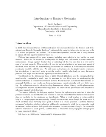
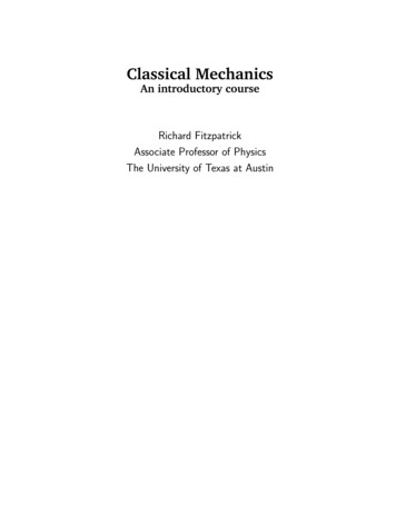
J. Weickenmeier et al. / Comput. Methods Appl. Mech. Engrg. 314 (2017) 180–195183Table 1Material parameters of the Mooney–Rivlin model in different tissue types.SubstructureParameter c1 [kPa]Parameter c2 [kPa]Cerebral gray matterCerebral white matterCerebellumCerebrospinal fluid0.280.560.280.03333.0666.0333.033.3The two Mooney–Rivlin parameters c1 and c2 are related to the shear modulus µ as 2 [c1 c2 ] µ. The Piola stressof the Mooney–Rivlin model follows from the Clausius Duhem inequality,P 111 U t ψ 2/3 [c1 I 1 c2 ] F 4/3 c2 F · Ft · F [ I 1 c1 2 I 2 c2 ] F t JF , F3 JJJ(7)and the Kirchhoff stress, τ P · Ft , is the partial push-forward of the Piola stress,τ 2 ψ111 U· b 2/3 [c1 I 1 c2 ] b 4/3 c2 b2 [ I 1 c1 2 I 2 c2 ] I JI. b3 JJJ(8)In the case of quasi-incompressibility, we could make a volumetric ansatz, U (J ) 21 κ [J 1]2 , and the derivative U/ J κ [J 1] p would take the interpretation of the pressure p. In the case of incompressibility, whereJ 1, p no longer follows from a constitutive equation; instead, p becomes an arbitrary Lagrange multiplierassociated with the incompressibility constraint, J 1. Given the incompressible nature of brain tissue, we followthe second approach and use a hybrid element formulation and evaluate the volumetric term using a mixed elementapproach to enforce the incompressibility constraint [36].Table 1 summarizes the material parameters of the different tissue types. We adapt the Mooney–Rivlin parametersc1 0.28 kPa and c2 333 kPa reported for brain tissue in the literature [37]. We assume that the cerebral whitematter is twice as stiff as the cerebral gray matter and the cerebellum [38]. For simplicity, we model the cerebrospinalfluid as an ultrasoft solid with a ten times lower stiffness than the cerebral gray matter. We assume that the individualtissue types display neither directional [26] nor regional [39] variations, and model them as isotropic [32] andhomogeneous [40]. We further assume all soft tissues as incompressible, and discretize them using hybrid lineartetrahedral C3D4H elements [36].To model brain swelling, we multiplicatively decompose the total Jacobian J J e J s into an elastic part J e and aswelling part J s . We gradually increase the amount of swelling from J s 1.0 to J s 1.10 to model a volumetricswelling of 10%. In the case of swelling, we re-parameterize the volumetric term U (J ) in the definitions of the freeenergies (4)–(6), the Piola stress (7), and Kirchhoff stress (8) in terms of the elastic Jacobian U (J e ) U (J/J s ).Within our computational model, we represent volumetric swelling via volumetric thermal expansion [36], and onlyallow selected regions of the cerebral white matter tissue to swell, while all other substructures remain purely elastic.2.2. Magnetic resonance imagingFig. 1 shows representative transverse, sagittal, and coronal slices of an adult female head that form the basis of ourmodel. We acquired these images at the Stanford University Center for Cognitive and Neurobiological Imaging usinga 3 T scanner (GE MR750, Milwaukee, WI) with a 32-channel radiofrequency receive head coil (Nova Medical, Inc.,Wilmington, MA). The collected data set contains 190 slices in the sagittal plane taken at a spacing of 0.9 mm. Eachimage slice has a matrix representation of 256 256 pixels with an in-plane resolution of 0.9 mm 0.9 mm [41].From the magnetic resonance images, we create a personalized high-resolution finite element model of the brain.The individual gray scales allow us to generate anatomically detailed and geometrically accurate three-dimensionalreconstructions of the individual substructures including the cerebral gray and white matter, the cerebrospinal fluid,the cerebellum, the skin, and the skull.2.3. Finite element modelFig. 2 illustrates the results of image processing, segmentation, and model reconstruction using the ScanIPsoftware environment (Simpleware Ltd, Exeter, UK). ScanIP features gray-scale filtering to differentiate between
184J. Weickenmeier et al. / Comput. Methods Appl. Mech. Engrg. 314 (2017) 180–195Fig. 1. Magnetic resonance images of an adult female brain. The image set contains 190 slices in the sagittal plane taken at a spacing of 0.9 mm.Each image slice has a matrix representation of 256 256 pixels with an in-plane resolution of 0.9 mm 0.9 mm. Images were acquired using a3 T scanner with a 32-channel radiofrequency receive head coil.Fig. 2. Personalized finite element model of the head including the skull (gray), the skin (brown), the cortical gray matter (red), the inner whitematter (pink), the cerebellum (green), and the cerebrospinal fluid (beige). The top left image shows the specific relative locations of these individualsubstructures and their dimensions within the skull. The six images on the top right visualize mesh contours and the geometric outlines of cerebraland cerebellar white matter, the cerebral gray matter, and the skull. The images in the lower row show coronal sections across the entire brainmodel.individual tissue layers [42]. This semi-automatic process iteratively produces an optimal segmentation of all relevantsubstructures [39]. In particular, we segmented a homogenized layer of skin, the skull, and the soft tissues that make upthe brain. Our model features a high resolution representation of the brain’s gray and white matter tissue. In addition,it contains the cerebellum in the posterior part of the brain and the cerebrospinal fluid which fills all cavities insidethe skull. Fig. 2 documents multiple representations of the geometric reconstruction, with the skull shown in gray, theskin layer in brown, the cortical gray matter in red, the inner white matter in pink, the cerebellum in green, and thecerebrospinal fluid in beige. The top layer shows the finite element mesh of the model.
J. Weickenmeier et al. / Comput. Methods Appl. Mech. Engrg. 314 (2017) 180–195185Table 2Personalized finite element model of the head with individual substructures and number of elementsand nodes.SubstructureSkullSkinCerebral gray matterCerebral white matterCerebellumCerebrospinal fluidWhole head modelNumber of elementsNumber of nodesElement HC3D4H1,275,808241,845C3D4/C3D4HC3D4 linear tetrahedral element, C3D4H hybrid linear tetrahedral element.In the cerebrum, our model differentiates between cerebral gray and white matter tissue for three mechanicallyand medically relevant reasons: First, gray and white matter possess clearly distinct microstructures that give rise toa different constitutive response [31,38]. Second, during neurodevelopment, the interplay between gray and whitematter plays a critical role in modulating the characteristic folding pattern of our brain, and our model should besufficiently general to be applicable in such situations [43]. Third, during most human neuropathologies, alterationsaffect either the gray or white matter tissue and to properly represent the etiology and disease progression, it isimportant to represent cerebral gray and white matter as individual substructures.In the cerebellum, our model simplifies the discretization to reduce model complexity. The human cerebellumis the most folded structure of our brain, mainly because of its unique function during sensory acquisition, motorcontrol, and regulation [44]. The significant folding enables high sensitivity and short signaling pathways, but iscomplex to account for in the computational model. In view of the complex interaction between the cerebellumand the cerebrospinal fluid as well as the cerebellum and cerebral gray matter we model the cerebellum as a singlehomogenized material.Fig. 2 shows multiple representations of our finite element model generated using our finite element meshing tool(Simpleware Ltd, Exeter, UK). The full head model consists of 1,275,808 linear tetrahedral elements with 241,845nodes. Of these, the skull consists of 43,614 elements and 14,591 nodes, the skin of 33,821 elements and 11,250nodes, the cerebral gray matter of 666,570 elements and 99,124 nodes, the cerebral white matter of 338,346 elementsand 53,719 nodes, the cerebellum of 10,208 elements and 3466 nodes, and the cerebrospinal fluid of 185,249 elementsand 59,595 nodes.Table 2 summarizes our personalized brain model with the individual substructures, the number of elements, and thenumber of nodes. We import our brain model into the commercial finite element software package Abaqus (DassaultSystèmes Simulia Corp., Providence, RI, USA) where we prescribe the material models, the boundary conditions, andthe interaction constraints for the individual substructures.2.4. Decompressive craniectomy modelWe use the brain model to simulate the effects of a unilateral decompressive craniectomy. Our finite element modelprovides a personalized, anatomically detailed representation of the head to investigate the mechanical strains andradial and tangential stretches upon brain tissue swelling as a potential source of diffuse axonal injury. We simulatethe surgical intervention of skull opening and allow the brain to bulge outward to reduce the intracranial pressure. Inthe clinical setting, a decompressive craniectomy usually takes four to six hours [45]. The actual opening of theskull involves the successive creation and removal of the bone flap and the gradual opening of the dura, whichtakes approximately half an hour [10]. This suggests that we can neglect effects of acceleration and simulate thedecompressive craniectomy as a quasi-static procedure. On these time scales, we also neglect porous and viscouseffects, which typically decay within three to five minutes [32], and simulate brain tissue as purely hyperelastic [46].The challenge of this procedure is to identify an optimal opening location and an optimal opening size to maximizethe control of the elevated intracranial pressure and, at the same time, minimize the risk of axonal damage.Fig. 3 illustrates our decompressive craniotomy model, which we create from the whole head model by introducinga circular opening with a diameter of 10 cm in the left posterior skull. To generate the opening, we semi-manually
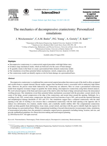
186J. Weickenmeier et al. / Comput. Methods Appl. Mech. Engrg. 314 (2017) 180–195Fig. 3. Personalized decompressive craniectomy model. We remove 2494 skull elements from the initial whole head model to introduce a circularopening with a diameter of 10 cm in the left posterior skull. This allows the swelling brain to bulge outward and release the elevated intracranialpressure. Whole brain without skull (left), decompressive craniectomy model with frontal and lateral brain regions removed for visualizationpurposes (middle), and decompressive craniectomy model of whole brain with surgically opened skull (right).Fig. 4. Boundary and loading conditions of our decompressive craniectomy model. Homogeneous Dirichlet boundary conditions at the lowerouter surface of the cerebrospinal fluid (red region) limit the motion of the inferior soft tissue regions (left). Frictionless contact at the remainingskull–fluid interface (pink region) enables a free sliding of the brain along the skull (second from left). Prescribed expansion mimics brain swelling.We compare the effects of decompressive craniectomy for three distinct cases: swelling of the white matter tissue in both hemispheres (middle),exclusively in the left hemisphere (second from right), and exclusively in the right hemisphere (right).remove a total of 2494 skull elements. Upon swelling, the brain can now bulge outward and release the elevatedintracranial pressure. Fig. 3 shows the whole brain without skull, the decompressive craniectomy model with frontaland lateral brain regions removed for visualization purposes, and the decompressive craniectomy model of the wholebrain with the surgically opened skull.Fig. 4 shows the boundary and loading conditions of our decompressive craniectomy model. To limit the motionof the inferior soft tissue regions, we apply homogeneous Dirichlet boundary conditions at the lower outer surfaceof the cerebrospinal fluid shown in red [14]. To reduce the computational time, we ignore the skin layer and modelthe skull as a rigid body. We assume a tight contact between all substructures, i.e., between the cerebral gray andwhite matter, the cerebellum, and the cerebrospinal fluid [14]. Only at the interface between the cerebrospinal fluidand the skull, we apply frictionless contact to enable a free sliding of the brain along the skull shown in pink. Wesimulate the swelling of the brain by prescribing a local volumetric expansion in a predefined white matter region.To explore the effects of decompressive craniectomy, we investigate three different scenarios of brain swelling: amaximum volumetric expansion of 10% in the white matter tissue of both hemispheres, exclusively in the collateralleft hemisphere, and exclusively in the contralateral right hemisphere. In all three scenarios, the circular skull openingis located in the posterior left hemisphere of the brain. We quantify and compare the mechanical response in terms ofthe overall deformation field, local stretches and strains, and the midline shift. The midline shift is a common clinicalindicator to characterize the degree of subcortical swelling and axonal damage.3. Results3.1. Progressive unilateral brain swellingFig. 5 shows the evolution of progressive unilateral swelling of the left hemisphere. The individual columns areassociated with the time points at 2%, 4%, 6%, 8%, and 10% volume expansion of the left white matter tissue. Theindividual rows show the deformed geometry, displacement, maximum principal strain, radial stretch, and tangential
J. Weickenmeier et al. / Comput. Methods Appl. Mech. Engrg. 314 (2017) 180–195187Fig. 5. Timeline of progressive swelling of the left hemisphere. Columns illustrate the time points at 2%, 4%, 6%, 8%, and 10% volumetricexpansion in the white matter tissue of the left hemisphere; rows show the deformed geometry, displacement, maximum principal strain, radialstretch, and tangential stretch. Strain and stretch are largest along the edge of the skull opening suggesting that axons in this region are mostvulnerable to damage and diffuse axonal injury.stretch. The displacement field illustrates the gradual outward herniation of the gray and white matter tissues resultingin a maximum displacement on the order of 6 mm. Strains and stretches take extreme values along the edge of the skullopening. These high values suggest that axons in this region are most vulnerable to damage and diffuse axonal injury.The maximum principal strain field shows a notable asymmetry: The average maximum principal strain of 5% in theswelling left hemisphere is twice as large as the strain in the non-swelling right hemisphere, which is squeezed againstthe outer right skull. Inside the brain, the average axonal stretch differs by about 10% between both hemispheres inaccordance with the prescribed amount of swelling. Axons in the swelling left hemisphere are stretched more thanaxons in the non-swelling right hemisphere. Along the edge of the skull opening increased swelling causes a severecompression in the contact region. In the gray matter tissue, this compression of up to 24% could trigger severe damageof dendritic connections in the cortex and cause axonal injury in the white matter layer underneath. Simultaneously,we observe shearing of up to 28% in the gray and white matter tissue. These high shear strains in both tissue types
188J. Weickenmeier et al. / Comput. Methods Appl. Mech. Engrg. 314 (2017) 180–195Fig. 6. Sagittal and transverse sections of the computational simulation of decompressive craniectomy in response to swelling of the lefthemisphere. Columns show the sagittal and transverse sections of the deformed geometry, displacement, maximum principal strain, radial stretch,and tangential stretch.could trigger axonal damage and lead to cortical and subcortical ischemic cell death due to restricted blood flow,restricted oxygen supply, and fiber rupture.Fig. 6 provides insight into the deformed geometry, displacement, maximum principal strain, radial stretch, andtangential stretch inside the brain. Inspired by the magnetic resonance images in Fig. 1, which provide sagittal, coronal,and transverse sections of the brain, we visualize the mechanical behavior inside the brain in response to swelling ofthe left hemisphere in representative sagittal and transverse sections. Especially transverse sections are widely used inmedial diagnostics to quantify the state of the swollen brain since they clearly visualize the in-plane deformation field,the dimension of the outward bulge, and the midline shift. The displacement field clearly highlights the magnitude ofoutward bulging on the open side of the skull. The maximum principal strain takes maximum values on the order of30% in a wide circular region around the opening. Across the gyri of the outward bulging region, maximum principalstrains are on the order of 10% indicating a slight compression of the outer surface across the bulge. The radialstretch takes maximum values o
knowledge of brain mechanics increases, our computational models become more sophisticated. Since the first computational craniectomy model was presented less than a decade ago [18], significant progress has been made towards making the model anatomically more realistic [19]. To date, however, the most frequent application of