Transcription
Huang et al. Stem Cell Research & Therapy (2017) 8:264DOI 10.1186/s13287-017-0719-7RESEARCHOpen AccessPellet coculture of osteoarthriticchondrocytes and infrapatellar fat padderived mesenchymal stem cells withchitosan/hyaluronic acid nanoparticlespromotes chondrogenic differentiationShu Huang1, Xiongbo Song1, Tao Li1, Jingfang Xiao2, Yemiao Chen2, Xiaoyuan Gong1, Weinan Zeng1,Liu Yang1* and Cheng Chen1*AbstractBackground: Cell source plays a key role in cell-based cartilage repair and regeneration. Recent efforts in cell coculturehave attempted to combine the advantages and negate the drawbacks of the constituent cell types. The aim of thisstudy was to evaluate the chondrogenic outcome of articular chondrocytes (ACs) and infrapatellar fat pad (IPFP)derived mesenchymal stem cells (MSCs) in direct coculture.Methods: ACs and IPFP MSCs from the same patients with knee osteoarthritis (OA) were cocultured in monolayer andin pellets. The monocultures of each cell type were also used as controls. Morphological and histologic analysis,immunofluorescence staining, reverse transcription-polymerase chain reaction, and enzyme-linked immunosorbentassay were performed to characterize the chondrogenic differentiation of cocultures. Furthermore, the effects ofchitosan/hyaluronic acid (CS/HA) nanoparticle exposure on the chondrogenesis of cocultures were examined.Results: In both monolayer and pellet coculture, the hypertrophy of MSCs and the inflammatory activities of ACs wereinhibited, although the chondrogenic production in coculture was not promoted compared with that in monoculture.In addition, the exposure of CS/HA nanoparticles to pellet coculture improved the production of type II collagen andaggrecan.Conclusions: We demonstrate for the first time that pellet coculture of ACs and IPFP MSCs with CS/HA nanoparticlescould promote chondrogenic outcome while preventing the inflammatory status of ACs and the hypertrophicdifferentiation of MSCs. These findings suggest that the combination of ACs, IPFP MSCs, and CS/HA might beuseful in cartilage repair in knee OA.Keywords: Msenchymal stem cells, Chondrocytes, Chondrogenesis, Osteoarthritis* Correspondence: jointsurgery@163.com; cclljjff@163.com1Center for Joint Surgery, Southwest Hospital, Third Military MedicalUniversity, Chongqing, ChinaFull list of author information is available at the end of the article The Author(s). 2017 Open Access This article is distributed under the terms of the Creative Commons Attribution 4.0International License (http://creativecommons.org/licenses/by/4.0/), which permits unrestricted use, distribution, andreproduction in any medium, provided you give appropriate credit to the original author(s) and the source, provide a link tothe Creative Commons license, and indicate if changes were made. The Creative Commons Public Domain Dedication o/1.0/) applies to the data made available in this article, unless otherwise stated.
Huang et al. Stem Cell Research & Therapy (2017) 8:264BackgroundOsteoarthritis (OA) is the most prevalent form of jointdisease [1], and is the leading global cause of disability[2]. A common hallmark of OA is the progressive loss ofarticular cartilage due to its avascular nature and lowmitotic activity of articular chondrocytes (ACs) embedded within the dense cartilage extracellular matrix(ECM) [3]. As such, significant research efforts areaimed at producing engineered cartilage as a cell-basedapproach for articular cartilage repair [4–6]. The mostcommonly used cell sources in cartilage tissue engineering and regenerative medicine are native cartilage cells,or autologous chondrocytes, but they undergo rapiddedifferentiation during in-vitro expansion, and result inthe formation of fibrocartilage with inferior mechanicalproperties [7, 8]. Mesenchymal stem cells (MSCs) havebeen widely investigated as an alternative cell source incartilage regeneration due to their great chondrogenicpotential [9, 10]. However, MSCs with the current chondrogenic differentiation protocol usually result in thehypertrophic phenotype and calcification [11, 12].To avoid the need for autologous chondrocyte expansion, and to enhance MSC differentiation to the chondrogenic lineage, the coculture approach [13, 14] of bothcell types has been developed recently and has shownpromising results. By coculturing rabbit chondrocyteswith MSCs, Shi et al. found that the proliferation ofchondrocytes is promoted and the chondrogenic differentiation of MSCs is enhanced [15]; Xu et al. furtherconfirmed that the proliferation of rabbit chondrocytesis provoked by the paracrine factors of MSCs [16]. Studies with human cells have also been encouraging; forexample, Li et al. showed that human umbilical cordblood-derived MSCs contribute to chondrogenesis in coculture with chondrocytes [17], and Zhang et al. cocultured human osteoarthritic chondrocytes with bonemarrow-derived MSCs and found that the proliferationof chondrocytes is increased and the inflammatory activity of chondrocytes is inhibited [18]. Although theseintensive research efforts have generated positive findings concerning coculture of MSCs and ACs, the clinicaltranslation of the coculture approach remains challenging. First, it requires extra invasive procedures to isolateMSCs from other tissues, which increases pain and costfor the patients; second, the success of some in-vitro coculture studies relies on the indirect contact of MSCsand ACs, which is difficult to implement in vivo.Here, we seek to develop a new strategy that avoidsnot only the isolation of normal chondrocytes from nonload bearing areas, but also the extra procedure forobtaining MSCs from other tissues. Following this idea,we cocultured human infrapatellar fat pad (IPFP)-derived MSCs and osteoarthritic ACs since both cellscould be obtained from a single arthroscopy. Our dataPage 2 of 12demonstrated that pellet coculture in vitro inhibits boththe hypertrophy of IPFP MSCs and the inflammatory activity of osteoarthritic chondrocytes. Moreover, thechondrogenesis of cells in pellet coculture is promotedin the presence of chitosan/hyaluronic acid (CS/HA)nanoparticles (NPs). Results of this study indicate thepossibility of developing a one-step cartilage repair byusing a combination of IPFP MSCs, ACs, and CS/HANPs for patients with knee OA.MethodsHuman IPFPs and cartilage tissues were obtained intraoperatively from total knee arthroplasties after informedconsent and approval from the Ethics Committee of Southwest Hospital (Chongqing, China). Only patients with primary knee OA (grade IV in The Kellgren Lawrence gradingsystem) were selected, and patients with inflammatory arthritis or with a history of prior knee surgery were excluded.Cell isolation and characterizationDonor-matched infrapatellar fat pad and articular cartilage were obtained from four OA patients (two male andtwo female, average age 65 3 years). For isolation ofIPFP MSCs, infrapatellar fat pad was harvested, washedin phosphate-buffered saline (PBS) supplemented with1% penicillin/streptomycin (P/S; Beyotime), and thendiced into 2-mm pieces. The diced tissues were digestedin 0.1% type I collagenase (Sigma) for 2 h, and theresulting cell suspension was filtered through a 40-μmcell strainer (BD Bioscience). The collected cells werecentrifuged (400 g for 5 min) and resuspended in RedCell Lysis Buffer (Beyotime) at room temperature in thedark for 10 min. The cells were then centrifuged again,and resuspended in Dulbecco’s modified Eagle’s medium(DMEM)/F12 (Invitrogen) supplemented with 10% fetalbovine serum (FBS; Gibco) and 1% P/S. The mediumwas changed every 2 days.For isolation of ACs, cartilage specimens were washedin PBS supplemented with 1% P/S and then diced. ACsfrom the diced tissues were isolated by digesting thematrix overnight in high-glucose DMEM (Invitrogen)supplemented with 0.2% type II collagenase (Sigma) and1% P/S. The resulting cell suspension was filteredthrough a 40-μm cell strainer; collected cells werecentrifuged (400 g for 5 min), and resuspended in highglucose DMEM supplemented with 10% FBS and 1% P/S. The medium was changed every 2 days.IPFP MSCs were characterized using flow cytometryand the Human MSC Analysis Kit (BD Biosciences).Briefly, cells were resuspended at a concentration of 1 107 cells/ml in 0.5% bovine serum albumin in PBS. Cellsaliquots (100 μl per tube) were stained with mesenchymal cell markers (CD90, CD105, CD73, and CD44) andimmunoglobulin (Ig)G1 and IgG2a isotype controls.
Huang et al. Stem Cell Research & Therapy (2017) 8:264Cell coculture and cell labelingIPFP MSCs at passage 2 and ACs at passage 0 from a single patient were used in coculture. For monolayer coculture, IPFP MSCs (2 105 cells) and ACs (2 105 cells) in2 ml chondrogenic medium were seeded onto each well ofa six-well plate. IPFP MSCs alone (4 105 cells per well)and ACs alone (4 105 cells per well) were used as controls. For three-dimensional pellet coculture, a cell suspension containing 1 106 IPFP MSCs and 1 106 ACswas centrifuged at 1000 rpm for 5 min in 15-ml polypropylene conical tubes. The supernatant was then removed,and 2 ml chondrogenic medium was gently added intoeach tube without disturbing the cell sediment. IPFPMSCs alone (2 106 cells per tube) and ACs alone (2 106 cells per tube) were used as controls. For both twoand three-dimensional coculture, the chondrogenicmedium used was Advanced DMEM (Gibco) supplemented with 1% P/S, 40 μg/ml L-proline (Biosharp),50 μg/ml insulin-transferrin-selenium-A supplement(Gibco), 10 ng/ml recombinant human transforminggrowth factor (TGF)-β3 (Peprotech), 0.1 mM ascorbicacid 2-phosphate (Sigma), and 100 nM dexamethasone(Sigma). Cells were maintained at 37 C and 5% CO2, andthe chondrogenic medium was replaced every 3 days.To distinguish these two cell types and to evaluate cellsurvival during coculture, IPFP MSCs and ACs wereincubated with the fluorescent dyes ine perchlorate (DiI;10 μM, Beyotime) and 3,3′-dioctadecyloxacarbocyanineperchlorate (Dio; 10 μM, Beyotime), respectively, beforecoculture. After 1 week of coculture, fluorescent imagesof the cells were taken and the cells were counted.Hematoxylin and eosin staining and Alcian blue stainingAfter 3 weeks of coculture, hematoxylin and eosin(H&E) staining for cell and matrix distribution andAlcian blue staining for proteoglycan were performed.For H&E staining, a staining kit (Beyotime) was used.For Alcian blue staining, samples were equilibrated in3% glacial acetic acid for 30 min, stained with 0.1%Alcian blue (Biosharp) dissolved in 3% glacial acetic acid(pH 2.5) for 30 min with constant agitation, and rinsedwith 3% glacial acetic acid three times for 30 min each.Immunofluorescence stainingAfter 3 weeks of coculture, cells in the monolayer werefixed in 4% paraformaldehyde, and cells in pellet werefrozen-sectioned. Non-specific bindings were blockedwith 1% bovine serum albumin in PBS for 60 min. Fixedcells were incubated with primary antibody (COL I,ab6038; COL II, ab185430; Aggrecan,ab3778; SOX9,ab185230; 1:100; Abcam) overnight at 4 C, followed byincubation with secondary antibody (Ms-488 or Rb-488;ZSGB-BIO) for 60 min at room temperature. Nuclei werePage 3 of 12stained with 0.1 μg/ml 4’6-diamidino-2-phenylindole(DAPI; Sigma) in PBS for 1 min. Immunostained sampleswere observed by an Olympus IX71 microscope.Reverse transcription-polymerase chain reaction (RT-PCR)RT-PCR was used to analyze the gene expression of chondrogenic markers. Total RNA was isolated from the cellmonolayer or pellets using TRIzol (Takara) following themanufacture’s protocol, and a RevertAid First StrandcDNA Synthesis Kit (Thermo Fisher Scientific). The targetgene primers were designed as follows: COL1A1, forward,CCTGGATGCCATCAAAGTCT, reverse, AATCCATCGGTCATGCTCTC; COL2A1, forward, TGCTGCCCAGATGGCTGGAGGA, reverse, TGCCTTGAAATCCTTGAGGCCC; COL10A1, forward, GGGAGTGCCATCATCG, reverse, GAGGCTTCACATACGTTT; ACAN, forward,TCGAGGACAGCGAGGCC, reverse, TCGAGGGTGTAGCGTGTAGAGA; SOX9, forward, GACTTCCGCGACGTGGAC, reverse, GTTGGGCGGCAGGTACTG; GAPDH,forward, GAGAACGGGAAACTTGTCAT, reverse, GGCAGGTCAGGTCAACAA. Real-time PCR was performedusing the PCR kit PowerUp SYBR Green Master Mix(Thermo Fisher Scientific). Assays were performed intriplicate.Enzyme-linked immunosorbent assay (ELISA)The supernatant of the culture medium was collected atdays 7, 14, and 21 of coculture. The expression of metallopeptidase (MMP)-13 (CSB-E04674h), interleukin (IL)1β (CSB-E08053h), and a disintegrin and metalloproteinase with thrombospondin motifs (ADAMTS)5 (CSBEL001312HU) were detected using ELISA (CUSABIO).According to the kit instructions, 100 μl standard orsample was added to each well and incubated for 2 h at37 C. Then 100 μl Biotin antibody (1 ) was added toeach well and incubated for 1 h at 37 C. After threewashes, 100 μl HRP-avidin (1 ) was added to each welland incubated for 1 h at 37 C. After five washes, 90 μlTMB Substrate was added to each well and incubatedfor 20 min at 37 C in the dark. Finally, 50 μl Stop Solution was added to each well. The optical density (OD)value of each well was measured at a wavelength of450 nm. This was positively correlated to its respectiveconcentration of MMP-13, IL-1β, and ADAMTS 5. Theexperiment was repeated three times.Fabrication and characterization of CS/HA NPsCS/HA NPs were prepared as previously described withminor modification [19, 20]. Briefly, CS (100 kDa) was dissolved in 2% acetic acid at a concentration of 1% (w/v),and HA (10 kDa) was dissolved in water at a concentrationof 0.1% (w/v). Both CS and HA solutions were mixed at aratio of 1:1 (v/v) and sonicated for 15 min at 25 C to formNPs. Eight different mixtures were prepared with CS/HA
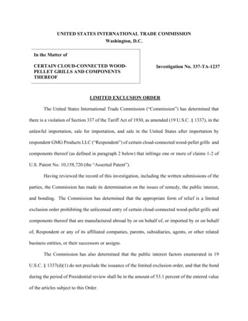
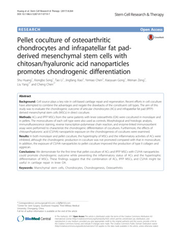
Huang et al. Stem Cell Research & Therapy (2017) 8:264weight ratios at 1:2, 1:1, 2:1, 3:1, 4:1, 5:1, 6:1, and 7:1. Accordingly, a constant HA concentration of 50 μg/ml wasused in all mixtures, and the CS concentration was variedas 25, 50, 100, 150, 200, 250, 300, and 350 μg/ml.To measure the size and charge of the CS/HA NPs, dynamic light scattering and zeta-potential measurement ofNPs in aqueous solution were performed with the MalvernZetasizer Nano ZS instrument (Malvern, UK) at 25 C. Thesamples were dried at room temperature after being stainedon copper grids, and then transmission electron microscopy (TEM) was performed on a TECNAI-10 microscope(Philips) at an acceleration voltage of 50 kV to observe theparticle morphology. FITC-conjugated CS/HA NPs wereused to confirm the cellular intake of these NPs.Page 4 of 12(Fig. 1a). Flow cytometry analysis (Fig. 1b) showed thatthese cells were positive for the surface markers CD90(88.39%), CD105 (83.43%), and CD73 (88.43%). Althoughexpressing weakly for CD44 (4.32%), these cells conformedto the minimal identification of human MSCs proposed bythe International Society for Cellular Therapy [21].The primary ACs had a polygon, spindle, or irregularshape (Fig. 1c, left). After immunolocalization of type IIcollagen, brown-yellow granules were found in the cells(Fig. 1c, middle). Alcian blue staining revealed that thechondrocyte cytoplasm and matrix were stainedgreenish-blue (Fig. 1c, right), indicating the deposition ofglycosaminoglycans (GAGs).Preliminary observation of cocultureStatistical analysisData were expressed as mean standard deviation (SD).Statistically significant differences between test groups weredetermined by one-way analysis of variance (ANOVA) at aconfidence interval of 95%. In addition, pairwise comparisons were made after ANOVA using the Holm-Sidak test.SigmaStat (Systat Software) was used for performing all thestatistical analysis.ResultsCell characterizationThe isolated IPFP MSCs based on plastic adhesion displayed a spindle-shaped morphology typical of MSCsAfter 1 week of coculture in chondrogenic medium, weobserved the cell morphology and viability. In monolayerculture, the morphology of both IPFP MSCs (Fig. 2a,left) and ACs (Fig. 2a, middle) did not change comparedwith cells in the growth medium; in the coculture image(Fig. 2a, right), most ACs could be distinguished fromIPFP MSCs. In pellet culture (Fig. 2b), cell depositionwas clearly observed at the bottom of the 15-ml tube.After being taken out of the tube, the pellets from theIPFP MSC group and coculture group were both about2 mm in size (Fig. 2b, inserts), while the pellet from theAC group collapsed due to inadequate strength. Thenumber of labeled cells in the monolayer (Fig. 2c, left)abcFig. 1 Characterization of infrapatellar fat pad-derived mesenchymal stem cells (MSCs) and articular chondrocytes (ACs). a Phase contrast imagesof P0, P1, and P2 MSCs. b MSCs were tested for mesenchymal surface markers (CD90, CD105, and CD73) by flow cytometry. c Phase contrastimage, immunohistochemistry (IHC) of type II collagen (Col II), and Alcian blue staining of P0 ACs
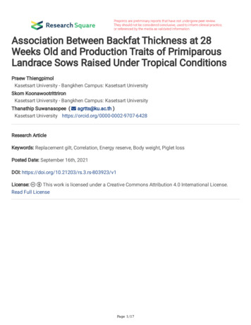
Huang et al. Stem Cell Research & Therapy (2017) 8:264Page 5 of 12abcFig. 2 Cell morphology and cell labeling after 1 week of chondrogenic coculture. a Phase contrast images of IPFP mesenchymal stem cells(MSCs) (left), articular chondrocytes (ACs) (middle), and coculture of MSCs and ACs (right) in monolayer. The green arrow points to ACs, and the redarrow points to MSCs in coculture. b Macroscopic observation of MSCs (left), ACs (middle), and coculture of MSCs and ACs (right) in pellets. Arrowspoint to cell sediments after 1 week of culture; inserts illustrate the size of the pellets. c MSCs were labeled with DiI (red) and ACs were labeledwith Dio (green) in monolayer (left) and pellet (middle) coculture. After 1 week of coculture, the labeled cells were counted, and the ratio of ACnumber to MSC number was calculated (right). Data are presented as mean SD. *P 0.05or in the pellet (Fig. 2c, middle) was calculated usingImageJ (Fig. 2c, right); in monolayer coculture, the ratioof AC number to IPFP MSC number was 0.88 0.04,indicating more loss of ACs; in pellet coculture, the ratioof AC number to IPFP MSC number was close to 1,indicating a balanced viability of both cells.Histologic analysis and immunofluorescence stainingAfter 3 weeks of coculture, H&E staining and Alcianblue staining were performed. In general, cells in pelletsshowed a better matrix deposition and GAG productioncompared to cells in monolayer. Specifically, for H&Estaining (Fig. 3a), cells in the monolayer coculture groupformed a large cell cluster; cells in the pellet coculturegroup formed a denser pellet compared to pellet cultureof IPFP MSCs or ACs alone. In particular, for Alcianblue staining (Fig. 3b), the coculture group did not showmuch difference compared with the other groups ineither monolayer or pellet culture.The immunofluorescence of SOX9 and major ECMproteins (type I, II, and X collagen) were also detected.In monolayer culture (Fig. 4a), the IPFP MSC group wasabundant in type X collagen, the AC group was abundant in type I collagen, and the coculture group wasabundant in both type I and type II collagen. Moreover,the staining of the chondrogenic markers aggrecan andSOX9 in the coculture group was much stronger thanthat in the IPFP MSC group or the AC group. In pelletsections (Fig. 4b), the staining of both type I and type IIcollagen was stronger in the coculture group than thatin the IPFP MSC group or the AC group, while thestaining of aggrecan and SOX9 was relatively similaramong the groups.Effects of CS/HA on the chondrogenesis of cocultureThe relationship between the weight ratio of CS/HA andthe size of the NPs is shown in Fig. 5a. There was a significant increase in nanoparticle size when the CS/HAratio increased from 1:2 to 1:1; with the increasingamount of CS, the size of the NPs decreased to 100 nmless. The smallest particle size (74.6 7.6 nm) was obtained at the CS/HA weight ratio of 4:1. The averagezeta potential became more positive with the increase inthe CS amount within the polyelectrolyte complex. TEMimage (Fig. 5a, insert) showed that the CS/HA NPs werespherical in shape and well dispersed. For subsequentexperiments, CS/HA NPs with a CS/HA weight ratio of4:1 were used. The fluorescent images (Fig. 5b) indicatedthat many FITC-conjugated NPs were taken by the cells.
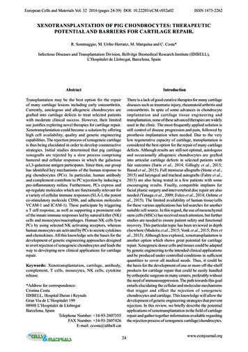
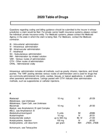
Huang et al. Stem Cell Research & Therapy (2017) 8:264Page 6 of 12abFig. 3 Histological analysis of monoculture of IPFP mesenchymal stem cells (MSCs), monoculture of articular chondrocytes (ACs), and coculture ofMSCs and ACs after 3 weeks of chondrogenic induction. a H&E staining showing cell and matrix distribution. b Alcian blue staining showingGAG depositionAfter 3 weeks of exposure to HA/CS NPs, the morphology and histology of the coculture group was examined. In monolayer coculture (Fig. 5c, top), the cells lostadherence to the culture dish and formed spheres. Inpellet coculture (Fig. 5c, bottom), the pellet was about4 mm in size; H&E staining showed that the coculture NP group was comparable in cell density to the pelletcoculture group, while Alcian blue staining indicated ahigher production of GAGs after NP intake.Fluorescent staining of the marker proteins was alsoperformed. In monolayer coculture (Fig. 5d, top), the intensity and distribution of the marker proteins could notbe determined because the cells formed spheres; in pellet coculture (Fig. 5d, bottom), fluorescent staining indicated a higher expression of aggrecan and type IIcollagen in the presence of HA/CS NPs (compared withthe lower-right images in Fig. 3a and b).Quantitative analysis of chondrogenic markers andinflammatory markersThe mRNA expression of the chondrogenic markersCOL2A1, SOX9, and ACAN, the fibrotic marker COL1A1,and the hypertrophic marker COL10A1 in both monolayerand pellet culture was analyzed after 3 weeks of culture. Inmonolayer culture (Fig. 6a), the MSC group had significantly lower expression of COL1A1 but significantly higherexpression of COL10A1 than the other groups, indicatingthat two-dimensional coculture did not weaken the fibrosisof chondrocytes but could inhibit the hypertrophy of IPFPMSCs. Furthermore, the coculture NP group had higherCOL2A1 expression than the AC group or the MSC groupalone, and lower ACAN expression than the coculturegroup. In pellet culture (Fig. 6b), the coculture NP grouphad significantly higher COL2A1 expression than the othergroups, and higher ACAN expression than the coculturegroup or the MSC group, suggesting that the addition ofNPs greatly improves the production of type II collagenand aggrecan in pellet coculture.To determine the level of cytokines and enzymes (IL1β, ADAMTS5, and MMP-13) during coculture, themedium at 1 week, 2 weeks, and 3 weeks was collectedand analyzed using ELISA. In general, there was a downward trend in concentration of IL-1β and MMP-13 asculture time increased in both monolayer culture andpellet culture. In monolayer culture (Fig. 7a), the ACgroup kept expressing a higher level of IL-1β than theother groups, and only expressed higher expression ofMMP-13 than the other group in the first week. In pellet
Huang et al. Stem Cell Research & Therapy (2017) 8:264AggrecanCol-ICol-IICol-XSOX9MSCs ACsACsMSCsaPage 7 of 1250 µmMSCs ACsACsMSCsb50 µmFig. 4 Immunofluorescence assay of monoculture of IPFP mesenchymal stem cells (MSCs), monoculture of articular chondrocytes (ACs), andcoculture of MSCs and ACs after 3 weeks of chondrogenic induction. a Fluorescent images of two-dimensional monolayers. b Fluorescent imagesof three-dimensional pellet sections. Col collagenculture (Fig. 7b), a significant difference was only detected at 2 weeks, and there was no difference of cytokine concentration among the groups at 1 week or at3 weeks. Note that there was no difference in cytokine/enzyme concentration between the coculture group andthe coculture NP group, indicating that the presence ofNPs neither stimulated nor restrained the release ofthese cytokine/enzymes.DiscussionThe success of cell-based therapies for cartilage-relatedinjury and disease requires fine control of cell differentiation. However, chondrocytes alone usually end upshowing the fibrotic phenotype, while MSCs alone leadto the hypertrophic phenotype in cartilage tissue engineering. For this reason, chondrocyte-MSC coculture systemshave been recently employed in several studies [22] to limitchondrocyte dedifferentiation and MSC hypertrophy. Thetissue sources of MSCs used in those studies are distinctfrom articular cartilage; thus, multiple procedures areneeded to isolate chondrocytes and MSCs for coculture. Inthis study, we selected human IPFP MSCs to coculture withosteoarthritic chondrocytes. The biggest advantage of IPFPMSCs over other stem cells is that IPFP MSCs along withchondrocytes can be extracted from the same knee jointduring a single knee arthroscopy, thereby avoiding multipleinvasive procedures. The safety and suitability of IPFPMSCs from OA patients has also been proven in vitro [23–25] and in clinical studies [26, 27]. Furthermore, it has beenshown that IPFP MSCs have a higher capacity for chondrogenic differentiation than MSCs from body fat, bone marrow, and Wharton’s jelly of the umbilical cord [28].Here, we cocultured IPFP MSCs and ACs from thesame osteoarthritic knee in both monolayer and pelletculture. We did not examine the effects of cell ratio inthis study, but instead simply chose a 50:50 ratio ofMSCs to ACs since it has been shown that this ratioprovides optimal type II collagen expression and GAGproduction in several studies [29–31]. However, themaximum ratio of MSCs to ACs still needs investigationin further studies. In terms of culture condition, the pellet culture, a well-established three-dimensional culturemethod without scaffolding materials, exhibited morematrix deposition compared with monolayer culture, asevidenced by our H&E and Alcian blue staining results.In particular, when we cocultured ACs with IPFP MSCs
Huang et al. Stem Cell Research & Therapy (2017) 8:264aPage 8 of 12bNCMSCsACsMSCs ACs30Zeta Potential70020600105000400-10300500 nm200-20100-300Zeta Potential(m V)Mean nanoparticles size(nm)Mean nanoparticles size800-401/21/12/13/14/15/16/110 µm7/1C S /H A (W /W )MorphologyMonolayercH&E50 µm50 µm50 µm50 µmPellet50 µmAlcian BlueAggrecanCol-ICol-IICol-XSOX950 µmPelletMonolayerd50 µmFig. 5 Effects of chitosan/hyaluronic acid (CS/HA) NP exposure on IPFP mesenchymal stem cell (MSC) and articular chondrocyte (AC) cocultures. aEffect of the weight ratio of CS to HA on the size and zeta potential of NPs. Insert: TEM micrograph of CS/HA NPs. b Fluorescent images of cellsexposed to FITC-conjugated CS/HA NPs for 1 h. c Morphology, hematoxylin and eosin (H&E), and Alcian blue staining of monolayer (top) andpellet (bottom) culture of MSCs ACs NPs. d Immunofluorescence assay of monolayer (top) and pellet (bottom) culture of MSCs ACs NPs. Colcollagen, NC negative controlin monolayer in chondrogenic medium for 1 week, therewas a significant loss of chondrocyte numbers. This resultcontradicts a previous study [32] in which the human chondrocytes had a fourfold increase in cell numbers after 7 daysof monolayer culture in chondrogenic medium. Despite thisdiscrepancy, our major finding and that of the other studyboth exhibit the advantage of three-dimensional culture forchondrogenesis.Interestingly, our PCR analysis revealed that, whetherin monolayer or pellet culture, there was no significantdifference in expression of the marker genes COL2A1,ACAN, and SOX9 between the coculture group and themonoculture control, indicating that coculture did notchange the chondrogenic capacity of both cells. Inaddition, there was no significant difference in COL1A1expression between the AC monoculture group and thecoculture group, indicating that the fibroblastic differentiation of ACs was not reduced in coculture; however,there was a significant difference in COL10A1 expression between the MSC monoculture group and thecoculture group, indicating that hypertrophy of MSCswas inhibited in coculture. This inhibition of MSC
Huang et al. Stem Cell Research & Therapy (2017) 8:264Page 9 of 12abFig. 6 Expression profile of COL1A1, COL2A1, COL10A1, ACAN, and SOX9 after 3 weeks of chondrogenic induction in monolayer (a) and pellet(b) culture. Gene expression of each group was normalized to the AC group. Data are presented as mean SD. *P 0.05. AC articularchondrocyte, MSC mesenchymal stem cell, NP nanoparticlehypertrophy might be attributed to the parathyroidhormone-related protein (PTHrP) secreted by ACsthroughout the coculture [33, 34].We further added CS/HA NPs into the AC-MSC coculture. Both CS and HA have been widely used inclinics; in particular, HA injection has been used as atreatment for knee OA [35]. Moreover, the CS/HA NPsare usually used as drug delivery vehicles [36, 37].Therefore, the use of CS/HA NPs might add beneficialeffects to coculture, and would facilitate drug loading(such as growth factors and anti-inflammatories) ifnecessary. In monolayer coculture, the cells lost adherence to the culture dish and formed spheres after theaddition of CS/HA NPs. Similarly, in another study,MSCs formed spheres after transwell coculture withchondrocytes in the presence of TGF-β [38]. Therefore,we speculate that, given strong chondrogenic stimuli(HA, TGF-β, and so forth), MSCs in monolayer culturefavor cellular aggregation rather than plastic adhesion.In pellet coculture, the addition of CS/HA NPs increasedthe expression level of the marker genes COL2A1 andACAN, indicating that the chondrogenesis of pelletcoculture was promoted by CS/HA NPs. This could beattributed to the presence of HA, since it has beenshown that HA causes a fourfold increase in chondrogenic differentiation of IPFP MSCs [28].Since the ACs and IPFP MSCs were both from OA patients, the inflammatory state of the cells should be aconcern. Here, we examined the concentration of IL-1β(a major cytokine in OA [39]), AD
drogenic differentiation protocol usually result in the hypertrophic phenotype and calcification [11, 12]. To avoid the need for autologous chondrocyte expan-sion, and to enhance MSC differentiation to the chon-drogenic lineage, the coculture approach [13, 14] of both cell types has been developed recently and has shown promising results.