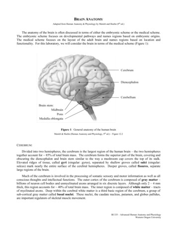
Transcription
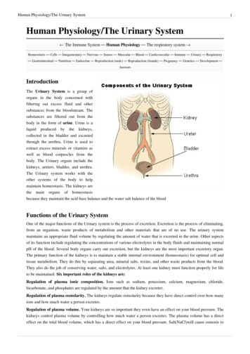
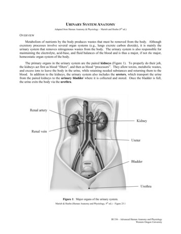
URINARY SYSTEM ANATOMYAdapted from Human Anatomy & Physiology – Marieb and Hoehn (9th ed.)OVERVIEWMetabolism of nutrients by the body produces wastes that must be removed from the body. Althoughexcretory processes involve several organ systems (e.g., lungs excrete carbon dioxide), it is mainly theurinary system that removes nitrogenous wastes from the body. The urinary system is also responsible formaintaining the electrolyte, acid-base, and fluid balances of the blood and is thus a major, if not the major,homeostatic organ system of the body.The primary organs in the urinary system are the paired kidneys (Figure 1). To properly do their job,the kidneys act first as blood “filters”, and then as blood “processors”. They allow toxins, metabolic wastes,and excess ions to leave the body in the urine, while retaining needed substances and returning them to theblood. In addition to the kidneys, the urinary system also includes the ureters, which transport the urinefrom the paired kidneys to the urinary bladder where it is collected and stored. Once the bladder is full,the urine exits the body via the urethra.Figure 1: Major organs of the urinary system.Marieb & Hoehn (Human Anatomy and Physiology, 9th ed.) – Figure 25.1BI 336 – Advanced Human Anatomy and PhysiologyWestern Oregon University
FUNCTIONAL ANATOMYKidneyThe paired kidneys lie in a retroperitoneal position (between the dorsal body wall and the parietalperitoneum) in the superior lumbar region. Extending approximately from T 12 to L3, the kidneys receivesome protection from the lower part of the rib cage. The right kidney is slightly lower than the left kidneybecause it is “crowded” by the liver. In a living person, fat deposits (call the adipose capsule) hold thekidneys in place against the muscles of the posterior trunk wall (Figure 1).Each kidney is fed by a renal artery which branches off the descending aorta. A renal vein drainsblood from each kidney, entering into the inferior vena cava. These vessels enter / exit the kidney in theindented medial region of the kidney called the renal hilum.Externally, each kidney is surrounded by a smooth transparent membrane called the fibrous capsule(Figure 2). Internally, the kidney is divided into several regions. The renal cortex is the most superficialregion. It appears lighter in color and is the site of glomerular filtration and a majority of tubularreabsorption / secretion. Deep to the cortex is located the renal medulla. The medulla, appearing muchdarker in color, is segregated into triangular areas called the medullary (renal) pyramids. The base of eachpyramid faces toward the cortex with the pointed apex, termed the renal papilla, directed toward theinnermost region of the kidney. The renal pyramids are separated by the renal columns, which arecomposed of tissue similar in appearance to the renal cortex.The innermost region of the kidney, medial to the renal hilus, consists of a relatively flat, basin-likecavity called the renal pelvis. Finger-like extensions of the pelvis form cup-like areas called calyces thatenclose the renal papilla of the medullary pyramids. Minor calyces immediately surround each renalpapilla with the minor calyces then joining to form major calyces which drain into the renal pelvis.Figure 2: Internal anatomy of the kidney.Marieb & Hoehn (Human Anatomy and Physiology, 9th ed.) – Figure 25.3BI 336 – Advanced Human Anatomy and PhysiologyWestern Oregon University
Exercise 1Utilizing the kidney model, locate the following structures: fibrous capsule, renal hilum,renal cortex, renal medulla, medullary pyramid, renal column, renal papilla, renal pelvismajor calyx, and minor calyx.Kidney Blood SupplyThe kidneys continuously cleanse the blood and adjust its composition, so it is not surprising that theyhave a rich blood supply. Under normal resting conditions, the large renal arteries deliver one-fourth of thetotal cardiac output ( 1200 ml) to the kidneys each minute.The renal arteries exit at right angles from the abdominal aorta. As each renal artery approaches akidney, it divides into five segmental arteries (Figure 3). Within the renal sinus, each segmental arterybranches further to form several interlobar arteries. At the cortex-medulla junction, the interlobararteries branch into the arcuate arteries that arch over the bases of the medullary pyramids. Small corticalradiate arteries radiate outward from the arcuate arteries to supply the cortical tissue.Afferent arterioles branching from the cortical radiate arteries begin a complex arrangement ofmicroscopic blood vessels. These vessels are key elements of kidney function which will be examinedshortly during the description of the nephron.Figure 3: Blood vessels of the kidney.Marieb & Hoehn (Human Anatomy and Physiology, 9th ed.) – Figure 25.4BI 336 – Advanced Human Anatomy and PhysiologyWestern Oregon University
Veins trace the pathway of the arterial supply in reverse (Figure 3). Blood leaving the renal cortexdrains sequentially into the cortical radiate veins, arcuate veins, interlobar veins, and finally the renalveins.Exercise 2Utilizing the kidney and nephron models, locate the following vessels: renal artery,segmental artery, interlobar artery, arcuate artery, cortical radiate artery, cortical radiatevein, arcuate vein, interlobar vein, and renal vein.Microanatomy of KidneyAt the microscopic level, each kidney is composed of upwards of a million nephrons, the anatomicalunit responsible for forming urine (Figure 4). Each nephron consists of a glomerulus (a capillary cluster)and a renal tubule. Each renal tubule begins as a blind-ended sac that gradually surrounds and encloses anadjacent glomerulus. The enlarged end of the tubule encasing the glomerulus is the glomerular(Bowman’s) capsule, and its inner wall consists of specialized cells with long branching processes calledpodocytes. Podocytes cling to each other and to the endothelial wall of the glomerular capillaries, forming avery porous membrane around the glomerulus.Figure 4: Location and structures of the nephron.Marieb & Hoehn (Human Anatomy and Physiology, 9th ed.) – Figure 25.5BI 336 – Advanced Human Anatomy and PhysiologyWestern Oregon University
The anatomical areas of the rest of the renal tubule, in order from the glomerular capsule, are theproximal convoluted tubule, loop of Henle, and the distal convoluted tubule. The proximal convolutedtubules are formed by cuboidal epithelial cells with their luminal (exposed) surfaces bearing densemicrovilli – this is where a large part of the filtrate reabsorption occurs. The distal convoluted tubules aresimilar in structure to the proximal convoluted tubules except the cells appear smaller and lack microvilli.The U-shaped loops of Henle are either composed of simple squamous epithelium (thin segment) or simplecuboidal / columnar (thick segment), depending on whether the region is involved in the passive diffusionof water or the active transport of salts.Most nephrons, called cortical nephrons, are located entirely within the cortex. However, parts of theloops of Henle of the juxtamedullary nephrons penetrate well into the medulla. The collecting ducts,each of which receives urine from many nephrons, run downward through the medullary pyramids to emptythe urine product into the calyces and ultimately the pelvis of the kidney.Nephron function depends on some unique features of the renal circulation. There are two distinctcapillary beds, the glomeruli (described earlier) and the peritubular capillaries. The glomerulus, fed by theafferent arteriole and drained by the efferent arteriole, is responsible for forming the filtrate which isprocessed by the renal tubule. Peritubular capillary beds arise from the efferent arterioles and cling to therenal tubules. This capillary bed is responsible for absorbing solutes and water that are reclaimed from thefiltrate by the tubule cells. Specialized peritubular capillaries, called vasa recta, are found around the deeploops of Henle associated with the juxtamedullary nephrons. These vessels play an important role informing concentrated urine.Exercise 3Utilizing the model showing a kidney nephron, located the following structures:glomerulus, glomerular capsule, proximal convoluted tubule, loop of Henle (thin / thicksegments), distal convoluted tubule, collecting duct, afferent arteriole, efferent arteriole,peritubular capillaries, vasa recta, cortical nephron, and juxtamedullary nephron.Exercise 4On the class webpage (www.wou.edu/ lemastm/Teaching/BI336) there is a PowerPointfile containing histological images taken from the urinary system. Find the image labeled‘Kidney’ and properly label renal cortex, renal medulla, glomerulus, glomerular capsule,podocyte, proximal convoluted tubule, distal convoluted tubule, thin segment of loop of Henle,and thick segment of loop of Henle. Next, examine an actual histological slide (see slide boxat your bench) taken from a kidney and visually identify the structures listed above.BI 336 – Advanced Human Anatomy and PhysiologyWestern Oregon University
UreterThe ureters are slender tubes that convey urine from the kidneys to the bladder. Each ureter begins atthe level of L2 as a continuation of the renal pelvis (Figure 1). From there, it descends behind theperitoneum and runs obliquely through the posterior bladder wall. This arrangement prevents backflow ofurine because any increase in bladder pressure compresses and closes the distal ends of the ureters.Exercise 5Utilizing the model showing the complete urinary system, locate the ureters.Histologically, the ureter wall has three layers. The mucosa contains a transitional epithelium that iscontinuous with the mucosae of the kidney pelvis superiorly and the bladder medially. The muscularis iscomposed chiefly of two smooth muscle sheets – the internal longitudinal layer and the external circularlayer. The adventitia covering the ureter’s external surface is typical fibrous connective tissue.Exercise 6Find the image from the PowerPoint file containing histological images labeled ‘Ureter’and properly label mucosa, transitional epithelium, muscularis, and adventitia. Next,examine an actual histological slide (see slide box at your bench) taken from a ureter andvisually identify the structures listed above.Urinary Bladder:The urinary bladder is a smooth, collapsible, muscular sac that stores urine temporarily (Figure 5).The bladder is located retroperitoneally on the pelvic floor just posterior to the pubic symphysis. Theinterior of the bladder has openings for both ureters (ureteric orifices) and the urethra. The smooth,triangular region of the bladder based outlined by these three openings is the trigone, important clinicallybecause infections tend to persist in this region.Exercise 7Utilizing the model showing the complete urinary system, located the bladder, trigone,and ureteric orifices.The wall of the bladder is composed of three layers: a mucosa containing a transitional epithelium, athick muscular layer called the detrusor muscle, and a fibrous adventitia. When empty, the bladdercollapses into its basic pyramidal shape and its walls are thick and thrown into folds. When full, the bladderexpands, becomes pear-shaped, and rises superiorly in the abdominal cavity.BI 336 – Advanced Human Anatomy and PhysiologyWestern Oregon University
Figure 5: Structure of the urinary bladder and urethra (female).Marieb & Hoehn (Human Anatomy and Physiology, 9th ed.) – Figure 25.20Exercise 8Find the image from the PowerPoint file containing histological images labeled ‘UrinaryBladder’ and properly label mucosa, transitional epithelium, detrusor, and adventitia. Next,examine an actual histological slide (see slide box at your bench) taken from a urinary bladderand visually identify the structures listed above.Urethra:The urethra is a thin-walled muscular tube that drains urine from the bladder and conveys it out of thebody (Figure 5). At the bladder-urethra junction, the detrusor smooth muscle thickens to form the internalurethral sphincter. This involuntary sphincter, controlled by the autonomic nervous system, keeps theurethra closed when urine is not being passed and prevents leaking between voiding. The external urethralsphincter surrounds the urethra as it passes through the urogenital diaphragm. This sphincter is formed ofskeletal muscle and is voluntarily controlled.The length and functions of the urethra differ in the two sexes. In females, the urethra is only 3 – 4 cmlong and fibrous connective tissue binds it tightly to the anterior vaginal wall. In males the urethra isapproximately 20 cm long and has three distinct regions: the prostatic urethra, which runs through theprostate gland, the membranous urethra, which runs through the urogenital diaphragm, and the spongyurethra, which runs through the penis. The male urethra serves two functions – it carries semen as well asurine out of the body.BI 336 – Advanced Human Anatomy and PhysiologyWestern Oregon University
Exercise 9Utilizing the model showing the complete urinary system, located the urethra, internalurethral sphincter, and external urethral sphincter.The epithelium of the urethral mucosa is mostly pseudostratified columnar epithelium with shortstretches of transitional epithelium near the urinary bladder and stratified squamous epithelium near theexternal opening. Otherwise, the muscularis is composed chiefly of two smooth muscle sheets – theinternal longitudinal layer and the external circular layer – and the adventitia covering the urethral surfaceis typical fibrous connective tissue.Exercise 10Find the image from the PowerPoint file containing histological images labeled ‘Urethra’and properly label mucosa, pseudostratified columnar epithelium, muscularis, and adventitia.Next, examine an actual histological slide (see slide box at your bench) taken from a urinarybladder and visually identify the structures listed above. (Note: The actual histological slide maycontain different epithelial types depending on where along the urethra the section was taken)BI 336 – Advanced Human Anatomy and PhysiologyWestern Oregon University
Marieb & Hoehn (Human Anatomy and Physiology, 9th ed.) - Figure 25.5 . BI 336 - Advanced Human Anatomy and Physiology Western Oregon University . collapses into its basic pyramidal shape and its walls are thick and thrown into folds. When full, the bladder expands, becomes pear-shaped, and rises superiorly in the abdominal cavity. .