Transcription
(2021) 13:93Cao et al. Clin Open AccessREVIEWThe role of DNA methylation in syndromicand non‑syndromic congenital heart diseaseJiali Cao1†, Qichang Wu2†, Yanru Huang3, Lingye Wang4, Zhiying Su2* and Huiming Ye1,4*AbstractCongenital heart disease (CHD) is a common structural birth defect worldwide, and defects typically occur in the wallsand valves of the heart or enlarged blood vessels. Chromosomal abnormalities and genetic mutations only accountfor a small portion of the pathogenic mechanisms of CHD, and the etiology of most cases remains unknown. The roleof epigenetics in various diseases, including CHD, has attracted increased attention. The contributions of DNA methylation, one of the most important epigenetic modifications, to CHD have not been illuminated. Increasing evidencesuggests that aberrant DNA methylation is related to CHD. Here, we briefly introduce DNA methylation and CHDand then review the DNA methylation profiles during cardiac development and in CHD, abnormalities in maternalgenome-wide DNA methylation patterns are also described. Whole genome methylation profile and important differentially methylated genes identified in recent years are summarized and clustered according to the sample type andmethodologies. Finally, we discuss the novel technology for and prospects of CHD-related DNA methylation.Keywords: Congenital heart disease, DNA methylation, Genome methylation level, Differentially methylated regions,Maternal factorsIntroductionCongenital heart disease (CHD) is characterized byimproper heart development manifesting as defects inthe heart walls, valves or main blood vessels. The incidence of CHD in different studies varies from approximately 4/1000 to 50/1000 live births, and CHD is themost common type of congenital malformation [1, 2].Unlike the lungs or kidneys, the heart is not composedof repeating functional units. The heart is an asymmetrical complex organ that is mainly composed of cardiomyocytes, fibroblasts, endothelial cells and perivascularcells [3]. The development of the heart is controlled by*Correspondence: dyyyszy@126.com; yehuiming@xmu.edu.cn†Jiali Cao and Qichang Wu contributed equally to this work1Department of Clinical Laboratory, Women and Children’s Hospital,School of Medicine, Xiamen University, Xiamen 361003, People’s Republicof China2Prenatal Diagnosis Centre, Women and Children’s Hospital, Schoolof Medicine, Xiamen University, Xiamen, Fujian 361003, People’s Republicof ChinaFull list of author information is available at the end of the articleseveral overlapping morphogenetic systems that regulate complex cardiac transcriptional networks. Chromosomal abnormalities account for approximately 10–12%of all CHDs in live births [4, 5]; exome sequencing identified a set of de novo mutations in congenital heart disease, single mutations in specific morphogens or cardiacgenes and related transcription factors have been shownto independently cause about 10% of severe CHDs [6, 7];however, the etiology for the majority of cases remainsunknown [2, 8]. A multifactorial pathogenesis for thisdisease with interplay between inherited and environmental causes has been proposed. To further improvethe diagnosis, understanding and treatment of CHD,researchers must identify extragenomic factors that areessential for heart development.Epigenetics refers to heritable processes that alter geneactivity without changing the DNA sequence [9]. Threecanonical mechanisms of epigenetic regulation are DNAmethylation, histone modifications, and noncodingRNA activity [8]. The best known epigenetic process isDNA methylation, partially due to its mature detection The Author(s) 2021. Open Access This article is licensed under a Creative Commons Attribution 4.0 International License, whichpermits use, sharing, adaptation, distribution and reproduction in any medium or format, as long as you give appropriate credit to theoriginal author(s) and the source, provide a link to the Creative Commons licence, and indicate if changes were made. The images orother third party material in this article are included in the article’s Creative Commons licence, unless indicated otherwise in a credit lineto the material. If material is not included in the article’s Creative Commons licence and your intended use is not permitted by statutoryregulation or exceeds the permitted use, you will need to obtain permission directly from the copyright holder. To view a copy of thislicence, visit http:// creat iveco mmons. org/ licen ses/ by/4. 0/. The Creative Commons Public Domain Dedication waiver (http:// creat iveco mmons. org/ publi cdoma in/ zero/1. 0/) applies to the data made available in this article, unless otherwise stated in a credit line to the data.
Cao et al. Clin Epigenet(2021) 13:93methods [9]. As a switch to regulate gene expression, epigenetic regulation plays an essential role in differentiationand the maintenance of cell fate and is thus required fornormal development and health, but epigenetic regulation is also responsible for some diseases [10]. Epigeneticchanges that contribute to CHD have been summarizedpreviously [2, 8, 11–13], and the correlations betweendysregulated DNA methylation and CHD are describedin detail here.DNA methylationAlthough the genomes of major cell types in each individual are identical and fixed throughout the life cycle,epigenetic modifications are plastic and affect the spatiotemporal pattern of gene expression [14]. DNA methylation, which refers to the addition of methyl groups toDNA molecules, is one of the most important epigeneticmechanisms. This modification establishes and stabilizes cellular phenotypes by maintaining gene expressionstates [15, 16]. DNA methylation is required in manyprocesses, such as embryogenesis, gametogenesis, Xchromosome inactivation, imprinting, silencing of repetitive DNA elements, and regulation of gene expressionand, therefore, cell function [17]. DNA methylation is anattractive topic in the study of complex diseases becauseit may be restored by environmental factors and dietaryinterventions [18–23]. DNA methylation present in genepromoters, gene bodies and repeated sequences has different effects. Abnormal methylation in promoter regionscan inhibit transcriptional activity by preventing transcription factors from binding to target genes, which cancause gene silencing and diseases [24]. CpG islands havealso been identified outside of promoter regions; in contrast, methylation of these islands sometimes increasestranscription [25]. The development of DNA methylation detection technology has substantially promoted theunderstanding of the connection between DNA methylation and related diseases [26].Cardiac development and diseaseThe heart is the first organ to form during embryonicdevelopment. Heart development is a complex processthat requires precise spatiotemporal control of multiplesignals. Congenital heart diseases (CHDs) are malformations of the heart that appear during cardiac development [11]. Familial aggregation of CHD suggests thatgenetic factors play a role in CHD development. However, the majority of congenital heart malformations donot segregate in Mendelian ratios [27]. Other factors,such as the environment and epigenetics, also contributeto CHD development [2].Page 2 of 18Stages of heart developmentMajor insights into the cardiac development process were gained in studies of animal models, such aszebrafish, chickens and mice. The most primitive heart inthe cephalochordate Amphioxus is just a contractile vessel, also called the tubular heart. Fish have two chambers:an atrium and a ventricle; amphibians and reptiles havethree chambers. The hearts of humans, other mammalsand birds consist of four separate chambers: left and rightatria (upper half ) and left and right ventricles (lower half )[28]. The development of the four-chamber heart has 4main stages.Myocardial cells are derived from the mesoderm.Myocardial markers are first detected between humanembryonic days 12 and 15 (E12–E15), and two subsequent populations of cardiac mesoderm, termed the firstheart field (FHF) and second heart field (SHF), migratebilaterally away from the primitive streak and coalesceat the ventral midline of the embryo [29]. Is1/Gata4and Foxh1/Nkx2.5 are at the top layer of the hierarchical order controlling SHF development [30]. The endocardial tubes fuse to form the primary linear heart tubeat E20-E22, consisting of an inner layer of endocardiumand an outer layer of myocardium [8]. The heart tubeinitially elongates linearly when new myocardial, endocardial and smooth muscle cells are added from the SHFto the two poles of the heart tube. During the third andfourth weeks, the linear heart tube undergoes rightwardlooping, and the poles of the heart tube continue to growlinearly, while the middle of the tube begins radial growth[31]. The heart begins to beat around this time. By E28,four heart regions—the atrium, atrioventricular canal(AVC), ventricle, and outflow tract (OFT)—become distinguishable. By approximately E50, the four heart chambers separate with the formation of four cardiac valves:the truncus arteriosus divides into the aorta and pulmonary artery, and the pulmonary veins and vena cava formand fuse with the left and right atria [32]. In addition tothe complex morphological changes, the transformationof a heart tube to a mature heart requires extensive myocardial growth, including trabeculation, compaction, andthickening of the compact myocardium [33]. These stagesare effectively summarized by Jarrell [8], Günthel [34],and Margaret Buckingham [29], and each stage has alsobeen reviewed in detail [33, 35–40]. Minor errors at anystage may contribute to CHDs.Common types of congenital heart diseases (CHDs)Dozens of distinct types of congenital heart defectsare recognized, and the classification of CHDs can befound in previous reviews [1, 41]. These conditionscan be divided into three main categories: heart valve
Cao et al. Clin Epigenet(2021) 13:93defects, heart wall defects and blood vessel defects,depending on the site of the defect. Alternatively, theycan be classified into cyanotic congenital heart diseaseand acyanotic congenital heart disease according tothe levels of oxygen in the blood. In this section, fivetypes of CHDs are briefly introduced.The most common congenital heart defect is thebicuspid aortic valve (BAV). A bicuspid aortic valvehas two instead of three cusps between the left ventricle and aorta. Approximately 1–2% of all people havea bicuspid aortic valve, as stated by Bo Yang, M.D., anassistant professor of cardiac surgery at the University of Michigan Frankel Cardiovascular Center, butmost are not affected by valve problems until they areadults. Treatment may be required when the conditionchanges, such as valve problems or an enlarged aorta.Ventricular septal defects (VSDs), another frequentCHD, exist in a hole in the wall separating the twolower chambers (ventricles) of the heart, which arisesdue to malalignment of the ventricular and OFT septa,and four types of VSDs can be distinguished anatomically, namely, muscular, central perimembranous, andoutlet/inlet perimembranous VSDs [42]. Similar toVSDs, atrial septal defect (ASD) is a birth defect of theheart in which there is a hole in the wall (septum) thatdivides the atria of the heart. The hole lets blood fromthe left atrium mix with blood in the right atrium.Approximately 1 in every 1,859 babies born in theUnited States each year are born with ASD [43], andit comprises 30 to 40% of all congenital heart diseasesfound in adults [44].TOF is an outflow tract (OFT or conotruncal) defectresulting from the incorrect alignment of the ascending aorta and pulmonary trunk with the left and rightventricles. The entire OFT is derived from the SHF,and impairment of SHF cell deployment results inshortening of the OFT, which then results in TOF [45].TOF accounts for 10% of all congenital heart defects[46].Double outlet right ventricle (DORV), in which bothgreat arteries originate from the morphological rightventricle in a heart, is a rare form of CHD, accounting for 1–3% of all CHD cases [47–49]. In DORV, leftventricular drainage is usually achieved through different locations of ventricular septal defects and different relationships with the pulmonary artery and aorticoutflow tract. Insufficiency of mitral valve and atrialseptal defects (ASDs) can be found in DORV cases[50].Abnormal development of any cardiac structure canlead to CHD. Improvements in diagnosis and surgical techniques have greatly improved the prospects ofinfants born with CHD.Page 3 of 18Causes of congenital heart diseaseFamilies with autosomal dominant, recessive andX-linked CHDs have been reported [51–53]; however,genetic mechanisms have not been identified in themajority of cases. It is postulated that sporadic CHD,which accounts for approximately 80% of all cases, arisesfrom an interaction of genetic and environmental factors[1]. Dysregulation of multiple genes and transcriptionalpathways due to chromosome abnormalities, geneticmutations or dysregulated epigenetics contributes tocongenital heart disease.Chromosomal aberrations are frequently observed inCHD patients, especially those with syndromic CHD(CHD is one phenotype of the syndrome) [54]. The frequency of chromosomal abnormalities was approximately12.9–23.1% [55]. Numerical and structural chromosomalabnormalities both contributed to CHDs. The most common numerical chromosomal abnormality in CHDs is trisomy of chromosome 21, which is also the cause of Downsyndrome. Approximately 40–50% of patients with Downsyndrome present with CHD [56]. Trisomy of chromosome 18 was the second most frequent trisomy. TrisomyX and monosomy X have also been observed. Structuralchanges in chromosomes, such as deletion of the shortarm of chromosome 6 and duplication of the short armof chromosome 17, were observed in septal defects [55].CHD patients also show a higher incidence of manyother syndromes existing chromosomal abnormalities orgenetic mutations, including 22q11 deletion syndrome,Turner syndrome (complete or partial absence of the Xchromosome), Williams syndrome (7q11.23 microdeletion), Heterotaxy syndrome (mutations in genes such asDNAH5, ZIC3, CFC1, NODAL, ACVR2B, DNAI1, andLEFTY2), Noonan syndrome (mutations in genes associated with the RAS-MAPK signaling pathway, such asPTPN11, SOS1, RAF1, KRAS, NRAS, BRAF, SHOC2,and CBL), Marfan syndrome (mutations in FBN1 gene),Alagille syndrome (mutation in Notch signaling pathwaygenes JAG1 and NOTCH2), CHARGE syndrome (mutation in CHD7 gene) [56].Genetic studies have identified a set of transcriptionfactors that maintain the identity of differentiated myocytes. For example, COUP-TFII is an orphaned nuclearhormone receptor expressed in atrial myocytes but notin ventricular myocytes [57]. This molecule is involved inrestricting ventricular identity and promoting atrial identity by controlling a large range of genes important foratrial and ventricular identity [58]. Conversely, the transcription factors NKX2.5, HEY2 and IRX4 are importantfor maintaining ventricular chamber identity. The criticalsignaling pathways and transcription factor networks thatregulate cardiomyocyte lineage specification are summarized by Sharon L. Paige [59] and Benoit G. Bruneau [60],
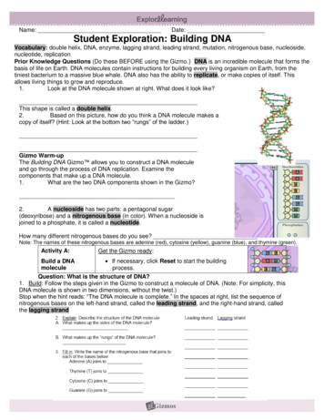
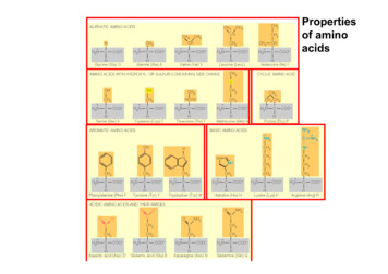
Cao et al. Clin Epigenet(2021) 13:93and the genetic basis of congenital cardiovascular malformations is also highlighted by Seema R. Lalani [5] andTroels Askhøj Andersen [61]. Dysregulated expression ofthese genes hampered the proper formation of cardiacstructure. Exome sequencing conducted by AlejandroSifrim et al. identified distinct genetic architectures forsyndromic and nonsyndromic CHD. In syndromic CHD,a significant enrichment of de novo protein-truncatingvariants, but not inherited protein-truncating variants, inknown CHD-associated genes was observed. Conversely,significant enrichment of protein-truncating variantsinherited from unaffected parents in nonsyndromic CHD[62].More than 55 human genes have been shown to beinvolved in CHDs [61]. For example, the NOTCH pathway is a paradigmatic endocardial-derived signal thatregulates cell fate specification and tissue patterning todefine chamber versus valve domains in the early vertebrate heart [63]. Defective endocardial NOTCH signalingaffects cardiac valve and chamber development, leading to diseases such as bicuspid aortic valve (BAV) andleft ventricular noncompaction (LVNC) cardiomyopathy [63–65]. Loss-of-function mutations in TBX20 cancause dilated cardiomyopathy, atrial septal defects, ormitral valve disease, while gain-of-function mutationsin TBX20 have been reported in patients with tetralogyof Fallot [66–70]. According to the function of genes,these pathogenic genes can be divided into 5 categories:genes associated with laterality defects (e.g., ACVR2B,CFC1, CITED2, GDF1, LEFTY2, NODAL and ZIC3),genes encoding components of signaling pathways (typically the NOTCH pathway and the MAPK pathway),genes encoding cardiac transcription factors (e.g., BMP2,GATA4, NKX2-5, TBX20, TBX2 AND TBX5), genesencoding components of the cardiac sarcomere (e.g.,ACTC1, MYH6, MYH7 and MYH11) and genes encoding chromatin modifiers (e.g., CHD7, KMT2D, EP300, CREBBP and EHMT1) [61].Despite significant progress in understanding heartdevelopment and diagnosing heart disease, the strikingcomplexity of the heart continues to limit our ability toidentify the molecular mechanisms of CHD. In additionto genetic factors, epigenetics and maternal factors affectthe development of the heart, which will be discussed inthe following sections. For many cases, the pathogenesisremains to be explored.Maternal DNA methylation patterns and congenitalheart defectsIt is known that maternal factors have maximum influence on fetal growth. Maternal age, gestational diabetes,obesity, dietary deficiencies, cigarette smoking, alcoholintake, exposure to teratogenic medications, and viralPage 4 of 18infections have been reported as maternal-environmental risk factors for the development of CHD [71]. Howthese environmental factors contribute to CHD is a difficult question that remains to be solved. Changes in DNAmethylation patterns may be one mechanism for theserisky environmental factors.Prognostic and predictive significance of long interspersed nucleotide element-1 (LINE-1) methylation hasbeen proposed in several diseases, such as advancedstage colorectal cancer and high-grade cervical intraepithelial neoplasia [72, 73]. Maternal LINE-1 DNAhypomethylation has been found to be associated withan increased occurrence of nonsyndromic CHDs. Measurement of maternal DNA methylation in a case–controlstudy of CHDs assessed the relationship between LINE-1DNA methylation and the risk of CHDs, and the findingsindicated that maternal LINE-1 hypomethylation is associated with an increased risk of CHD-affected pregnancies [74]. Significantly lower levels of LINE-1 methylationin the mothers of children with Down syndrome compared to mothers of healthy children were also observed[75]. However, the association between maternal LINE-1methylation and CHD in children with Down syndromewas not found in a study consisting of 44 mothers of children with Down syndrome and CHD and 46 mothers ofchildren with DS without CHD (P 0.997) [76] (Table 1).Shimul Chowdhury et al. conducted another study todetermine if alterations in gene-specific methylationwere associated with CHDs. Genome-wide maternalDNA methylation analysis of these samples identified 425CpG sites encompassing 415 genes that were differentially methylated between cases and controls (P 0.005).Case mothers showed hypermethylation in most differentially methylated loci (379 sites, 89.2%) comparedto hypomethylated loci (46 sites, 10.8%) [77]. Multiplegenes involved in the mitogen-activated protein kinase(MAPK) pathway and EGFP, GATA4, and Wnt5a, whichare implicated in heart development, were differentiallymethylated in this study [74, 78–80].The roles of maternal nutritional epigenetics in congenital heart disease have been well summarized byRadha O Joshi et al. The influence of diet quality onthe incidence of heart defects were observed in mouse,rat and population-based study. The maternal dietduring the gestation period is considered to alter theDNA methylation status globally or at specific loci byaffecting direct provision of methyl donors or by providing cofactors of DNMTs and enzymes that regulatethe one-carbon cycle [81]. Some related studies suggest the linkage of maternal risk factors and abnormalDNA methylation, and further CHD. Folate is a dietary B vitamin that provides methyl groups for DNAmethylation and other methylation reactions in cells.
non-syndromic CHDMothers, BloodMothers, BloodMothers, Blood[74][76][77]non-syndromic CHDCHD with Down syndromeCohortStudyStudy(reference) populationSample typeLINE-1 DNA methylation was analyzed by quantificationof LINE-1 methylation using the MethyLight methodCase: 180, Control:187 Maternal gene specific methylation in over 27,000 CpGsites was assessed using the Illumina Infinium HumanMethylation27 BeadChipCase: 44, Control: 46Methylation measures425 CpG sites encompassing 415 genes were differentiallymethylated between cases and controls (P 0.005).The majority of differentially methylated CpG sites werehypermethylated in casesLINE-1 methylation was not significantly different betweenDS-CHD and DS-CHD mothers (P 0.997)DNA methylation changeCase: 180, Control:187 Methylation of LINE-1 assessed with Methylight was used LINE-1 DNA methylation was significantly lower in casesas a surrogate marker of global DNA methylation statuscompared with controls ( P 0.049)Sample sizeTable 1 Differently Maternal DNA methylation patterns and congenital heart defectsCao et al. Clin Epigenet(2021) 13:93Page 5 of 18
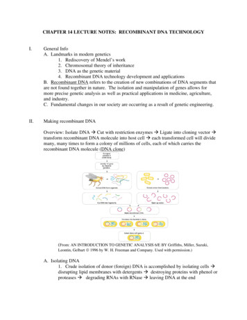
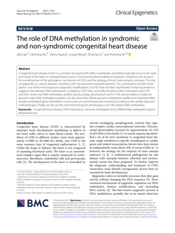
Cao et al. Clin Epigenet(2021) 13:93Genomic DNA methylation directly correlates withfolate levels. Folate deficiency is known to cause hyperhomocysteinemia, which is a risk factor for the development of CHD. A population-based case–controlstudy of 572 women with CHD-affected pregnanciesand 363 control women indicated that functional polymorphisms in folate-related genes increase the risk ofhaving a fetus with CHD when maternal lifestyle factors that alter folate metabolism are present. Methylene tetrahydrofolate reductase (MTHFR) is an enzymeimportant for folate metabolism [82]. Retrospectivestudies have shown that the MTHFR C677T mutationis associated with up to 50% of certain types of CHD[83]. The effect of maternal obesity on the developingheart appears to be greatest among women who carrythe MTHFR C677T or BHMT G742A polymorphism.Maternal smoking and/or alcohol use in combinationwith the TCII C776G polymorphism may increase therisk of CHDs [84]. Another study also found that theMTHFR genotype/diet combination and BMI weresignificantly associated with LINE-1 methylation inmothers of children with Down syndrome and CHD[76]. These findings are consistent with the role ofDNA methylation in the development of CHD.Early diagnosis and noninvasive diagnosis of fetaldiseases using maternal samples are aspirational;although the complicated physical conditions of pregnant women make these processes highly difficult, theyare still a valuable research direction. Alterations inmaternal DNA methylation may exert effects throughdirect interactions with the fetus or indirectly affectthe fetus through changes in the intrauterine environment. Changes in the maternal environment can resultin direct changes in gene expression in the developingfetus [77, 85, 86]. Based on knowledge of genetic susceptibility, lifestyle factors and their interactions, riskstratification of women of childbearing age may playan important role in reducing adverse pregnancy outcomes, including heart defects and other structuralmalformations. Alterations in maternal DNA methylation can be achieved via nutritional intervention,making DNA methylation an attractive potential therapeutic target [74].DNA methylation orchestrates cardiomyocytedevelopment, maturation and diseaseThe development of the heart is a complicated process.DNA methylation plays a key role in the fine-tuned spatiotemporal expression of cardiac genes during development. Cardiac DNA methylation differs in differentembryonic stages, neonates and adults, healthy personsand patients.Page 6 of 18Dynamic DNA methylation profiling in the developmentof heartDuring mammalian differentiation and development,DNA methylation undergoes remodeling to producea cell type-specific methylation pattern found in adultsomatic cells. DNA is demethylated within each developing germ cell and then specifically remethylated duringgametogenesis [87–89]. Several reports have shown thatthe paternal genome undergoes genome-wide DNA demethylation before replicating its DNA during the development of mouse zygotes [90–92]. It was also observedin human zygotes that paternal chromatin underwentactive demethylation soon after fertilization [93]. Singlecell DNA methylation sequencing of human preimplantation embryos revealed that thousands of genomic lociexhibit de novo DNA methylation [94]. These reportsindicate that genome-wide DNA methylation reprogramming during embryo development is a dynamic balancebetween global demethylation and focused methylation. The epiblast genome is remethylated rapidly afterimplantation [95, 96]. Whole-genome surveys conductedin mice and humans have shown that there are extensive and focal differences in DNA methylation patternsamong various adult tissues, which indicate that discrepancies in de novo DNA methylation and demethylationoccur during tissue differentiation [97–99].DNA methylation is dynamic during cardiomyocytedevelopment. Several studies on dynamic DNA methylation profiling in the development of heart was summarized in Table 2. A genome-wide DNA methylationprofiling of mouse embryos showed that global methylation of 1.64 million ACGT sites in developing heartsremained stable between embryonic day (E) 11.5 andE14.5, and a small fraction (2901) of them exhibited differential methylation (1946 showed increased methylation and 955 showed decreased methylation in late-stagehearts); these sites were enriched in genes involved inheart development, including ERBB4, GATA6, FOXP1,FGF2, FGF9, HAS2, INVS, MEF2C, ROBO2 and WNT2.Of the 350 genes analyzed by quantitative real-time PCR,79 genes had correlative changes between methylationand expression [100]. Cardiomyocytes contained shortregions of low DNA methylation, which showed characteristics of cis-regulatory elements compared withthose of ES cells. Five percent of CpGs were differentiallymethylated between cardiomyocytes of healthy adultmice and undifferentiated ES cells. Among them, 90% ofthese CpGs were hypomethylated and 10% were hypermethylated in cardiomyocytes versus ES cells, whichindicated that differential CpG methylation showed celltype specification during embryonic development. Demethylated regions of adult cardiomyocytes were significantly enriched for upstream transcription factor binding
Study population SampletypeHeart tissue (mouse)Heart tissue (mouse)Heart tissue (mouse)Heart tissue (mouse)Study(reference)[100][102][103][101]Chronic pressureoverload in adultmiceHealthyHealthyHealthyCohortnewborn, adult healthy and failinghearts (n 3–6)P1, P7, P14, P28, P84 (n 3 per group)E14.5, E17.5, NB, P7, P14, adultE11.5, E14.5 (n 3 per group)SampleTable 2 Dynamic DNA methylation profiling in the development of heartDNA methylome were analyzed bywhole-genome bisulfite sequencingFor analysis of global DNA methylation, 5‐methylcytosine levels weremeasured using the MethylFlashMethylated DNA Quantification KitThe DNA methylation levels of CpGdinucleotides region in ssTnI genepromoter were detected using MSPand BSPGenome‐wide DNA methylation profiling using MSFE/MPSMethylation measuresIdentified large genomic regions that aredifferentially methylated during cardiomyocyte development and maturationMajority of DMRs ( 80%) were hypermethylated between P1 and P14, andhypermethylated regions were associated with transcriptional shut downof important developmental signalingpathwayDNA methylation levels of the CpG dinucleotides region in ssTnI gene promoterwere increased with the developmentof mouse heartGlobal methylation in developing heartsremained stable between E11.5 andE14.5, a small fraction AGCT sites exhibited differential methylation, whichwere enriched in genes involved inheart developmentDNA methylation changeCao et al. Clin Epigenet(2021) 13:93Page 7 of 18
Cao et al. Clin Epigenet(2021) 13:93motifs (TF motifs) of known cardiac transcription factors, including MEF2C and GATA1-4 [101]. A comparation of heart from embryonic, newborn and adult mousefound that DNA methylation levels of the CpG dinucleotides region in ssTnI gene promoter were increasedwith the development of mouse heart, corresponding toa decreased expression of ssTnI gene. These results indicate that DNA methylation is involved in regulation ofmyofibril gene expression during heart development inmice[102].DNA methylation is also highly dynamic during postnatal maturation. Genome-wide sequencing of methylated DNA conducted by Choon Boon Sim et al. identifieddynamic changes in the cardiac methylome during postnatal development (2545 DMRs from P1 to P14 in themouse). Majority of DMRs ( 80%) were hypermethylated between P1 and P14, and these hypermethylatedregions were associated with transcriptional shut downof important developmental signaling pathways, including Hedgehog, bone morphogenetic protein, TGF-β,fibroblast growth factor, and Wnt/β-catenin signaling[103]. A substantial number of CpGs were differentiallymethylated in newborn and adult cardiomyocytes [101].Gene body demethylation starting at the 5′ end andextending to the 3′ end was observed during postnata
Congenital heart disease (CHD) is a common structural birth defect worldwide, and defects typically occur in the walls and valves of the heart or enlarged blood vessels. Chromosomal abnormalities and genetic mutations only account . Full list of author information is available at the end of the article. Cao .