Transcription
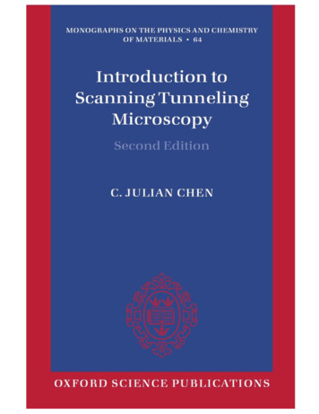
Introduction to ScanningTunneling MicroscopySecond EditionC. Julian ChenDepartment of Applied Physics and Applied MathematicsColumbia University, New YorkOXFORDUNIVERSITY PRESS
MONOGRAPHS ON THE PHYSICS AND CHEMISTRY OF MATERIALSTheory of dielectrics H. FrohlichStrong solids (Third edition) A. Kelly and N. H. MacmillanOptical spectroscopy of inorganic solids B. Henderson and G. F. ImbuschQuantum theory of collective phenomena G. L. SewellPrinciples of dielectrics B. K. P. ScaifeSurface analytical techniques J. C. RivièreBasic theory of surface states Sydney G. Davison and Maria SteslickaAcoustic microscopy G. A. D. BriggsLight scattering: principles and development W. BrownQuasicrystals: a primer (Second edition) C. JanotInterfaces in crystalline materials A. P. Sutton and R. W. BalluffiAtom probe field ion microscopy M. K. Miller, A. Cerezo, M. G. Hetherington, andG. D. W. SmithRare-earth iron permanent magnets J. M. D. CoeyStatistical physics of fracture and breakdown in disordered systems B. K. Chakrabartiand L. G. BenguiguiElectronic processes in organic crystals and polymers (Second edition) M. Pope andC. E. SwenbergNMR imaging of materials B. BlümichStatistical mechanics of solids L. A. GirifalcoExperimental techniques in low-temperature physics (Fourth edition) G. K. White andP. J. MeesonHigh-resolution electron microscopy (Third edition) J. C. H. SpenceHigh-energy electron diffraction and microscopy L.-M. Peng, S. L. Dudarev, andM. J. WhelanThe physics of lyotropic liquid crystals: phase transitions and structural propertiesA. M. Figueiredo Neto and S. SalinasInstabilities and self-organization in materials, Volume 1: Fundamentals of nanoscience,Volume 2: Applications in materials design and nanotechnology N. Ghoniem andD. WalgraefIntroduction to scanning tunneling microscopy (Second edition) C. J. Chenistm: “prelims” — 2007/7/19 — 17:54 — page ii — #2
Preface to the Second EditionIn a 1959 speech entitled There’s Plenty of Room at the Bottom [1], RichardFeynman invited scientists to a new field of research: to see individual atomsdistinctly, and to arrange the atoms the way we want. Feynman envisionedthat, by achieving those goals, one could synthesize any chemical substancethat the chemist writes down, resolve many central and fundamental problems in biology at the molecular level, and dramatically increase the densityof information storage. Some 20 years later, those goals began to be achievedthrough the invention and application of the scanning tunneling microscope(STM) [2, 3] and the atomic force microscope (AFM) [4]. The inventors ofSTM, two physicists at IBM Research Division, Gerd Binnig and HeinrichRohrer, shared the 1986 Nobel Prize in physics [5, 6].At that time, I was fortunate to be in the Department of Physical Sciences of IBM Research Division, and had the opportunity to design, build,and run those fascinating instruments. Partially based on my personal experience and understanding, in 1993, the first edition of this book was published [7]. In the decade following, Feynman’s foresight has grown into avast field of research, nanoscience and nanotechnology. As a result, tremendous advances have been achieved in the understanding of the basic physicsas well as instrument design and operation of STM and AFM. It is time topublish a second edition to include those recent advances, and to satisfy theurgent need for an updated, unified, accurate, and pedagogically assessabletextbook and reference book on STM and AFM.During the years of 1994 to 2003, I was concentrating on the researchof human voice and languages, which were my favorite subjects ever sincemy college years. And I received more corporate recognition than for mybasic research in physics [8]. However, the news about the advancements inSTM and AFM constantly called me to come back to nanoscience and nanotechnology. In December 2003, I received a kind invitation from ProfessorRoland Wiesendanger, the Director of the Institute of Applied Physics atHamburg University, to become a guest scientist. This is one of the largestand most productive centers of STM and AFM research, especially in spinpolarized STM and non-contact AFM. And for the first time in my life,I could concentrate 100% of my time on nanoscience and nanotechnologyresearch. In the summer semester of 2005, in a graduate-level course innanostructure physics jointly given to the Department of Physics and theDepartment of Chemistry at Hamburg University, Professor Roland Wiesendanger lectured the analyses of various nanostructures, and I lectured theprinciples and instrumentation of STM and AFM. The lecture notes on STMand AFM then became the blueprint of the second edition of the STM book.Following are some examples of the additions to the second edition:Atomic force microscopy, with a refined frequency-modulation mode,
iiChen: Introduction to Scanning Tunneling Microscopyhas achieved true atomic resolution in the attractive atomic force regime,often referred to as the non-contact AFM. In some cases, its resolutionhas even surpassed that of STM. The observed bias-dependence of atomicforces provides information about the details of electronic structure. Thisnew technique enables atomic-scale imaging and characterization not onlyfor conductors, but also for insulators.Significant breakthrough in spin-polarized STM has enabled the observation of local magnetic phenomena down to atomic scale. Such advancementwas to drive the development of nanomagnetism, which would have deepimpact on the technological applications of magnetism.Inelastic electron tunneling spectroscopy (IETS), initially discovered inmetal-insulator-metal tunneling junctions to observe vibrational frequencies of embedded molecules, was advanced to STM junctions, enabling theobservation of vibrational states of individual molecules. The successfuldemonstration of STM-IETS elevated the field of single-molecule chemistryto an unprecedented level.At the time that the first edition was written, atom manipulation wasstill a highly specialized personal art. In the later years, the underlyingphysics has gradually been discovered, and the atom-manipulation process isbecoming a precise science. Besides single atoms, molecules are also subjectto manipulation. It was often said that STM is to nanotechnology whatthe telescope was to astronomy. Yet STM is capable of manipulating theobjects it observes, to build nanoscale structures never existed in Nature.No telescope is capable of bringing Mars and Venus together.In the process of further improving the resolution of STM and AFM,the understanding of its basic physics has been advanced. Numerous convincing theoretical and experimental studies have shown that the imagingmechanism of both STM and AFM at atomic resolution can be understoodas a sequence of making and breaking of partial covalent bonds betweenthe anisotropic quasi-atomic orbitals on the tip and those on the sample.The nature of STM and AFM, including those with spin-polarized tips, canbe understood with a unified perspective based on Heisenberg’s concept ofresonance in quantum mechanics.Commercialization of STM and AFM has been greatly advanced. Owingto the rapidly expanding research in nanotechnology, especially in molecularbiology and in materials science, AFM with tapping mode operating in airor in liquid now constitutes the largest market share. Therefore, a briefpresentation of its basic principles is included.In spite of the availability of commercial STMs and AFMs, researchgroups worldwide continue to design and build customized instruments toachieve advanced features and to serve special experimental needs. Often,those new designs are then adapted by instrument manufacturers to becomeproducts. Although the basic principles of the design and constructionof STM and AFM was laid down in the second part of the first edition,Copyright 2008 Oxford University Press
Chen: Introduction to Scanning Tunneling MicroscopyiiiInstrumentation, new trends and ideas have been added.The basic organization of the second edition is essentially identical tothe first edition. All the materials in the first edition proven to be useful arepreserved. Some of the less important materials are eliminated or convertedto Problems at the ends of various chapters. The first chapter, Overview,is preserved but updated. Several recent applications of STM and AFM,probably of interest to general readers, are added. The title of the first Partis changed to Principles from Imaging Mechanism because of the inclusionof the physics of atom and molecule manipulation. The Gallery of STMImages is updated, with more historical photos and AFM images added,which is now entitled simply Gallery. To preserve the classical style of thefirst edition and to reduce the cost of printing, all photographs and imagesare in black-and-white. Similar to the first edition, only a few illustrativeapplications of STM and AFM are presented, because there are alreadymany excellent books on various applications. For example, the monographScanning Probe Microscopy and Spectroscopy: Methods and Applications byR. Wiesendanger [9]; the book series Scanning Tunneling Microscopy I, II,and III edited by R. Wiesendanger and H.-J. Güntherodt [10]; ScanningTunneling Microscopy edited by J. A. Stroscio and W. J. Kaiser [11]; thesecond edition of the monograph Scanning Tunneling Microscopy and itsApplications by C. Bai [12]; and the second edition of Scanning Probe Microscopy and Spectroscopy: Theory, Techniques, and Applications edited byD. Bonnell [13]. Similar to the first edition, to ensure pedagogical soundness,the focus is on simple but useful theories, with every derivation presented infull detail. Many new figures are added to illustrate the concepts in physics.In the second edition, care has been taken to use SI units as much as possible. That would provide a unified order-of-magnitude mental picture ofthe physical quantities involved. Because of the enormous growth of thesize of literature, the reference list at the back of the second edition onlyincludes those cited by the text, selected and arranged automatically by LaTeX. In the age of the Internet, exhaustive reference lists can be obtainedby searching on the web.The author is deeply grateful to H. Rohrer for commenting extensively onthe manuscript of the first edition, and publicly recommending the book tonewcomers as well as experts. The author is equally grateful to Ch. Gerber,an architect of both the first STMs and the first AFMs, for comments on thesecond edition, and especially for providing a number of precious originalphotographs and images of historical interest. An early manuscript of thesecond edition was thoroughly reviewed by a number of experts in that field.Corrections and improvements were made upon their comments. Followingis an incomplete list. The entire book by K.-H. Rieder of Swiss FederalLaboratories for Materials Research. Part I and Chapter 15 by R. Pérezof Universidad Autonoma de Madrid. Part II, especially Chapter 15, byF. Giessibl of University of Regensburg. All chapters related to scanningCopyright 2008 Oxford University Press
ivChen: Introduction to Scanning Tunneling Microscopytunneling spectroscopy and spin-polarised STM by O. Pietzsch of HamburgUniversity. The main part of Chapter 1 and Chapter 12 by F. Besenbacher,J. V. Lauritsen, and E. Laegsgaard of University of Aarhus. Chapter 9by W. Coburn and P. Stokes of EBL Products, Inc., and M. Ordillas ofMorgan Electro Ceramics, Inc. Chapter 14 by R. Feenstra of CarnegieMelon University. Chapter 15, especially Section 15.4, by C. Prater of VeecoInstruments Inc. Section 1.5.2 and Section 13.6 by the group of J. Ulstrupof Technical University of Denmark. Section 1.5.4 by J. Hu of JiaotongUniversity, Shanghai. Section 13.3.5 by R. A. Wolkow of University ofAlberta. Last but not least, Sections 5.2.1, 5.2.2, and 5.2.3 by W. Hofer ofUniversity of Liverpool.The Gallery is an integrated part of the book. The author is indebtedto the following authors who contributed high-resolution black-and-whiteoriginal photographs and images for the publication of this book: Plates 2,3, and 4, Ch. Gerber. Plate 5, R. Wiesendanger. Plates 7, 11, 12, and 16,J. Boland. Plates 8, 10, and 15, R. Feenstra. Plate 9, J. V. Barth. Plate 13,D. J. Thomson. Plate 14, J. Repp and G. Meyer. Plate 17, M. Bode. Plate18, L. Berbil-Bautista. Plate 19, O. Pietzsch. Plate 20, A. R. Smith. Plates21 and 22, S. Fölsch. Plate 23, K. F. Braun. Plates 24, P. Weiss. Plate 25,C. F. Hirjibehedin. Plate 27, F. J. Giessibl. Plate 28, J. V. Lauritsen and F.Besenbacher. Plate 29, R. Pérez and Ó. Custance. Plate 30, Ó. Custance.C. Julian ChenColumbia Universityin the City of New YorkJuly 2007Copyright 2008 Oxford University Press
GalleryHistorical photographsPlate 1. The IBM Zurich Laboratory soccer teamPlate 2. A humble gadget that shocked the science communityPlate 3. The creators of the atomic force microscopePlate 4. The first atomic force microscopeSTM studies of surface structuresPlate 5. ‘Stairway to Heaven’ to touch atomsPlate 6. Zooming into atomsPlate 7. Underneath the Si(111)-7 7 surfacePlate 8. STM image of a GaN(0001̄) surface Plate 9. Large-scale image of the Au(111)-22 3 structurePlate 10. Large-scale image of the Ge(111) surfacePlate 11. Details of the Ge(111)-c(2 8) surfaceMolecularPlate 12.Plate 13.Plate 14.Plate 15.Plate 16.orbitals and chemistryIndividual π and π molecular orbitals observed by STMOrganic molecules observed by STMObservation of the HUMO and LOMO of an organic moleculeVoltage-dependent images of the Si(111)-2 1 surfaceChemical vapor deposition of the Si(100) surfaceSpin-polarized STMPlate 17. SP-STM images of an Fe islandPlate 18. SP-STM images of Dy filmsPlate 19. SP-STM studies of nanoscale Co islands on Cu(111)Plate 20. SP-STM studies of antiferromagnetic crystal Mn3 N2Atom manipulationPlate 21. Construction of Cun chains by atom manipulation using STMPlate 22. Quantum states observed on Cun chainsPlate 23. The triangular quantum corralPlate 24. Manipulating hydrogen atoms underneath palladium surfacePlate 25. An artificial atomic-scale rock gardenAtomic force microscopyPlate 26. Si(111)-7 7 structure resolved by AFMPlate 27. Current images and higher-harmonics force images on graphitePlate 28. Tip dependence of non-contact AFM images of TiO2Plate 29. Chemical identification of individual surface atoms using AFMPlate 30. Atom manipulation using AFM
2Chen: Introduction to Scanning Tunneling MicroscopyPlate 1. The IBM Zurich Laboratory soccer team. On October 15,1986, the soccer team of IBM Zurich Laboratory and Dow Chemical playeda game which had been arranged earlier. To everyone’s surprise, a few hoursbefore the game, the Swedish academy announced the Nobel Prize for GerdBinnig (right, holding flowers) and Heinrich Rohrer (left, holding flowers).Newspaper reporters rushed in for a press conference.Towards the end of the press conference, Binnig and Rohrer said thatthey must leave immediately because both were members of the laboratorysoccer team. The reporters followed them to the soccer field. A photographer for the Swiss newspaper Blick took this photograph before the gamestarted. At the center of the photograph, holding a soccer ball is ChristophGerber, responsible for building the first scanning tunneling microscope aswell as the first atomic force microscope.(Original photograph by courtesy of IBM Zurich Laboratory.)Copyright 2008 Oxford University Press
Chapter 1Overview1.1The scanning tunneling microscopeThe scanning tunneling microscope (STM) was invented by Binnig andRohrer and implemented by Binnig, Rohrer, Gerber, and Weibel [2, 3].Figure 1.1 shows its essential elements. A probe tip, usually made of Wor Pt–Ir alloy, is attached to a piezodrive, which consists of three mutuallyperpendicular piezoelectric transducers: x piezo, y piezo, and z piezo. Uponapplying a voltage, a piezoelectric transducer expands or contracts. By applying a sawtooth voltage on the x piezo and a voltage ramp on the y piezo,the tip scans on the xy plane. Using the coarse positioner and the z piezo,the tip and the sample are brought to within a fraction of a nanometer eachother. The electron wavefunctions in the tip overlap electron wavefunctionsin the sample surface. A finite tunneling conductance is generated. By applying a bias voltage between the tip and the sample, a tunneling currentis generated. The concept of tunneling will be presented in Section 1.2.Fig. 1.1.The scanning tunneling microscope in a nutshell. The scanningwaveforms, applying on the x and y piezos, make the tip raster scan on the samplesurface. A bias voltage is applied between the sample and the tip to induce a tunnelingcurrent. The z piezo is controlled by a feedback system to maintain the tunneling currentconstant. The voltage on the z piezo represents the local height of the topography. Toensure stable operation, vibration isolation is essential.
24Chen: Introduction to Scanning Tunneling MicroscopyThe most widely used convention of the polarity of bias voltage is thatthe tip is virtually grounded. The bias voltage V is the sample voltage. IfV 0, the electrons are tunneling from the occupied states of the tip intothe empty states of the sample. If V 0, the electrons are tunneling fromthe occupied states of the sample into the empty states of the tip.The tunneling current is converted to a voltage by the current amplifier,which is then compared with a reference value. The difference is amplifiedto drive the z piezo. The phase of the amplifier is chosen to provide anegative feedback: if the absolute value of the tunneling current is largerthan the reference value, then the voltage applied to the z piezo tends towithdraw the tip from the sample surface, and vice versa. Therefore, anequilibrium z position is established. As the tip scans over the xy plane, atwo-dimensional array of equilibrium z positions, representing a contour plotof the equal tunneling-current surface, is obtained, displayed, and stored inthe computer memory.The topography of the surface is displayed on a computer screen, typically as a gray-scale image, see Fig. 1.2(a). The gray-scale image is similarto a black-and-white television picture. Usually, the bright spots representhigh z values (protrusions), and the dark spots represent low z values (depressions). The z values corresponding to the gray levels are indicated bya scale bar. For a more quantitative representation of the topography, acontour plot along a given line is often provided, as shown in Fig. 1.2(b).The most convenient unit for x and y is nanometer (nm, 10 9 m), and themost convenient unit for z is picometer (pm, 10 12 m).To achieve atomic resolution, vibration isolation is essential. This isachieved by making the STM unit as rigid as possible, and by reducing theinfluence of environmental vibration to the STM unit. See Chapter 10.Fig. 1.2.Gray-scale image and contour plot. (a) A 5nm 5nm gray-level topographic image of Si(111)7 7. The bright spots represent protrusions, and the darkspots represent depressions. The z values corresponding to the gray levels are indicatedby a scale bar. (b) The topographic contour along a line in (a), for a more quantitativerepresentation. By courtesy of F. Giessibl.Copyright 2008 Oxford University Press
Chen: Introduction to Scanning Tunneling Microscopy1.525Illustrative applicationsOver the last decades, the applications of STM and AFM have grown sofast and so vast that a thorough review would take many volumes. Andthere are already many good books as well as review articles publishedon applications of STM and AFM in various fields. In this section, a fewexamples are presented as illustrations.1.5.1Catalysis researchCatalysis is the backbone of synthetic chemistry. It is a basic process inthe production of fabrics, fuels, fertilizers and pharmaceuticals. Catalysisis also essential in environmental protection. For example, required by law,each automobile must have a catalytic converter to remove toxic exhaustgases, notably CO and NOx . Although it is well known that catalyticreactions often take place at specific active atomic sites on the catalystsurface, the research in catalysis have been mainly relying on test-anderror experimentation involving macroscopic parameters. The inventionof STM enables detailed studies of the electronic structures of the activesites at catalyst surfaces. STM can even operate in an atmosphere withactual reactants, thus to observe the transformation of molecules at activeatomic sites on real time. Therefore, STM research can help facilitate a fullunderstanding and control of the constituents at the molecular and atomiclevels, and to open up the possibility of designing the catalysis process atthe single molecule level [14]. Here are two examples.Ni-Au catalyst for steam reformingSteam reforming is a method in the chemical industry to produce hydrogenfrom hydrocarbons. For example, in the presence of a catalyst, alkanes reactwith steam (overheated water vapor) to generate H2 and CO:Cn H2n 2 nH2 O n CO (2n 1) H2 .(1.1)The most important case is the reaction of methane with steam,CH4 H2 O CO 3 H2 .(1.2)It is the most common and least expensive method to produce hydrogen. The typical catalyst is nickel nanoclusters supported on a MgAl2 O4substrate. Nevertheless, in parallel with the steam reforming process, nickelalso catalyzes the production of graphite, which impedes the activity due tothe formation of a blocking carbon layer on the nickel surface (coking) andmay eventually lead to the breakdown of the catalyst. An existing solutionto that problem is by mixing a minute amount of H2 S to the reactants,which poisons the nickel catalyst. Because sulfur poisons the formation ofCopyright 2008 Oxford University Press
26Chen: Introduction to Scanning Tunneling Microscopygraphite more than it poisons the steam formation reaction, the lifetime ofthe catalyst is prolonged. However, the overall rate is lower, and the inclusion of H2 S is harmful to the subsequent processes and the environment.A new idea comes from the STM studies of the Ni-Au system [15]. Niand Au are immiscible in the bulk. No binary alloy can be formed. However, STM studies showed that by mixing a few percents of Au to Ni, afterannealing at 800 K, a surface binary alloy is formed: many Ni atoms on thesurface are substituted by Au atoms, see Fig. 1.3. The topographic STMimage shows that as if the Au atoms are darker (deeper), and the Ni atomsimmediately adjacent to an Au atom are brighter (higher). It is not becausethe Au atoms are depressed and the adjacent Ni atoms are protruded, butis an electronic effect inherent to STM. According to the Tersoff-Hamannmodel of STM imaging [16, 17] (see Chapter 6), a topographic STM imageis the contour of equal Fermi-level local density of states of the sample,measured at the center-of-curvature of the tip r0 , ρ(EF , r0 ). On the Auatoms, the value of ρ(EF , r) is lower than that of the normal Ni atoms. Onthe other hand, the value of ρ(EF , r) of a Ni atom adjacent to a Au atomis higher owing to the perturbation by the Au atom. For a Ni atom withtwo Au neighbors, the perturbation is even stronger, see Fig. 1.3. Becausethe Fermi-level local density of states is closely related to catalytic reactivity, those Ni atoms provide a higher reactivity than the normal Ni atoms.Furthermore, the carbon atoms are much less likely to adsorb on the Niatoms adjacent to a Au atom. The results are: by mixing a few percentsof Au to Ni, the reactivity of the steam formation process is enhanced, andFig. 1.3. STM topographical images of the Ni-Au system. (a) An image ofNi(111) with 2% Au. The Au atom appears as depressions because of the LDOS at theFermi level is lower. The Ni atoms surrounding an Au atom appear as protrusions becauseof the enhancement of LDOS at the Fermi level, indicating a higher chemical reactivity.For the Ni atom between two Au atoms, the LDOS enhancement is even higher. (b) Animage of Ni(111) surface with 7% of Au. The number of Ni atoms with doubly enhancedFermi-level LDOS is increased. (Original figure in black-and-white with high resolutionby courtesy of F. Besenbacher and J. V. Lauritsen. Reproduced with permission [15].Copyright 1998 the American Association for the Advancement of Science.)Copyright 2008 Oxford University Press
Chen: Introduction to Scanning Tunneling Microscopy27Fig. 1.4. Conversion rates of the Ni catalyst and the Ni-Au catalyst. The conversion rates of steam reformation of n-butane and water vapor using different catalysts.Gray curve: pure Ni catalyst. the rate deteriorates with time as a result of the parasiticgraphite formation process. Black curve: Ni-Au catalyst, which shows no deteriorationof reaction rate. (Original figure in black-and-white with high resolution by courtesy ofF. Besenbacher and J. V. Lauritsen. Reproduced with permission [15]. Copyright 1998the American Association for the Advancement of Science.)the graphite formation process is reduced. While the conversion rate of thepure Ni catalyst decreases markedly after an hour of operation, the conversion rate of Ni-Au catalyst remains constant. Therefore, the Ni-Au catalystcould be active for a much longer time, see Fig. 1.4.Based on the STM study, a new catalyst, with about 16.5 weight percentsof Ni and 0.3 weight percents of Au on a MgAl2 O4 matrix, is designed, whichsubstantially improves the steam formation process.Understand and improve the MoS2 catalystPetroleum contains a wide range of sulfur compounds, for example, alkanethiol (Cn H2n 1 SH) and thiophene (C4 H4 S). Sulfur is harmful for thechemical processings and for the environment. The process to remove sulfur,hydrodesulphurization (HDS), is an essential step in petrochemical industry.The typical catalyst is molybdenum disulfate (MoS2 ). In the presence ofhydrogen, the HDS reaction converts alkanethiol into alkane,Cn H2n 1 SH H2 Cn H2n 2 H2 S,(1.3)and the gas-phase H2 S is easily removed.MoS2 is a layered material, similar to graphite. The bulk of MoS2 consists of thin sheets of S-Mo-S sandwiches, and those sheets are held togetherby van der Waals forces. The basal planes of MoS2 are chemically inert.Therefore, catalytic reactivity must reside in the rims of the sheets. However, before the application of STM, the preferred structure and the activesites of MoS2 crystallites, as well as the reaction paths of the HDS process,were only vaguely guessed based on macroscopic evidences.Copyright 2008 Oxford University Press
28Chen: Introduction to Scanning Tunneling MicroscopyA systematic study of the structure, active sites, and reaction pathsis carried out with STM experiments and DFT computations [18]. In theindustry, MgAl2 O4 is usually used as substrates for the MoS2 clusters. However, it is not appropriate for STM studies because it is an insulator. ForSTM studies, single crystal gold is chosen as the substrate, especilly theAu(111) surface. Gold is relatively inert and allows for investigation of theintrinsic properties of the catalysts.The nanoclusters of MoS2 were prepared by depositing molybdenum onAu(111) surface in an atmosphere of H2 S at a pressure of 1 10 6 mbar, thenthe substrate was annealed to 673–723K for 15 minutes. A fairly uniformdistribution of crystalline MoS2 nanoclusters was formed. Most of the MoS2nanoclusters appear to have a triangular shape, with an average area of 5nm2 . The side length of each triangular nanocluster is about 3 nm. From acombination of STM experiments and first-principles computations, it wasconcluded that the edges are saturated with sulfur atoms [18].By exposing the MoS2 nanoclusters with various substances, the reactivity of MoS2 is studied with STM. It was found that MoS2 neither reactswith organic molecules containing sulfur, such as alkanethiol (Cn H2n 1 SH)Fig. 1.5. Reaction of H and thiophene with a MoS2 nanocluster. (a) Thetriangular MoS2 nanocluster, after exposed to atomic hydrogen and then thiophene atelevated temperature, is cooled down and characterized by STM. Image size: 5 5 nm2 .Bean-like structures, identified as the intermediate product of thiophene, cis-but-2-enethiolate (CH3 -CH CH-CH2 -S ), are found on the bright rim. Also, there is a prominentchange in the outermost rim protrusions, identified as sulfur vacancies. Part of the rimis highlighted in (b). Details of a line scan of the rim is shown in (c). Solid curve (I):STM line scan along the dashed line in (b). Dashed curve (II): the line scan of thecorrespondent unreacted area, shown for comparison. (Original high-resolution image inblack-and-white by courtesy of J. V. Lauritsen. See [14, 18] for details.)Copyright 2008 Oxford University Press
Chen: Introduction to Scanning Tunneling Microscopy29and thiophene (C4 H4 S), nor reacts with H2 . Only after exposing MoS2with predissociated (atomic) hydrogen, visible changes are observed. Sulfurvacancies are created at the rims.By further exposing the MoS2 nanoclusters already treated with atomichydrogen with thiophene (C4 H4 S), a strong reaction is found, as shown inFig. 1.5. As shown, parts of the rim are significantly altered. The outermostprotrusions of the rims are markedly shifted, and bean-like structures appearadjacent to the bright rim.The STM study revealed the mechanism of catalytic reaction. First, ahydrogen atom reacts with the rim of a MoS2 nanocluster, strip off a sulfuratom and expose a molybdenum atom. The molybdenum atom becomesan active site. A alkanethiol molecule can chemically adsorb on the activesite by forming a S-Mo bond. After picking up a H atom, the hydrocarbonmolecule becomes free, and the sulfur atom occupies the S vacancy at therim of the MoS2 nanocluster. The desulphurization of thiophene is morecomplex. At an active site of MoS2 , it is first converted to an intermediateproduct
principles and instrumentation of STM and AFM. The lecture notes on STM and AFM then became the blueprint of the second edition of the STM book. Following are some examples of the additions