Transcription
OCTOPUS 900Flexibility and reliabilityTradition and innovation – Since 1858, visionary thinking and afascination with technology have guided us to develop innovative productsof outstanding reliability: Anticipating trends to improve the quality of life.
02 03 PERIMETRY YOU CAN TRUSTOCTOPUS 900Goldmann Perimetry for the futureIn 1945, Professor Hans Goldmann of the University of Berne, together withHaag-Streit, developed the Manual Goldmann perimeter. This instrument isstill the reference for kinetic perimetry today and all of its capabilities andspecifications are built into the Octopus 900. Furthermore, Haag-Streit haspioneered many significant innovations, including automated static perimetry, the G-Program and the direct projection system, as well as fast strategies and outstanding software for visual field analysis.Addressing all major perimetry needs, the Octopus 900 is the perimeter ofchoice for anyone who wants diagnostic flexibility to analyse, assess andtrack patients’ visual fields.It makes it possible to run standard central and peripheral visual fields withminimum test duration, seamlessly integrated into your practice environment. This supports high patient throughput and effective practice management. With its built-in reliability features, the Octopus 900 is easy-to-use anddelivers results you can trust.
Full field standardwhite-on-white perimetryThe Octopus 900 performs standard white-on-whitethreshold testing in just 2–4 minutes in the central visual field. Due to its 90 degree cupula, full 180-degreeperipheral testing is possible. This allows for disabilitytesting, including ptosis examination, as well as binocular driving tests.Reliable resultsmade easyWorry less about patient compliance. The Octopus900 automatically recognises any fixation losses andadjusts patients accordingly until optimal test conditions are achieved. Thus, the Octopus 900 producesresults you can always trust.True Goldmann KineticperimetryWith the Octopus 900, kinetic vectors can be definedanywhere and with all the characteristics availablein the Manual Goldmann perimeter. Additionally, user-defined templates help generate fast and reproducible results when defining standard isopters andmapping the blind spot, as well as for ptosis, drivingand disability examinations.
04 05 FASCINATING VERSATILITYOCTOPUS 900Full field standard white-on-whiteperimetryThe Octopus 900 offers a wide range of static test patterns, including 32, 30-2, 24-2, and 10-2. In addition, there are two uniquephysiology-based patterns: the G-Program (a 30-degree field forglaucoma assessment) and the M-Program (a 10-degree fieldfor analysing the macula). They are both correlated with a nervefibre bundle map and thus make it possible to test the pointswhich are most important for a structure-function correlation.These examination patterns offer a higher density of stimuli inthe centre, which supports the discovery of paracentral scotomas that are often missed by the common 32 pattern.The Octopus 900, with its 90-degree radius Goldmann bowlpermitting 180-degree full field testing, allows additional testingin the periphery for any kind of driving and disability examination. Common tests like monocular and binocular Estermanntests as well as a ptosis test are already built into the device forease of use.Further test patterns can be created and saved using the Custom Test function built into the Octopus 900. For low vision patients, testing with the larger size V Goldmann stimulus not onlyextends the dynamic testing range, but also decreases the levelof fluctuation between tests1,2.
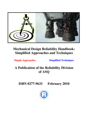
PROGRESSION ANALYSISImmediately identify levels of changeINTUITIVE TRAFFIC LIGHT SYMBOLSEyeSuite Perimetry software is included as standard, featuring the most advanced EyeSuite Progression analysis for following up visual fields. As recommended by the International Glaucoma Society, the global progression rate iscalculated in dB per year, including the probability level. Areas for normal range(grey band), impaired vision (15 dB) and legal blindness (25 dB) provide a starting point for further investigation.Often, progression is local and not noticeable on global progression analysis.No more counting of single points and looking for clusters. EyeSuite does thework for you! The EyeSuite Cluster Trend analysis is based on specific “clusters”of test points that are matched to the nerve fibre bundles, while the Polar Trendanalysis allows direct comparison with structural findings. With these two localprogression analyses, even small local changes that are not visible at a globallevel can be easily detected and followed up6,7.Intuitive colour codes save time by immediately identifying levels of change.A red triangle will always indicate significant worsening and a green trianglesignificant improvement.EYESUITE CLUSTER TREND ANALYSISVARIOUS PRINTOUTSIntuitive interpretation of visualfield resultsConfigure your favorite printout and graphics representation, in order to reduce the time necessary to interpret the results. Choose either the provenOctopus 7-in-1 printout containing the cumulative defect curve (Bebie curve)or the HFA-style printout. Furthermore, the 4-in-1 printout or the series reportcan also be customised.Don’t want a paper copy? Save the report as an image or PDF and view it onyour screen or export it to your electronic medical record (EMR) system.VARIOUS PRINTOUTS
Visual field evaluationis made simple with thewidely-used Octopus7-in-1 printout.
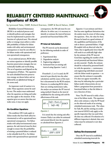
TOP FAST-THRESHOLDING STRATEGYIncreased efficiencyTendency Oriented Perimetry (TOP) presents a further optimisation in fastthreshold testing by reducing the examination time by nearly 80% to just2–4 minutes3,4 compared to 6–8 minutes (Dynamic strategy) or 10–12 minutes (Normal strategy). The TOP algorithm is a systematic method whichtakes the correlation of the threshold values in neighbouring locations intoaccount.Since the first test points are presented at a supra-threshold level, eveninexperienced patients quickly understand the nature of the test.TIME COMPARISON BETWEEN TEST STRATEGIESCLUSTER ANALYSISProviding meaningful resultsCluster analysis combines high sensitivity with good specificity5. Test locations are grouped (clustered) along nerve fibre bundles, to better analysechanges in crucial areas such as the nasal step or the macula. This eliminatesthe time-consuming method of counting isolated points. A combinedprobability/deviation graph highlights pathological regions.CLUSTER ANALYSISPOLAR ANALYSISCombining structure and functionCombining the results of both structure and function (see picture: A) is keyto obtaining a comprehensive assessment of the onset and progressionof Glaucoma. With the Octopus Polar Analysis, the nerve fibre bundles thatare in danger or defective are easily identified. Local defects are projectedalong the nerve fibres to the optic disk and are represented as red lines(B). The projected defects (C, D) are vertically mirrored and scaled withrings for 10, 20 and 30 dB deviation (E). The Octopus Polar Analysis allowsfor direct comparison with structural (F) findings6.POLAR ANALYSIS
08 09 RESULTS YOU CAN TRUSTFixation controlReliable results made easyFixation losses due to low patient compliance are a major reason for unreliable visualfields. The Octopus 900 gives you less reason to worry about these. Blink Control, PupilPosition Control and Automated Eye Tracking (AET) continously support the correct patientand eye position for a reliable result you can trust.
BLINK CONTROLPAUSEDRUNNINGNever miss a pointNormal blinking prevents dry eyes and helps the patient to relax and concentrate during examination.With Octopus Blink Control, you need never worryagain about missing a stimulus presented in staticperimetry. Stimuli interrupted by the patient’s blinking are automatically repeated later during the test.This means that every test location is tested reliably.BLINK CONTROLPUPIL POSITION CONTROLControlled positionPAUSEDRUNNINGMaintaining the correct pupil position during examination is essential for correct identification of thelocation of a defect. If the pupil position changesduring stimulus presentation, due either to shifitingof the head or eye movement, the Pupil PositionControl pauses the examination automatically untilthe pupil is recentred. This stimulus is automaticallyrepeated later during the test. The result is a visualfield that you can trust.PUPIL POSITION CONTROLAUTOMATED EYE TRACKINGMinimise artefactsRUNNINGRUNNINGPositioning the pupil in the centre of the trial lens isessential to preventing lens rim and anatomical artefacts. Automated Eye Tracking (AET)* recognises theposition of the pupil and keeps the pupil centred byautomatically moving the head and chin rest into theideal position. Thus, the Octopus 900 provides optimum conditions for reliable and undelayed results.AUTOMATED EYE TRACKING*Automated Eye Tracking checks the location of the pupil, but not the gaze direction.
10 11 THE ORIGINALTrue Goldmann Kinetic PerimetrySimplified operation andgreater reliabilityThe Octopus 900 is the true successor of the Manual Goldmann Kinetic perimeter, as thecomputer-assisted perimeter that retains the capabilities and specifications of the originalGoldmann standard. It performs equivalent testing times and attains the same results withgreater reliability 8, 9, 10.TRUE GOLDMANN KINETIC PERIMETRYUnparalleled flexibility with the vectors ofyour choiceAll of the basic functionality of the original Manual Goldmann Kinetic perimeter is includedin the Octopus 900. This includes: choice of standard stimulus sizes from I to V in a combination of 1a to 4e stimulus intensities, the ability to plot static points within thekinetic field, and manual vector selection to plot free-style vectors.CONTROLLED VECTOR SPEED & REACTION TIME COMPENSATIONStandardised operation for reliable resultsVector speed is now controlled and thereby repeatable. Additionally, the patient responseis immediately marked by pressing the response button. Furthermore, patient reactiontime can be measured and isopter size automatically adjusted. All of these features increase test-retest repeatability and reduce operator variability11,12.QUANTIFICATION OF ISOPTER AREASimple identification of progressionIdentifying change in consecutive kinetic visual fields is both time-consuming and challenging. A built-in function of the Octopus 900 allows calculation of isopter areas with onesimple click. This makes identifying progression both fast and easy and ensures increasedefficiency.USER-DEFINED TEMPLATESReproducible results with added flexibilityThe Octopus 900 allows for customised templates to match your current testing methodologies. This ensures that each examination is performed in exactly the same way forevery patient. In addition, a follow-up button allows high reproducibility of the patient’sprevious examination. The templates still allow the operator to add vectors, as necessary,in order to accommodate the patient’s needs.
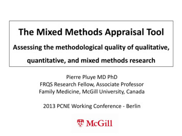
FAST AND REPRODUCIBLE RESULTSSemi-automated KineticPerimetryHigh resolution and fastperipheral testingTESTING FOR PTOSISIt takes a long time to examine peripheral defects, or areas that need highresolution, with static perimetry. Why not benefit from the advantages ofthe semi-automated Goldmann perimetry available with the Octopus 900?It helps generate fast and reproducible results in the periphery becauselarge areas can be covered quickly. Additionally, it provides high resolutionbecause answers can be marked anywhere on a vector.Pre-defined testing methodologies or even templates help to generate fastand reproducible results. Below are some examples that are easy to learnand use.PTOSIS TEMPLATEPtosis testing in just 2–4 minutesTesting for ptosis becomes fast and easy. With a line of simple, verticalvectors the edges can easily be mapped in a matter of 2–4 minutes. Thisis faster than with static automated perimetry13. Why spend more time onan equivalent result?BLIND SPOT TESTING METHODOLOGYDetailed outline of blind spotMAPPING OF THE BLIND SPOTUse kinetic perimetry to quickly map the blind spot in very high resolution.First, identify a point within the blind spot with some horizontal and vertical static points. Then use radial kinetic vectors to define the boundaries ofthe blind spot in high resolution. This fast and repeatable procedure provides you with far more detailed information than standard static programswhere test points are often several degrees apart.GENERAL BASELINE VECTOR TEMPLATEAutomate baseline peripheral testingYou want to check for neurological defects, glaucoma, diabetic diseaseand disability in a fast, patient-friendly and reproducible manner? By usinga pre-set template with a few radial vectors moving to the centre, you cangenerate your baseline isopters with minimal effort. Once the baseline isestablished, you can continue with your individual testing.BASELINE TESTING
VECTOR TEMPLATEINDIVIDUAL VECTOROriginal Goldmann Kineticperimetry to create isoptersthat are accurate, consistentand repeatable
14 15 CONNECTIVITY IS KEYEyeSuite PlatformFlexible interfaces for easy integrationinto your networkThe EyeSuite software is designed for optimum patient flows inbusy practices. It is very easy to use, making the Octopus 900fully networkable both with other Haag-Streit devices and yourpractice network. EyeSuite does not require any proprietarythird-party software to provide connectivity.Furthermore, the EyeSuite Script Language or standardised interfaces, such as GDT or DICOM, connect easily to almost anyelectronic medical record (EMR) system. Patient orders can bereceived from the EMR system and the measured results arethen automatically sent back to the EMR system.If the Octopus 900 is connected to an EyeSuite Server, all of itsdata can be accessed remotely from any number of viewing stations connected to the same database. This truly means goingbeyond a pre-defined printout and provides you with real-timeaccess to your data from anywhere in your network.With all these features availabe, you can save valuable staff timeand eliminate the risk of transcription errors.
Technical specificationsOctopus 900Octopus 900 Basic Octopus 900 ProStimulus generationPeripheral range (distance)Background illumination (asb)Stimulus size (Goldmann)Stimulus duration (ms)Stimulus intensity(asb, dynamic range)Fixation controlNetworkingData ImportMeasures (W x L x H)WeightTest methodsTest strategiesMirror projection system180 (30 cm radius Goldmann bowl)0/4/31/314I, II, III, IV, V100, 200, 500, 1000, infinite0.2–10000 (47 dB)Blink Control, Pupil Position Control, AET (Automated Eye Tracking)DICOM, EMR, EthernetOctopus 101, 123, 300 and 600; HFA II and III648 mm x 519 mm x 796 mm; 25.5’’ x 20.4’’ x 31.3’’25 kg; 55 lbsStandard white-on-white perimetry SAPBlue/yellow perimetry SWAP(DICOM optional)(DICOM optional)(Package Blue/YellowPerimetry)Flicker perimetry for early diagnosis(Package FlickerPerimetry)Red/white perimetry (custom tests only)(Package Scientific)Goldmann Kinetic Perimetry(Package GoldmannKinetic Perimetry)TOP (Tendency Oriented Perimetry,2–4 min)Dynamic (adaptive step size, 6–8 min)(Package TOP faststrategy)Normal (4-2-1 bracketing, 10–12 min)Test patternsOther (LV: Low Vision with Goldmann size V; 2-LT:2-Level Test, 1-LT: 1-Level Test; GST: GlaucomaScreening Test)General/Glaucoma 30 (G1-Program, 32, 30-2, 24-2)General/Glaucoma Periphery (G2-Program: 60 ;07-Program: 75 )Macula (M-Program (10 /30 ); 10-2)Screening (ST)Driving (Estermann monocular/binocular;FG: German Driving Licence)Disability (BT – Blepharoptosis;BG: German Blindness)Other Pathology(N1: Neurological; D1: Diabetes)Custom testsProgression analysisGlobal progression (MD, sLV)Cluster Trend/Polar Trend(Package Scientific)(Package Cluster/Polar Trend)IncludedSourcesOptionally available1 Wall M, Woodward KR, Doyle CK, Zamba G. The effective dynamic ranges of standard automated perimetry sizes III and V and motion and matrix perimetry. Archives ofophthalmology 2010; 128(5): 570-6 2 Wall M, Doyle CK, Zamba KD, Artes P, Johnson CA. The repeatability of mean defect with size III and size V standard automated perimetry. Invest Ophthalmol Vis Sci 2013; 54(2): 1345-51. 3 King AJ, Taguri A, Wadood AC, Azuara-Blanco A. Comparison of two fast strategies, SITA Fast and TOP, for the assessment of visual fields in glaucoma patients. Graefes Arch Clin Exp Ophthalmol. 2002 Jun;240(6):481-7. 4 Wadood AC, Azuara-Blanco A, Aspinall P, Taguri A, King A. Sensitivityand specificity of frequency-doubling technology, tendency-oriented perimetry, and Humphrey Swedish interactive threshold algorithm-fast perimetry in a glaucoma practice.Am J Ophthalmol. 2002 Mar;133(3):327-32. 5 Kovalska MP, Bürki E, Schoetzau A, Orguel SF, Orguel S, Grieshaber MC. Clinical evaluation of a novel population-based regression analysis for detecting glaucomatous visual field progression. Klin Monbl Augenheilkd. 2011 Apr;228(4):311-7. 6 Holló G, Naghizadeh F. Evaluation of Octopus Polar TrendAnalysis for detection of glaucomatous progression. Eur J Ophthalmol. 2014 Jun. DOI: 10.5301/ejo.5000504. 7 Naghizadeh F, Holló G. Detection of early glaucomatous progression with Octopus Cluster Trend analysis. J Glaucoma. 2014 Jun-Jul;23(5):269-75. 8 Rowe FJ, Hanif S. Uniocular and binocular fields of rotation measures: Octopus versus Goldmann. Graefes Arch Clin Exp Ophthalmol. 2011 Jun;249(6):909-19. 9 Nowomiejska K, Vonthein R, Paetzold J, Zagorski Z, Kardon R, Schiefer U. Comparison betweensemiautomated kinetic perimetry and conventional Goldmann manual kinetic perimetry in advanced visual field loss.Ophthalmology. 2005 Aug;112(8):1343-54. 10 VontheinR, Rauscher S, Paetzold J, Nowomiejska K, Krapp E, Hermann A, Sadowski B, Chaumette C, Wild JM, Schiefer U. The normal age-corrected and reaction time-corrected isopterderived by semi-automated kinetic perimetry. Ophthalmology. 2007 Jun;114(6):1065-72. 11 Nowomiejska K, Vonthein R, Paetzold J, Zagorski Z, Kardon R, Schiefer U. Reaction time during semi-automated kinetic perimetry (SKP) in patients with advanced visual field loss. Acta Ophthalmol. 2010 Feb;88(1):65-9. 12 Nowomiejska K, BrzozowskaA, Zarnowski T, Rejdak R, Weleber RG, Schiefer U. Variability in isopter position and fatigue during semi-automated kinetic perimetry. Ophthalmologica. 2012;227(3):166-72.13 Riemann CD, Hanson S, Foster JA. A comparison of manual kinetic and automated static perimetry in obtaining ptosis fields. Arch Ophthalmol. 2000 Jan;118(1):65-9.
HS-Art.No. 1511.7220444.02070Members of HAAG-STREIT GroupHAAG-STREIT AGGartenstadtstrasse 103098 KoenizSwitzerlandPhone 41 31 978 01 11Fax 41 31 978 02 IT Holding AGwww.haag-streit-holding.comCLEMENT CLARKE Ltd.www.clement-clarke.comHAAG-STREIT AG, Diagnosticswww.haag-streit.comHS DOMS GmbHwww.hs-doms.comHAAG-STREIT AG, Verkauf Schweizwww.haag-streit.chIPRO GmbHwww.ipro.deHAAG-STREIT Deutschland GmbHwww.haag-streit.deJohn Weiss Ltd.www.johnweiss.comHAAG-STREIT Far Eastwww.haag-streit-fareast.comMöller-Wedel GmbH & Co KGwww.haag-streit-surgical.comHAAG-STREIT Medtech AGwww.haag-streit-medtech.comMöller-Wedel Optical GmbHwww.moeller-wedel-optical.comHAAG-STREIT Surgical GmbHwww.haag-streit-surgical.comOptoMedical Technologies GmbHwww.haag-streit-surgical.comHAAG-STREIT UKwww.haag-streit-uk.comReliance Medical Inc.www.haag-streit-usa.comHAAG-STREIT USAwww.haag-streit-usa.comSPECTROS AGwww.spectros.ch HAAG-STREIT AG, 3098 Koeniz, Switzerland7. Edition / 2020 – 020297
Automated Eye Tracking (AET)* recognises the position of the pupil and keeps the pupil centred by automatically moving the head and chin rest into the ideal position. Thus, the Octopus 900 provides opti-mum conditions for reliable and undelayed results. BLINK CONTROL *Automated Eye Tracki