Transcription
GY 302 Crystallography and MineralogyImportant Optical Mineralogy Terms and ConceptsPart One: Basic StuffLight is a propagating wave front that moves fast. The velocity of light is a vacuum is one of themost important constants in science:Vc 2.988 x 10⁸ m/s (this constant is usually designated C)In any other medium beside a vacuum, the speed of light is less than C. When light travels fromone medium to another (vacuum to air, air to water), interesting effects occur. The physics behindthis is portrayed in the following sketch:The Index of Refraction (n) of a material is a ratio of C to the speed of light through a material(Vx).nx C/VxThe index of refraction for some common substances is given in the table .40-3.22The index of refraction of a mineral can be relatively easilymeasured in a laboratory, but it is important to minimize anydispersion of light that might occur. This is the prism effect thatoccurs when white light is split up into it’s component colors:
Light produced from an incandescent source (AKA light bulb) is emitted as waves that have nopreferred vibration direction (think of this as the orientation of the waveforms).In order to do petrography (which is complex enough as it is), you need to restrict the light thatpasses through your mineral specimens to waves that vibrate in a single direction. This is donewith the help of a polarizing lens:Do not confused polarized light with monochromatic light. The former is white light thatvibrates in a single direction. The latter is colored light of a single frequency (e.g. 550 nm is thepure red light you get in a laser pointer).When it comes to light transmission, most geologists recognize 3 classes of minerals:1) Transparent (minerals that transmit light and images through them) e.g., selenite2) Translucent (minerals that only transmit light) e.g. quartz3) Opaque (minerals that do not transmit light at all) e.g., galena, pyrite.Transmission light microscopy (AKA thin section microscopy or optical microscopy) studiestransparent and translucent minerals. The vast majority of minerals fall within these classes. Evenminerals that you might at first think are opaque (pyroxene, biotite) are translucent if they aresliced thinly enough.The way that light travels through minerals is entirely dependant on the crystalline structure ofthe minerals. In order to understand this, you need to recall the seven crystallographic systemsthat caused you so much hell in the crystallography component of this class.
Crystal SystemAxesAngles betweenaxes1Cubica b c" ( 90 2Tetragonala b c" ( 90 3Hexagonala a1 b c" 120 , ( 90 4Trigonala a1 b c" 120 , ( 90 5Orthorhombica b c" ( 90 6Monoclinica b c" 90 , ( 90 7Triclinica b c" ( 90 For some minerals (e.g., the cubic system), light is transmitted equally (in velocity orwavelength) in all directions through the crystal. The index of refraction is also uniform in alldirections. These minerals are said to be isotropic. In the rest of the mineral classes (termedanisotropic), the velocity of light is not consistent. There is a preferred orientation where light
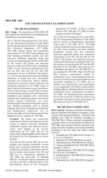
travels faster than in other directions. In order demonstrate the different ways that light can travelthrough minerals and to summarize the optical properties of minerals, microscopists came upwith the concept of an indicatrix. This is a geometrical figure that is produced around a pointusing the indices of refraction as radii. Isotropic minerals like the cubic mineral system havespherical indicatri (figure 1 below). Anisotropic minerals come in two major flavors. Minerals inthe tetragonal and trigonal/hexagonal classes are termed Uniaxial and the indicatrix is defined asa biaxial ellipsoid (2 different axes; figure 2). Minerals in the remaining classes (orthorhombic,monoclinic, triclinic) are termed Biaxial and the indicatrix is defined as a triaxial ellipsoid (3different axes; figure 3):
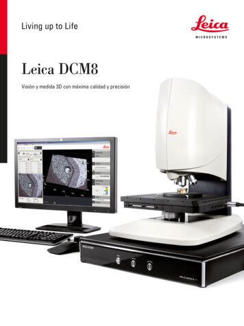
All anisotropic minerals do something interesting to light rays that pass through them. Theydivide the light (including polarized light) into two components which vibrate in mutuallyperpendicular planes. In the simplest case (that of the uniaxial minerals), one of these rays has aconstant velocity regardless of the direction that the light passes through the mineral. This ray iscalled the ordinary ray or o-ray for short. The other ray is called the extraordinary ray (or eray) and it’s velocity varies with direction. This splitting of rays is known as double refraction(or brirefringence1), and although it is difficult to envision, most geology students haveexperienced it’s effects in at least one mineral: optical calcite (or Icelandic spar). So pronouncedis the double refraction in calcite that it is possible to see a double image when a good crystal isplace on top of a piece of paper (think back to GY 111).In the same way that we can use the indicatrix to portray the various indices of refraction for themineral classes, it is possible to construct a graphic image summarizing the velocity and vibrationdirection differences of the o- and e-rays and in so doing, explain while double refraction occursin anisotropic minerals. The figures below shows the two possible situations that might occur inuniaxial minerals: (1) the o-ray is slower than the e-ray (uniaxial negative), (2) the o-ray is fasterthan the e-ray (uniaxial positive). Both figures are from Robinson and Bradbury (1992)1I don’t like to equate double refraction to birefringence. I prefer the more anal interpretation of birefringence whichis a measured difference between the indices of refraction of the o- and e- rays
There is only one orientation where the velocity of the o-ray and the e-ray is the same (the placeswhere the circle and the ellipse meet in the figure on the previous page). This orientation is calledthe optic axis and this is the only direction where double refraction does not occur. Theorientation of the optic axis frequently follows crystallographic axes (e.g., in quartz, the opticaxis is parallel to the c axis), but not always. In calcite, the optic axis is tilted relative to thecrystal faces which is one of the reasons why the double refraction is so pronounced.The phenomenon of double refraction in calcite also allows us to see the effects of polarizers ono-rays and e-rays2. Since each of these rays is perpendicular to each other, a sheet of polarizedglass placed on top of the calcite crystal as it rotates over a spot will induce predictable changesto the images.Part B: Minerals in thin-sectionThere are two different ways that thin-sections can be examined with a petrographic microscope.They can be examined with the upper polarizer (analyzer) in or out. Please note that the bottompolarizer is always in and that you are always using polarized light with these microscopes. If theupper polarizer is out, then the thin-section is being examined in plane polarized light. If theupper polarizer is in, then the thin-section is being examined under crossed polars or crossedNichols. Under crossed polars, all isotropic materials appear dark (this included the cubicminerals, non-crystalline minerals like opal and non-crystalline materials like glass. In fact, thefirst thing that you should always do when you first sit down at your microscope is to check thatthe polars are properly aligned. Before you start looking at thin-sections, first insert the upper2By the way, the terms ordinary and extraordinary rays were first used when discussing double refraction in calcite.The first image (the central spot in the diagram on this page) was termed “ordinary” because it stayed stationary asthe crystal was rotated over a spot on a piece of paper. The double image projected to the side rotated as the crystalwas rotated and it was called the extraordinary image. The e-ray image also appears to be lower in the crystal thanthe o-ray because of their different speeds through the crystal. Isn’t this a nice story?
polarizer (this is called crossing the polars). The field of view should be as black as possible. Ifnot, see Doug.Properties of minerals in plane polarized light (PPL)1) Colour. Most minerals are colourless under PPL, but some minerals intensely coloured. As ageneral rule, dark coloured minerals in hand specimen (e.g., pyroxene, amphibole, biotite etc.),are coloured in PPL. Light coloured minerals (quartz, fluorite, feldspars, muscovite etc.) arecolourless.2) Pleochroism. This is an interesting phenomenon where anisotropic minerals appear to changecolour as they are rotated in PPL. It has to do with variable indices of refraction and is related tothe crystal class of the minerals. Isotropic minerals are not pleochroic (light travels at the samevelocity regardless of orientation). Tetrahedral, trigonal and hexagonal minerals (unixialminerals) are said to be dichroic meaning that they can have up to two distinct colours. Theremaining mineral classes are trichroic and may have 3 colours. I say "may" because theorientation of the crystal in the thin-sectionwill determine which (if any) colourchanges might occur. Consider the biotitecrystal below. It is a biaxial mineral andtherefore has 3 possible colours dependingupon the orientation of the crystal in the thinsection and the orientation of the stageduring rotation.Some common pleochroic colour variationsare:Aegirine: dark green – light greenHornblende: yellow green - brownGlauconite: yellow-greenOne last thing about Pleochroism. Thepublished colours aren't always the ones you actually see. Biotite, for example, has advertisedcolours that go from brown to yellow to red. I've seen biotite also go green on rotation. Thismight just have been a weird mineral (or perhaps I mis-identified a hornblende as biotite).Whatever the reason, like all properties, don't assume that published data is always correct.3) Cleavage. Same property as seen in hand specimen. Just beware that now, you are more orless looking a 2 dimensional slices so ultimately, the orientation of your grains is important. Theonly time that you will see more than one cleavage direction is is you are looking down the c axisof the crystal (#3 for the pyroxene and amphibole examples given below). In this orientation, youwill see one strong cleavage plane and one that is significantly weaker. Do not confuse crackswith cleavage.
4) Relief. The official definition of relief is "the ratio of the index of refraction of a mineral tothe index of refractionof the materialimmediately adjacent toit (usually glass)." Amore practical way toregard the relief of amineral is how sharpand distinct the edge ofthe mineral looks. Theindex of refraction ofglass is about 1.50. If amineral is significantlyhigher or lower thanthis value, then the edge of the mineral looks very distinct and it is said to have high relief. If themineral has an index of refraction close to glass, then it has very indistinct edges and it is said tohave low relief.So the relief of a mineral tells you something about the index of refraction of the mineral. It isthis property that lets oil emersion testing identify unknown substances. Here's how this works. A
drop of an oil of known n is added to a slide containing fragments of an unknown substance. Ifthe relief of the mineral is high, you can tell that the index of refraction of the oil is way off whatthe mineral is. But how do you know if it is way below or way above the mineral? There is arather nice little procedure called the Becke Line Test that can tell you if the index of refractionof a mineral is higher or lower than the oil it is immersed in. The Becke Line is a bright band oflight that forms at the edge of crystals due to and edge effect. At the edge of crystals, taperingcauses the light rays to bend in toward the mineral if the mineral has a higher index of refractionthan the oil it is in, or out toward the oil if the mineral has a lower index of refraction than the oil.If you lower the stage while looking at the mineral in the microscope, the Becke Line will appearto move into the mineral (crystal high) or into the oil (crystal low) as the mineral defocuses. Thesaying goes; "Becke Line in, crystal high. Becke Line out, crystal low".The relief of a mineral is also affected by bubbles, inclusions and scratches. Also beware thatsome minerals (notably calcite and dolomites) have variable relief that like pleochroism, changesupon rotation of the stage.5) Crystallinity. This property is prettystraightforward. A crystal with sharp,geometric edges is said to be euhedral. Onethat has rounded edges (e.g., water abraded)is said to be anhedral.Properties of minerals under crossed-polars/crossed nichols (XN)Recap: Crossing the polars (inserting the upper polarizer) will result in a dark field of view forany substance that is non-crystalline (e.g., air, glass, non-crystalline materials like opal). Mineralsin the cubic class are also isotropic. They include garnet, halite, fluorite, spinel and sphalerite.
Anisotropic minerals will vary from bright to dark during rotation under crossed polars. There is,however, one orientation that will cause anisotropic minerals to appear isotropic. If you look rightdown the optic axis, you are looking right along the one direction where the e-rays and o-raystravel at the same speed. For all intents and purposes, the minerals is isotropic in this direction.For quartz, the optic axis is the same orientation as the c crystallographic axis. If you see ahexagonal quartz crystal, you are looking pretty much down the c axis and the optic axis. In thisorientation, the quartz crystal pretty much stays dark as you rotate the stage.While we are talking about the opticaxis, I should introduce anotherconcept that is used in opticalmineralogy. It is possible to see thepresence of o- and e-rays if you arelooking down the optic axis of amineral. In order to do this, you mustuse very high magnification (40Xobjective lens), have a well centeredmicroscope and insert the BertrandLens. This lens defocuses themicroscope and produces a crosspattern called an interference figureor an interference cross. The same typeof cross can frequently be seen in airbubbles, but for minerals, it tells you alot about the orientation of themineral's indicatrix. It also tells you ifthe mineral is uniaxial or biaxial.
The interference figurefor a quartz crystal with aproperly aligned opticaxis is shown at thebottom of the previouspage. The interferencefigure appears to stay putas the stage is rotated. Ifthe optic axis is a bit offof normal, theinterference figure willappear to orbit the centerpoint like the example tothe right. Uniaxialminerals (like quartz) areeasily identifiable if youcan see the interference figure. Biaxial minerals can also be identified if you can get aninterference figure that is centered in the eyepiece. As you rotate the stage for a biaxial mineral,you will see the cross separate into two curved components (gyres) that swing away from oneanother and then recombine again. The distance of separation (actually called the 2V angle) is adiagnostic characteristic of minerals that is useful in their identification.So if the interference figure is so great, why not just use it for mineral identification? Why do weneed to know all this other stuff? Simply put, the only time that you can ever really use theinterference figure is when you are looking right down the optic axis. This happens one in 1000times (if you are lucky). If you are trying to identify minerals in a rock (e.g., a thin section), youwill seldom get more than one crystal in the right orientation. So you need other options.1) Extinction. As you rotate the stage, you will observe that anisotropic minerals vary inbrightness/colour. At two points during a complete 360 degree rotation, anisotropic minerals godark (or nearly so). They are said to go extinct. Extinction occurs when the indicatrix aligns up
with the polars. For some minerals, extinction occurs in a sharp or sudden fashion. In someminerals, it is more like a curtain effect where part of the crystal fades out while other parts donot. Many minerals go extinct when the cleavage directions are parallel to polars. Others do not.The following diagram summarizes the types of extinction patterns that are recognized withinminerals:2) Birefringence. This is the most important property of minerals under crossed polars. It isdefined as the difference between the index of refraction of he minimum and maximum refractiveindices of a mineral. For uniaxial minerals, this is no – ne or ne - no. For biaxial crystals, this is
na – nc or nc - na. The main thing to note is that the greater the difference, the higher the colourthat is viewed. Please note that birefringence and extinction are kind of opposite properties.When a mineral goes extinct, it is at a minimum as far as birefringence is concerned. Themaximum birefringence occurs when the crystal is orientated exactly half way betweenextinctions (roughly 45 degrees from each extinction). The amount of birefringence is measuredmathematically, but it is best viewed via colour.Quartz has low birefringence (0.009). It's maximum colour is pale grey with a weak yellowishtinge. In contrast, olivine has high birefringence (0.036) and the maximum colour viewed is blue,green and/or pink. Calcite is said to have extreme birefringence (0.14). The birefringence coloursof calcite are so "high" that they are completely washed out and all you really see is white.The birefringence colours occur in a pattern similar to the colours of a rainbow and like arainbow, the colours repeat themselves. First order colours go from grey to white to yellow toorange to pink to red to blue. Second order colours are more vivid than first order colours and gofrom blue3 to green to yellow to red. Third order colours almost look fluorescent and go fromblue to green to yellow to pink. Fourth order colours are so faded that you can't really identifythem (but sometimes you catch a faded pink or green tint). Birefringence is very much affectedby thickness differences. Quartz has a maximum birefringence of 0.009 only when it is 30microns thick. If the thin section is 50 microns thick, quartz has orange birefringence. Thethickness of most thin sections is regulated at 30 microns to minimize the retardation4 effect.Reading the birefringence chart that occurs on the next page is a bit tricky at first because itactually summarizes 3 properties: (1) birefringence, (2) thickness variations and (3) minerals. Thediagonal lines that radiate from the bottom left corner lead to minerals at the top and right side of3The transition from first to second order colours occurs at blue. Some people refer to this first blue as first orderblue while others call it second order blue. I tend to do the latter.4Retardation is defined as thickness X birefringence
the chart. Quartz, for example, is located at a birefringence of 0.009. If you follow this diagonalline down toward the bottom of the chart, it eventually intersects the 0.003 mm thickness line thatruns horizontally across the chart. At this point, the vertical colour band is grey with a lightyellow tint. This is the maximum colour that quartz should be in a standard thin section.Once again, we need to be absolutely clear about the limitations of using birefringence to identifyminerals. Orientation is vital. In order to see the maximum birefringence colours, you need to belooking perpendicular to the optic axis of a crystal. This should make sense to you if you recallthat looking down the optic axis gives you a maximum (and constant) extinction. It stands toreason that if the birefringence and extinction are opposite properties, the maximum of oneshould be observed in a direction that is normal to the other.3) Optical Twinning. This is an interesting phenomenon that results when two or more crystalsof a single mineral grow together in a mathematically predicable pattern. This is the same type ofthing that happens in a crystallographic fashion and involves twin planes, twin axes etc. From anoptical point of view, the effects are quite frequently stunning. Optical twins are slightly out ofphase with one another. As you rotate the stage, one section goes extinct before the other does.Many minerals exhibit optical twins, but the all time leaders in this respect are the feldspars.Plagioclase feldspar exhibits polysynthetic twinning which resembles prison stripes. Microclinefeldspar exhibits tartan twinning which resembles the plaid of a Scottish kilt. Orthoclasedisplays Carlsbad twinning, but it is not as prominent as most of the other feldspars.By the way, it is possible to determine the composition of the plagioclase feldspars (percentage ofNa to Ca) by measuring the extinction angles of the twin sets. I'll show you how to do this in oneof the labs. If I fail to do this, Dr. Allison is likely to in GY 343.
4) Zonation. Some minerals change their composition has they grow, particularly those that thatform continuous series through solid solution during igneous processes (e.g., olivine,plagioclase). Because their core may be a different composition than their edges (e.g., plagioclaseusually starts off more Ca rich and later becomes more Na rich during crystallization in amagma), the core may go extinct slightly out of phase to the rest of the crystal. Sometimeszonation of crystals is very evident in some rocks (particularly in basalts). Most of the time it isnot. Still, when you see it, it really is quite impressive.
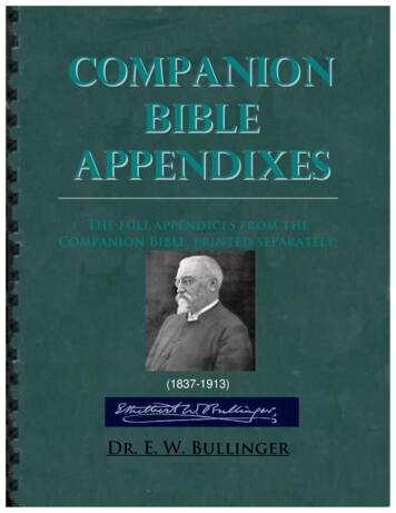
The Petrographic MicroscopeThe petrographic microscopes that you will be using in this class are pretty much the beststudent scopes currently available in the United States. They look much fancier than thoseusually portrayed in books (or in the figure below), but in many ways, they are no different thatthe microscopes Bowen used when he was experimenting with igneous petrology about 100 yearsago.Light Source. Our scopesuse a tungsten light bulbdesigned to burn at 6000degrees K (7-9 on your lightintensity knob). At thistemperature, the emissionspectra is yellow so a bluefilter is used to soften thelight. Do NOT use theintensity knob to lower thebrightness of the light. If it istwo bright for you, use theaperture or diaphragmCondenser assembly. On ourmicroscopes, this consists of adiaphragm, the bottom polarizer, and acondenser lens. The condenser focuseslight into a smaller (but brighter) beamwhich is necessary when looking atspecimens under very highmagnifications (25 to 40X objectives).The stage on a petrographicmicroscope is very different that theone that most of you are familiar with
on standard (biological) microscopes. As well as moving up and down via a rack and pinionsystem for focusing, this one also rotates. The amount of rotation can be measured via a scale onthe edge of the circular stage (this is useful for measuring the angle of extinction of a mineral).The stage has a hole through the middle on which, the thin section is placed. A thin section is acomplex thing consisting of a glass slide (the base) the rock specimen and a cover slip. Eachlayer is glued to the others with epoxy resins. The best resins (we use the best ones for our thinsections) are optically isotropic and have indices of refraction close to glass.The upper assembly of themicroscope is the most complexpart. It consists of a rotating turretthat houses 3, 4 or 5 objectivelens, a fixed barrel that houses theupper polarizer (the analyzer),the Bertrand lens, one or moreslots for accessory plates, and theeyepiece(s) (usually 10 X).Theeyepieces also contain a cross hairand/or scale for point counting andsize measurements (both of thesefun jobs will be part of some futurelabs).
Light is a propagating wave front that moves fast. The velocity of light is a vacuum is one of the . Crystal System Axes Angles between axes 1 Cubic a b c " ( 90 2 Tetragonal a b c " . 2 By the way, the terms ordinary and extraordinary rays were first used when discussing double refraction in calcite.