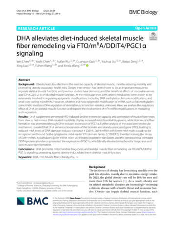
Transcription
Oatis CH04 045-068.qxd4/18/072:21 PMPage 45CHAPTERBiomechanics of Skeletal Muscle4CHAPTER CONTENTSSTRUCTURE OF SKELETAL MUSCLE . . . . . . . . . . . . . . . . . . . . . . . . . . . . . . . . . . . . . . . . . . . . . . . . . . . . . . . . . . . . . . . . . . . . . . .46Structure of an Individual Muscle Fiber . . . . . . . . . . . . . . . . . . . . . . . . . . . . . . . . . . . . . . . . . . . . . . . . . . . . . . . . . . . . . . . .46The Connective Tissue System within the Muscle Belly . . . . . . . . . . . . . . . . . . . . . . . . . . . . . . . . . . . . . . . . . . . . . . . . . . . .48FACTORS THAT INFLUENCE A MUSCLE’S ABILITY TO PRODUCE A MOTION . . . . . . . . . . . . . . . . . . . . . . . . . . . . . . . . . . . . . .48Effect of Fiber Length on Joint Excursion . . . . . . . . . . . . . . . . . . . . . . . . . . . . . . . . . . . . . . . . . . . . . . . . . . . . . . . . . . . . . . .48Effect of Muscle Moment Arms on Joint Excursion . . . . . . . . . . . . . . . . . . . . . . . . . . . . . . . . . . . . . . . . . . . . . . . . . . . . . . .50Joint Excursion as a Function of Both Fiber Length and the Anatomical Moment Arm of a Muscle . . . . . . . . . . . . . . .51FACTORS THAT INFLUENCE A MUSCLE’S STRENGTH . . . . . . . . . . . . . . . . . . . . . . . . . . . . . . . . . . . . . . . . . . . . . . . . . . . . . . . . .52Muscle Size and Its Effect on Force Production . . . . . . . . . . . . . . . . . . . . . . . . . . . . . . . . . . . . . . . . . . . . . . . . . . . . . . . . . .52Relationship between Force Production and Instantaneous Muscle Length (Stretch) . . . . . . . . . . . . . . . . . . . . . . . . . . .53Relationship between a Muscle’s Moment Arm and Its Force Production . . . . . . . . . . . . . . . . . . . . . . . . . . . . . . . . . . . . .56Relationship between Force Production and Contraction Velocity . . . . . . . . . . . . . . . . . . . . . . . . . . . . . . . . . . . . . . . . . . .58Relationship between Force Production and Level of Recruitment of Motor Units within the Muscle . . . . . . . . . . . . .60Relationship between Force Production and Fiber Type . . . . . . . . . . . . . . . . . . . . . . . . . . . . . . . . . . . . . . . . . . . . . . . . . . .61ADAPTATION OF MUSCLE TO ALTERED FUNCTION . . . . . . . . . . . . . . . . . . . . . . . . . . . . . . . . . . . . . . . . . . . . . . . . . . . . . . . . . .62Adaptation of Muscle to Prolonged Length Changes . . . . . . . . . . . . . . . . . . . . . . . . . . . . . . . . . . . . . . . . . . . . . . . . . . . . .62Adaptations of Muscle to Sustained Changes in Activity Level . . . . . . . . . . . . . . . . . . . . . . . . . . . . . . . . . . . . . . . . . . . . . .63SUMMARY . . . . . . . . . . . . . . . . . . . . . . . . . . . . . . . . . . . . . . . . . . . . . . . . . . . . . . . . . . . . . . . . . . . . . . . . . . . . . . . . . . . . . . . . . .64Skeletal muscle is a fascinating biological tissue able to transform chemical energy to mechanical energy. Thefocus of this chapter is on the mechanical behavior of skeletal muscle as it contributes to function and dysfunction of the musculoskeletal system. Although a basic understanding of the energy transformation from chemi-cal to mechanical energy is essential to a full understanding of the behavior of muscle, it is beyond the scope of thisbook. The reader is urged to consult other sources for a discussion of the chemical and physiological interactions thatproduce and affect a muscle contraction [41,52,86].Skeletal muscle has three basic performance parameters that describe its function:IMovement productionIForce productionIEnduranceThe production of movement and force is the mechanical outcome of skeletal muscle contraction. The factors thatinfluence these parameters are the focus of this chapter. A brief description of the morphology of muscles and the45
Oatis CH04 045-068.qxd4/18/072:21 PMPage 4646Part I BIOMECHANICAL PRINCIPLESphysiological processes that produce contraction needed to understand these mechanical parameters are also presentedhere. Specifically the purposes of this chapter are toIReview briefly the structure of muscle and the mechanism of skeletal muscle contractionIExamine the factors that influence a muscle’s ability to produce a motionIExamine the factors that influence a muscle’s ability to produce forceIConsider how muscle architecture is specialized to optimize a muscle’s ability to produce force or joint motionIDemonstrate how an understanding of these factors can be used clinically to optimize a person’s performanceIDiscuss the adaptations that muscle undergoes with prolonged changes in length and activitySTRUCTURE OF SKELETAL MUSCLEThe functional unit that produces motion at a joint consistsof two discrete units, the muscle belly and the tendon thatbinds the muscle belly to the bone. The structure of the muscle belly itself is presented in the current chapter. The structure and mechanical properties of the tendon, composed ofconnective tissue, are presented in Chapter 6. The musclebelly consists of the muscle cells, or fibers, that produce thecontraction and the connective tissue encasing the musclefibers. Each is discussed below.Structure of an Individual Muscle FiberA skeletal muscle fiber is a long cylindrical, multinucleatedcell that is filled with smaller units of filaments (Fig. 4.1). TheseBiceps brachii m.Muscle fascicleMuscle fiberSingle myofibrilWhole muscle(Biceps brachii m.)Actin myofilamentMyosin myofilamentFigure 4.1: Organization of muscle. A progressively magnified view of a whole muscle demonstrates the organization of the filamentscomposing the muscle.
Oatis CH04 045-068.qxd4/18/072:21 PMPage 47Chapter 4 BIOMECHANICS OF SKELETAL MUSCLE47filamentous structures are roughly aligned parallel to the muscle fiber itself. The largest of the filaments is the myofibril,composed of subunits called sarcomeres that are arranged endto end the length of the myofibril. Each sarcomere also contains filaments, known as myofilaments. There are two types ofmyofilaments within each sarcomere. The thicker myofilaments are composed of myosin protein molecules, and thethinner myofilaments are composed of molecules of the protein actin. Sliding of the actin myofilament on the myosinchain is the basic mechanism of muscle contraction.THE SLIDING FILAMENT THEORY OF MUSCLECONTRACTIONThe sarcomere, containing the contractile proteins actin andmyosin, is the basic functional unit of muscle. Contraction of awhole muscle is actually the sum of singular contraction eventsoccurring within the individual sarcomeres. Therefore, it isnecessary to understand the organization of the sarcomere.The thinner actin chains are more abundant than the myosinmyofilaments in a sarcomere. The actin myofilaments areanchored at both ends of the sarcomere at the Z-line and project into the interior of the sarcomere where they surround athicker myosin myofilament (Fig. 4.2). This arrangement ofmyosin myofilaments surrounded by actin myofilaments isrepeated throughout the sarcomere, filling its interior and giving the muscle fiber its characteristic striations. The amount ofthese contractile proteins within the cells is strongly related toa muscle’s contractile force [6,7,27].Contraction results from the formation of cross-bridgesbetween the myosin and actin myofilaments, causing the gure 4.2: Organization of actin and myosin within a musclefiber. The arrangement of the actin and myosin chains in twoadjacent sarcomeres within a fiber produces the characteristicstriations of skeletal muscle.chains to “slide” on the myosin chain (Fig. 4.3). The tension ofthe contraction depends upon the number of cross-bridgesformed between the actin and myosin myofilaments. The number of cross-bridges formed depends not only on the abundance of the actin and myosin molecules, but also on thefrequency of the stimulus to form cross-bridges.Contraction is initiated by an electrical stimulus from theassociated motor neuron causing depolarization of the musclefiber. When the fiber is depolarized, calcium is released intothe cell and binds with the regulating protein troponin. Thecombination of calcium with troponin acts as a trigger, causing actin to bind with myosin, beginning the contraction.Cessation of the nerve’s stimulus causes a reduction in calcium levels within the muscle fiber, inhibiting the crossbridges between actin and myosin. The muscle relaxes [86]. nedsarcomerelengthActinMuscle cellRELAXEDMuscle cellCONTRACTEDFigure 4.3: The sliding filament model. Contraction of skeletal muscle results from the sliding of the actin chains on the myosin chains.
Oatis CH04 045-068.qxd4/18/072:21 PMPage 4848Part I BIOMECHANICAL PRINCIPLESstimulation of the muscle fiber occurs at a sufficiently highfrequency, new cross-bridges are formed before prior interactions are completely severed, causing a fusion of succeeding contractions. Ultimately a sustained, or tetanic,contraction is produced. Modulation of the frequency andmagnitude of the initial stimulus has an effect on the force ofcontraction of a whole muscle and is discussed later in thischapter.The Connective Tissue System withinthe Muscle BellyThe muscle belly consists of the muscle cells, or fibers, and theconnective tissue that binds the cells together (Fig. 4.4). Theoutermost layer of connective tissue that surrounds the entiremuscle belly is known as the epimysium. The muscle belly isdivided into smaller bundles or fascicles by additional connective tissue known as perimysium. Finally individual fiberswithin these larger sheaths are surrounded by more connective tissue, the endomysium. Thus the entire muscle belly isinvested in a large network of connective tissue that then isbound to the connective tissue tendons at either end of themuscle. The amount of connective tissue within a muscle andthe size of the connecting tendons vary widely from muscle toMuscle gure 4.4: Organization of the connective tissue within muscle.The whole muscle belly is invested in an organized system ofconnective tissue. It consists of the epimysium surroundingthe whole belly, the perimysium encasing smaller bundles ofmuscle fibers, and the endomysium that covers individualmuscle fibers.muscle. The amount of connective tissue found within anindividual muscle influences the mechanical properties of thatmuscle and helps explain the varied mechanical responses ofindividual muscles. The contribution of the connective tissueto a muscle’s behavior is discussed later in this chapter.FACTORS THAT INFLUENCE A MUSCLE’SABILITY TO PRODUCE A MOTIONAn essential function of muscle is to produce joint movement.The passive range of motion (ROM) available at a jointdepends on the shape of the articular surfaces as well as onthe surrounding soft tissues. However the joint’s active ROMdepends on a muscle’s ability to pull the limb through a joint’savailable ROM. Under normal conditions, active ROM isapproximately equal to a joint’s passive ROM. However thereis a wide variation in the amount of passive motion availableat joints throughout the body. The knee joint is capable offlexing through an arc of approximately 140 , but themetacarpophalangeal (MCP) joint of the thumb usually iscapable of no more than about 90 of flexion. Joints thatexhibit large ROMs require muscles capable of moving thejoint through the entire range. However such muscles areunnecessary at joints with smaller excursions. Thus musclesexhibit structural specializations that influence the magnitudeof the excursion that is produced by a contraction. These specializations are The length of the fibers composing the muscle The length of the muscle’s moment arm.How each of these characteristics affects active motion of ajoint is discussed below.Effect of Fiber Length on Joint ExcursionFiber length has a significant influence on the magnitude ofthe joint motion that results from a muscle contraction. Thefundamental behavior of muscle is shortening, and it is thisshortening that produces joint motion. The myofilaments ineach sarcomere are 1 to 2 µm long; the myosin myofilaments are longer than the actin myofilaments [125,149].Thus sarcomeres in humans are a few micrometers inlength, varying from approximately 1.25 to 4.5 µm with muscle contraction and stretch [90–92,143]. Each sarcomerecan shorten to approximately the length of its myosin molecules. Because the sarcomeres are arranged in series in amyofibril, the amount of shortening that a myofibril and,ultimately, a muscle fiber can produce is the sum of theshortening in all of the sarcomeres. Thus the total shortening of a muscle fiber depends upon the number of sarcomeres arranged in series within each myofibril. The moresarcomeres in a fiber, the longer the fiber is and the more itis able to shorten (Fig. 4.5). The amount a muscle fiber canshorten is proportional to its length [15,89,155]. A fiber canshorten roughly 50 to 60% of its length [44,155], although
Oatis CH04 045-068.qxd4/18/072:21 PMPage 49Chapter 4 BIOMECHANICS OF SKELETAL MUSCLEBone49there is some evidence that fibers exhibit varied shorteningcapabilities [15].The absolute amount of shortening a fiber undergoes is afunction of its fiber length. Similarly, the amount a wholemuscle can shorten is dictated by the length of its constituentfibers. An individual whole muscle is composed mostly offibers of similar lengths [15]. However there is a wide variation in fiber lengths found in the human body, ranging from afew centimeters to approximately half a meter [86,146]. Thelength of the fibers within a muscle is a function of the architecture of that muscle rather than of the muscle’s total length.The following describes how fiber length and muscle architecture are related.BoneARCHITECTURE OF SKELETAL MUSCLEBoneBBoneAFigure 4.5: The relationship between fiber length and shorteningcapacity of the whole muscle. A muscle with more sarcomeres inseries (A) can shorten more than a fiber with fewer sarcomeres inseries (B).Although all skeletal muscle is composed of muscle fibers, thearrangement of those fibers can vary significantly amongmuscles. This fiber arrangement has marked effects on amuscle’s ability to produce movement and to generate force.Fiber arrangements have different names but fall into twomajor categories, parallel and pennate [42] (Fig. 4.6). Ingeneral, the fibers within a parallel fiber muscle are approximately parallel to the length of the whole muscle. These muscles can be classified as either fusiform or strap muscles.Fusiform muscles have tendons at both ends of the muscle sothat the muscle fibers taper to insert into the tendons. StrapFUSIFORMMULTIPENNATEUNIPENNATEBiceps brachii m.Subscapularis m.Flexor pollicis longus m.BIPENNATESTRAPASartorius m.BRectus femoris m.Figure 4.6: Muscle architecture. A. Muscles with parallel fibers include fusiform (biceps brachii) and strap (sartorius) muscles. B. Pennatemuscles include unipennate (flexor pollicis longus), bipennate (rectus femoris), and multipennate (subscapularis).
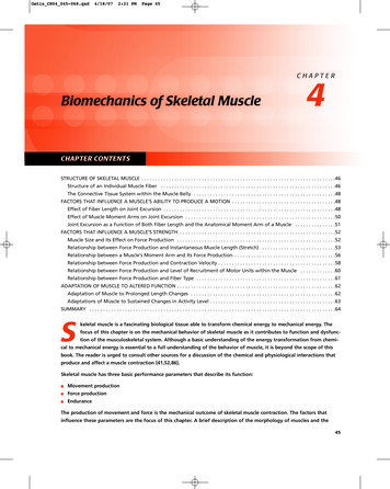
Oatis CH04 045-068.qxd4/18/072:21 PMPage 5050Part I BIOMECHANICAL PRINCIPLESmuscles have less prominent tendons, and therefore theirfibers taper less at both ends of the whole muscle. Parallelfiber muscles are composed of relatively long fibers, althoughthese fibers still are shorter than the whole muscle. Even thesartorius muscle, a classic strap muscle, contains fibers thatare only about 90% of its total length.In contrast, a pennate muscle has one or more tendons that extend most of the length of the whole muscle.Fibers run obliquely to insert into these tendons. Pennatemuscles fall into subcategories according to the number oftendons penetrating the muscle. There are unipennate,bipennate, and multipennate muscles. A comparison oftwo muscles of similar total length, one with parallel fibersand the other with a pennate arrangement, helps to illustratethe effect of fiber arrangement on fiber length (Fig. 4.7). Themuscle with parallel fibers has longer fibers than those foundin the pennate muscle. Because the amount of shorteningthat a muscle can undergo depends on the length of its fibers,the muscle with parallel fibers is able to shorten more thanthe pennate muscle. If fiber length alone affected joint excursion, the muscle with parallel fibers would produce a largerjoint excursion than the muscle composed of pennate fibersEffect of Muscle Moment Armson Joint ExcursionChapter 1 defines the moment arm of a muscle as the perpendicular distance between the muscle and the point ofrotation. This moment arm depends on the location of themuscle’s attachment on the bone and on the angle betweenthe line of pull of the muscle and the limb to which the muscle attaches. This angle is known as the angle of application(Fig. 4.8). The location of an individual muscle’s attachmenton the bone is relatively constant across the population.Therefore, the distance along the bone between the muscle’sattachment and the center of rotation of the joint can beestimated roughly by anyone with a knowledge of anatomyand can be measured precisely as well [57,81,95,151]. ThisθApproximatefiber lengthA Parallel[90]. However, a muscle’s ability to move a limb through anexcursion also depends on the length of the muscle’s momentarm. Its effect is described below.B PennateFigure 4.7: The relationship between muscle architecture andmuscle fiber length. The fibers in a muscle with parallel fibersare typically longer than the fibers in a muscle of similar overallsize but with pennate fibers.Figure 4.8: Angle of application. A muscle’s angle of applicationis the angle formed between the line of pull of the muscle andthe bone to which the muscle attaches.
Oatis CH04 045-068.qxd4/18/072:21 PMPage 51Chapter 4 BIOMECHANICS OF SKELETAL MUSCLE51θABFigure 4.9: The relationship between a muscle’s moment arm and excursion. The length of a muscle’s moment arm affects the excursionthat results from a contraction. A. Movement through an angle, , requires more shortening in a muscle with a long moment arm thanin a muscle with a short moment arm. B. The arc subtended by an angle, , is larger in a large circle than in a small circle.distance is related to the true moment arm by the sine of theangle of application, , which can also be estimated or measured directly.A muscle’s moment arm has a significant effect on thejoint excursion produced by a contraction of the muscle. Amuscle with a short moment arm produces a larger angularexcursion than another muscle with a similar shorteningcapacity but with a longer moment arm. Principles of basicgeometry help explain the relationship between musclemoment arms and angular excursion. Given two circles ofdifferent sizes, an angle, , defines an arc on each circle(Fig. 4.9). However, the arc of the larger circle is largerthan the arc of the smaller circle. Thus the distance traveled on the larger circle to move through the angle isgreater than that on the smaller circle. Similarly, a musclewith a long moment arm must shorten more to produce thesame angular displacement as a muscle with a shortmoment arm [76,77].Joint Excursion as a Function of BothFiber Length and the Anatomical MomentArm of a MuscleThe preceding discussion reveals that both a muscle’s fiberlength and its moment arm have direct effects on the amountof excursion a muscle contraction produces. These effects canbe summarized by the following: Because muscle fibers possess a similar relative shorteningcapability, longer fibers produce more absolute shorteningthan shorter fibers. Because muscles with parallel fibers generally have longerfibers than pennate muscles, whole muscles composed ofparallel fibers have a larger shortening capacity than wholemuscles of similar length composed of pennate fibers. Muscles with shorter anatomical moment arms are capable of producing greater angular excursions of a joint thanmuscles of similar fiber length with larger anatomicalmoment arms.It is interesting to see how these characteristics are combined in individual muscles. Muscles combine these seemingly opposing attributes in various ways, resulting in diversefunctional capacities. It appears that some muscles, like thegluteus maximus, possess both long fibers and relativelyshort moment arms. Such muscles are capable of producingrelatively large joint excursions [62]. Others, like the brachioradialis muscle at the elbow, combine relatively longmuscle fibers with large moment arms [89]. The long fibersenhance the muscle’s ability to produce a large excursion.However, the large moment arm decreases the muscle’sability to produce a large excursion. This apparent contradiction is explained in part by the recognition that the factors that influence production of movement, musclearchitecture and anatomical moment arm, also influenceforce production capabilities in a muscle. Muscles must findways to balance the competing demands of force productionand joint excursion. The ratio of a muscle’s fiber length to itsmoment arm is a useful descriptor of a muscle’s ability toproduce an excursion and its torque-generating capability[99]. This ratio helps surgeons determine appropriate donormuscles to replace dysfunctional ones.
Oatis CH04 045-068.qxd4/18/072:21 PMPage 5252Part I BIOMECHANICAL PRINCIPLESClinical RelevanceCONSIDERATIONS REGARDING TENDONTRANSFERS: Muscle fiber arrangement and musclemoment arms are inherent characteristics of a muscle andnormally change very little with exercise or functional use.However, surgeons commonly transfer a muscle or muscles toreplace the function of paralyzed muscles [15,16]. Successfulrestoration of function requires that the surgeon not onlyreplace the nonfunctioning muscle with a functional musclebut also must ensure that the replacement muscle has anexcursion-generating capacity similar to that of the originalmuscle. This may be accomplished by choosing a structurallysimilar muscle or by surgically manipulating the moment armto increase or decrease the excursion capability [155].For example, the flexor carpi radialis muscle at the wrist isa good substitute for the extensor digitorum muscle of thefingers in the event of radial nerve palsy. The wrist flexor haslong muscle fibers and, therefore, the capacity to extend thefingers through their full ROM. In contrast, the flexor carpiulnaris, another muscle of the wrist, has very short fibers andlacks the capacity to move the fingers through their fullexcursion. Thus the functional outcome depends on the surgeon’s understanding of muscle mechanics, including thosefactors that influence the production of motion.FACTORS THAT INFLUENCE A MUSCLE’SSTRENGTHStrength is the most familiar characteristic of muscle performance. However, the term strength has many differentinterpretations. Understanding the factors affecting strengthrequires a clear understanding of how the term is used. Thebasic activity of muscle is to shorten, thus producing a tensileforce. As noted in Chapter 1, a force also produces amoment, or a tendency to rotate, when the force is exertedat some distance from the point of rotation. The ability togenerate a tensile force and the ability to create a momentare both used to describe a muscle’s strength. Assessment ofmuscle strength in vivo is typically performed by determining the muscle’s ability to produce a moment. Such assessments include determination of the amount of manualresistance an individual can sustain without joint rotation,the amount of weight a subject can lift, or the direct measurement of moments using a device such as an isokineticdynamometer. In contrast, in vitro studies often assess muscle strength by measuring a muscle’s ability to generate a tensile force. Of course the muscle’s tensile force of contractionand its resulting moment are related by the following:MⴝrⴛF(Equation 4.1)where M is the moment generated by the muscle’s tensileforce (F) applied at a distance (r, the muscle’s moment arm)from the point of rotation (the joint axis). Therefore, musclestrength as assessed typically in the clinic by the measurement of the moment produced by a contraction is a functionof an array of factors that influence both the tensile force ofcontraction, F, and the muscle’s moment arm, r [54]. Toobtain valid assessments of muscle strength and to optimizemuscle function, the clinician must understand the factorsthat influence the output of the muscle. All of the followingfactors ultimately influence the moment produced by themuscle’s contraction. Some affect the contractile force, andothers influence the muscle’s ability to generate a moment.The primary factors influencing the muscle’s strength are Muscle sizeMuscle moment armStretch of the muscleContraction velocityLevel of muscle fiber recruitmentFiber types composing the muscleEach of the factors listed above has a significant effect on themuscle’s moment production. An understanding of each factor and its role in moment production allows the clinician touse these factors to optimize a person performance and tounderstand the alteration in muscle performance with pathology. The effects of size, moment arm, and stretch are mostapparent in isometric contractions, which are contractionsthat produce no discernable joint motion. Consequently, theexperiments demonstrating these effects usually employ isometric contractions. However, the reader must recognize thatthe effects are manifested in all types of contraction. Eachfactor is discussed below.Muscle Size and Its Effect on ForceProductionAs noted earlier in this chapter, the force of contraction isa function of the number of cross-links made between theactin and myosin chains [1,39]. The more cross-linksformed, the stronger the force of contraction. Therefore,the force of contraction depends upon the amount of actinand myosin available and thus on the number of fibers amuscle contains. In other words, the force of contraction isrelated to a muscle’s size [67,126]. In fact, muscle size isthe most important single factor determining the tensileforce generated by a muscle’s contraction [44,60].Estimates of the maximal contractile force per unit of muscle range from approximately 20 to 135 N/cm2 [15,22,120,155]. These data reveal a wide disparity in the estimates of the maximum tensile force that muscle can produce. Additional research is needed to determine if allskeletal muscle has the same potential maximum and whatthat maximum really is.Although the estimates presented above vary widely, theydo demonstrate that the maximum tensile force produced byan individual muscle is a function of its area. However, theoverall size of a muscle may be a poor indication of the number of fibers contained in that muscle. The relationship
Oatis CH04 045-068.qxd4/18/072:21 PMPage 53Chapter 4 BIOMECHANICS OF SKELETAL MUSCLEbetween muscle size and force of contraction is complicatedby the muscle’s architecture. The anatomical cross-sectional area of the muscle is the cross-sectional area at themuscle’s widest point and perpendicular to the length of thewhole muscle. In a parallel fiber muscle this cross-sectionalarea cuts across most of the fibers of the muscle (Fig. 4.10).However, in a pennate muscle the anatomical cross-sectionalarea cuts across only a portion of the fibers composing themuscle. Thus the anatomical cross-sectional area underestimates the number of fibers contained in a pennate muscleand hence its force production capabilities.The standard measure used to approximate the number offibers of a whole muscle is its physiological cross-sectionalarea (PCSA). The PCSA is the area of a slice that passesthrough all of the fibers of a muscle [15]. In a parallel fiberSartorius m.Rectusfemoris m.ABFigure 4.10: The relationship between muscle architecture andmuscle size. A. The anatomical cross-sectional area of a muscle isthe area of a slice through the widest part of the muscle perpendicular to the muscle’s length. It is similar in a parallel fibermuscle and a pennate muscle of similar overall size. B. Thephysiological cross-sectional area of a muscle is the area of a slicethat cuts across all of the fibers of the muscle. It is quite differentfor a parallel fiber muscle and a pennate muscle.53muscle the PCSA is approximately equal to the anatomicalcross-sectional area. However, in a pennate muscle the PCSAis considerably larger than its anatomical cross-sectional area.The PCSAs of two muscles of similar overall size demonstratethe influence of muscle architecture on force production.Although their anatomical cross-sectional areas are very similar, the pennate muscle has a much larger PCSA. Thus if allother factors are equal, the pennate muscle is capable of generating more contraction force than the muscle with parallelfibers [64,90,114].The angle at which the fibers insert into the tendon alsoinfluences the total force that is applied to the limb by a pennate muscle. This angle is known as the angle of pennation.The tensile force generated by the whole muscle is the vectorsum of the force components that are applied parallel to themuscle’s tendon (Fig. 4.11). Therefore, as the angle of pennation increases, the tensile component of the contraction forcedecreases. However, the larger the pennation angle is, thelarger the PCSA is [2]. In most muscles the pennation angleis 30 or less, and thus pennation typically increases the tensile force produced by contraction [86,146]. Resistance training increases the fibers’ angle of pennation (and the muscle’sPCSA). This increase appears to result from increases, orhypertrophy, in the cross-sectional area of individual musclefibers [2,13].Muscle architecture demonstrates how muscles exhibit specializations that enhance one performance characteristic oranother. Long fibers in a muscle promote the excursion-producing capacity of the muscle. However,
Biomechanics of Skeletal Muscle 4 CHAPTER CHAPTER CONTENTS 45 S keletal muscle is a fascinating biological tissue able to transform chemical energy to mechanical energy. The focus of this chapter is on the mechanical behavior of skeletal muscle as it contributes to function and dysfunc-tion of the musculoskeletal system.