Transcription
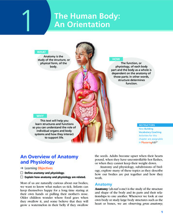
1The Human Body:An OrientationWHATAnatomy is thestudy of the structure, orphysical form, of thebody.HOWThe function, orphysiology, of each bodypart and the body as a whole isdependent on the anatomy ofthose parts; in other words,structure determinesfunction.WHYThis text will help youlearn structures and functionsso you can understand the role ofindividual organs and bodysystems and how they interactto support life.An Overview of Anatomyand Physiology Learning Objectives Define anatomy and physiology. Explain how anatomy and physiology are related.Most of us are naturally curious about our bodies;we want to know what makes us tick. Infants cankeep themselves happy for a long time staring attheir own hands or pulling their mother’s nose.Older children wonder where food goes whenthey swallow it, and some believe that they willgrow a watermelon in their belly if they swallowINSTRUCTORSNew BuildingVocabulary CoachingActivities for thischapter are assignableinthe seeds. Adults become upset when their heartspound, when they have uncontrollable hot flashes,or when they cannot keep their weight down.Anatomy and physiology, subdivisions of biology, explore many of these topics as they describehow our bodies are put together and how theywork.AnatomyAnatomy (ah-nat′o-me) is the study of the structureand shape of the body and its parts and their relationships to one another. Whenever we look at ourown body or study large body structures such as theheart or bones, we are observing gross anatomy;1
2Essentials of Human Anatomy and Physiologythat is, we are studying large, easily observablestructures. Indeed, the term anatomy, derivedfrom the Greek words meaning to cut (tomy) apart(ana), is related most closely to gross anatomicalstudies because in such studies, preserved animalsor their organs are dissected (cut up) to be examined. Microscopic anatomy, in contrast, is the studyof body structures that are too small to be seenwith the naked eye. The cells and tissues of thebody can only be seen through a microscope. Learning Objectives Name the six levels of structural organization thatmake up the human body, and explain how theyare related. Name the organ systems of the body, and brieflystate the major functions of each system. Identify and classify by organ system all organsdiscussed.PhysiologyPhysiology (fiz″e-ol′o-je) is the study of how thebody and its parts work or function ( physio nature; ology the study of). Like anatomy, physiology has many subdivisions. For example, neurophysiology explains the workings of the nervoussystem, and cardiac physiology studies the function of the heart.Relationship betweenAnatomy and PhysiologyAnatomy and physiology are always inseparable.The parts of your body form a well-organized unit,and each of those parts has a job to do to makethe body operate as a whole. Structure determineswhat functions can take place. For example, thelungs are not muscular chambers like the heartand so cannot pump blood through the body,but because the walls of their air sacs are verythin, they can exchange gases and provide oxygento the body. We stress the intimate relationshipbetween anatomy and physiology throughout thistext to make your learning meaningful.Did You Get It?1. Why would you have a hard time learning andunderstanding physiology if you did not alsounderstand anatomy?2. Kidney function, bone growth, and beating of theheart are all topics of anatomy. True or false? Levels of StructuralOrganizationFor answers, see Appendix A.CONCEPTLINKThroughout this text, Concept Links will highlight linksbetween concepts and/or organ systems. Keep in mindthat although discussions of the systems are separatedinto chapters for detailed study, the overall goal of thistext is for you not only to gain an understanding ofeach individual system, but also to learn how the bodysystems interact to sustain life. From Atoms to OrganismsThe human body exhibits many levels of structuralcomplexity (Figure 1.1). The simplest level of thestructural ladder is the chemical level (coveredin Chapter 2). At this level, atoms, tiny buildingblocks of matter, combine to form molecules suchas water, sugar, and proteins, like those that makeup our muscles. Molecules, in turn, associate inspecific ways to form microscopic cells, the smallest units of all living things. (We will examine thecellular level in Chapter 3.) All cells have somecommon structures and functions, but individualcells vary widely in size, shape, and their particular roles in the body.The simplest living creatures are composed ofsingle cells, but in complex organisms such astrees or human beings, the structural ladder continues on to the tissue level. Tissues consist ofgroups of similar cells that have a common function. There are four basic tissue types, and eachplays a definite but different role in the body. (Wediscuss tissues in Chapter 3.)An organ is a structure composed of two ormore tissue types that performs a specific functionfor the body. At the organ level of organization,extremely complex functions become possible.For example, the small intestine, which digestsand absorbs food, is composed of all four tissuetypes. An organ system is a group of organs thatwork together to accomplish a common purpose.For example, the heart and blood vessels of thecardiovascular system circulate blood continuouslyto carry nutrients and oxygen to all body cells.In all, 11 organ systems make up the livinghuman being, or the organism, which representsthe highest level of structural organization, theorganismal level. The organismal level is the sumtotal of all structural levels working together tokeep us alive. The major organs of each system
Chapter 1: The Human Body: An Orientation31Smooth muscle cellAtoms1 Chemical levelAtoms combine toform molecules.Molecules2 Cellular levelCells are madeup of molecules.Smoothmuscletissue3 Tissue levelTissues consist ofsimilar types of etissueConnectivetissueCardio–vascularsystem6 Organismal levelHuman organisms aremade up of many organsystems.Bloodvessel(organ)5 Organ system levelOrgan systems consist ofdifferent organs that worktogether closely.4 Organ levelOrgans are made up ofdifferent types of tissues.Figure 1.1 Levels of structural organization. In this diagram, components ofthe cardiovascular system are used to illustrate the levels of structural organizationin a human being.are shown in Figure 1.2 on pp. 5–6. Refer to thefigure as you read through the following descriptions of the organ systems.Organ System OverviewIntegumentary SystemThe integumentary (in-teg″u-men′tar-e) systemis the external covering of the body, or the skin,including the hair and fingernails (Figure 1.2a). Itwaterproofs the body and cushions and protectsthe deeper tissues from injury. With the help ofsunlight, it produces vitamin D. It also excretessalts in perspiration and helps regulate body temperature. Sensory receptors located in the skinalert us to what is happening at the body surface.Skeletal SystemThe skeletal system consists of bones, cartilages,and joints (Figure 1.2b). It supports the body and
4Essentials of Human Anatomy and Physiologyprovides a framework that the skeletal muscles useto cause movement. It also has protective functions (for example, the skull encloses and protectsthe brain), and the cavities of the skeleton are thesites where blood cells are formed. The hard substance of bones acts as a storehouse for minerals.in common is that they all secrete hormones,which regulate other structures. The body functions controlled by hormones are many and varied, involving every cell in the body. Growth,reproduction, and the use of nutrients by cells areall controlled (at least in part) by hormones.Muscular SystemCardiovascular SystemThe muscles of the body have only one function—to contract, or shorten. When this happens, movement occurs. The mobility of the body as a wholereflects the activity of skeletal muscles, the large,fleshy muscles attached to bones (Figure 1.2c).When these contract, you are able to stand erect,walk, jump, grasp, throw a ball, or smile. The skeletal muscles form the muscular system. Thesemuscles are distinct from the muscles of the heartand of other hollow organs, which move fluids(such as blood or urine) or other substances (suchas food) along definite pathways within the body.The primary organs of the cardiovascular system are the heart and blood vessels (Figure 1.2f).Using blood as a carrier, the cardiovascular systemdelivers oxygen, nutrients, hormones, and othersubstances to, and picks up wastes such as carbondioxide from, cells near sites of exchange. Whiteblood cells and chemicals in the blood help toprotect the body from such foreign invaders asbacteria, viruses, and tumor cells. The heart propels blood out of its chambers into blood vesselsto be transported to all body tissues.Nervous SystemThe role of the lymphatic system complements that of the cardiovascular system. Its organsinclude lymphatic vessels, lymph nodes, and otherlymphoid organs such as the spleen and tonsils(Figure 1.2g). When fluid is leaked into tissuesfrom the blood, lymphatic vessels return it to thebloodstream so that there is enough blood to continuously circulate through the body. The lymphnodes and other lymphoid organs help to cleansethe blood and house white blood cells involved inimmunity.The nervous system is the body’s fast-acting control system. It consists of the brain, spinal cord,nerves, and sensory receptors (Figure 1.2d). Thebody must be able to respond to stimuli coming from outside the body (such as light, sound,or changes in temperature) and from inside thebody (such as decreases in oxygen or stretchingof tissue). The sensory receptors detect changes intemperature, pressure, or light, and send messages(via electrical signals called nerve impulses) to thecentral nervous system (brain and spinal cord) sothat it is constantly informed about what is goingon. The central nervous system then assesses thisinformation and responds by activating the appropriate body effectors (muscles or glands, which areorgans that produce secretions).Endocrine SystemLike the nervous system, the endocrine (en′do-krin)system controls body activities, but it acts muchmore slowly. Endocrine glands produce chemical molecules called hormones and release theminto the blood to travel to relatively distant targetorgans.The endocrine glands include the pituitary,thyroid, parathyroids, adrenals, thymus, pancreas,pineal, ovaries (in the female), and testes (in themale) (Figure 1.2e). The endocrine glands are notconnected anatomically in the same way that theparts of other organ systems are. What they haveLymphatic SystemRespiratory SystemThe job of the respiratory system is to keep thebody supplied with oxygen and to remove carbon dioxide. The respiratory system consists ofthe nasal passages, pharynx, larynx, trachea, bronchi, and lungs (Figure 1.2h). Within the lungs aretiny air sacs. Gases are exchanged with the bloodthrough the thin walls of these air sacs.Digestive SystemThe digestive system is basically a tube runningthrough the body from mouth to anus. The organsof the digestive system include the oral cavity(mouth), esophagus, stomach, small and largeintestines, and rectum plus a number of accessoryorgans (liver, salivary glands, pancreas, and others) (Figure 1.2i). Their role is to break down foodand deliver the resulting nutrients to the blood fordispersal to body cells. The breakdown activities
nails(a) Integumentary SystemForms the external body covering;protects deeper tissue from injury;synthesizes vitamin D; location ofsensory receptors (pain, pressure,etc.) and sweat and oil glands.(b) Skeletal System(c) Muscular SystemProtects and supports body organs;provides a framework the musclesuse to cause movement; blood cellsare formed within bones; storesminerals.Allows manipulation of theenvironment, locomotion, and facialexpression; maintains posture;produces heat.Pineal glandBrainPituitary glandSensoryreceptorThyroid gland(parathyroid glandson posterior aspect)SpinalcordThymus glandAdrenal glandsNervesPancreasTestis (male)Ovary (female)(d) Nervous SystemFast-acting control system of thebody; responds to internal andexternal changes by activatingappropriate muscles and glands.Heart(e) Endocrine SystemGlands secrete hormones thatregulate processes such as growth,reproduction, and nutrient use bybody cells.Figure 1.2 The body’s organ systems.Bloodvessels(f) Cardiovascular SystemBlood vessels transport blood, whichcarries oxygen, nutrients, hormones,carbon dioxide, wastes, etc.; the heartpumps blood.(Figure continues on page 6.)
NasalcavityOral mphaticvessels(g) Lymphatic SystemPicks up fluid leaked from bloodvessels and returns it to blood;disposes of debris in the lymphaticstream; houses white blood cellsinvolved in immunity.(h) Respiratory SystemKeeps blood constantly suppliedwith oxygen and removes carbondioxide; the gaseous exchangesoccur through the walls of the airsacs of the lungs.KidneyUreter(i) Digestive SystemSeminalvesiclesBreaks food down into absorbablenutrients that enter the blood fordistribution to body cells; indigestiblefoodstuffs are eliminated as feces.ProstateglandUrinarybladderUrethra(j) Urinary inates nitrogen-containingwastes from the body; regulateswater, electrolyte, and acid-basebalance of the blood.Mammaryglands(in breasts)(k) Male Reproductive SystemVagina(l) Female Reproductive SystemOverall function of the reproductive system is production of offspring. Testesproduce sperm and male sex hormone; ducts and glands aid in delivery of viablesperm to the female reproductive tract. Ovaries produce eggs and female sexhormones; remaining structures serve as sites for fertilization and development ofthe fetus. Mammary glands of female breasts produce milk to nourish the newborn.Figure 1.2 (continued) The body’s organ systems.
Chapter 1: The Human Body: An Orientationthat begin in the mouth are completed in the smallintestine. From that point on, the major functionof the digestive system is to reabsorb water. Theundigested food that remains in the tract leavesthe body through the anus as feces. The liveris considered a digestive organ because the bileit produces helps to break down fats. The pancreas, which delivers digestive enzymes to thesmall intestine, has both endocrine and digestivefunctions.Urinary SystemA normal part of healthy body function is theproduction of waste by-products, which must bedisposed of. One type of waste contains nitrogen (examples are urea and uric acid), whichresults when the body cells break down proteinsand nucleic acids, which are genetic information molecules. The urinary system removes thenitrogen-containing wastes from the blood andflushes them from the body in urine. This system, often called the excretory system, is composed of the kidneys, ureters, bladder, and urethra(Figure 1.2j). Other important functions of this system include maintaining the body’s water and salt(electrolyte) balance, regulating the acid-base balance of the blood, and helping to regulate normalblood pressure.Reproductive SystemThe role of the reproductive system is to produce offspring. The male testes produce sperm.Other male reproductive system structures are thescrotum, penis, accessory glands, and the ductsystem, which carries sperm to the outside of thebody (Figure 1.2k). The female ovaries produceeggs, or ova; the female duct system consists ofthe uterine tubes, uterus, and vagina (Figure 1.2l).The uterus provides the site for the developmentof the fetus (immature infant) once fertilizationhas occurred.Did You Get It?3. At which level of structural organization is thestomach? At which level is a glucose molecule?4. Which organ system includes the trachea, lungs,nasal cavity, and bronchi?5. Which system functions to remove wastes and helpregulate blood pressure?For answers, see Appendix A.7Maintaining Life Learning Objectives List eight functions that humans must perform tomaintain life. List the five survival needs of the human body.Necessary Life FunctionsNow that we have introduced the structural levelscomposing the human body, a question naturallyfollows: What does this highly organized humanbody do? Like all complex animals, human beingsmaintain their boundaries, move, respond to environmental changes, take in and digest nutrients,carry out metabolism, dispose of wastes, reproduce themselves, and grow.Organ systems do not work in isolation; instead,they work together to promote the well-being ofthe entire body (Figure 1.3). Because this theme isemphasized throughout this text, it is worthwhile toidentify the most important organ systems contributing to each of the necessary life functions. Also,as you study this figure, you may want to refer tothe more detailed descriptions of the organ systemsjust provided (pp. 3–7 and in Figure 1.2).Maintaining BoundariesEvery living organism must be able to maintainits boundaries so that its “inside” remains distinctfrom its “outside.” Every cell of the human body issurrounded by an external membrane that separates its contents from the outside interstitial fluid(fluid between cells) and allows entry of neededsubstances while generally preventing entry ofpotentially damaging or unnecessary substances.The body as a whole is also enclosed by theintegumentary system, or skin. The integumentary system protects internal organs from dryingout (which would be fatal), from pathogens, andfrom the damaging effects of heat, sunlight, andan unbelievable number of chemical substances inthe external environment.MovementMovement includes all the activities promoted bythe muscular system, such as propelling ourselvesfrom one place to another (by walking, swimming, and so forth) and manipulating the external environment with our fingers. The skeletalsystem provides the bones that the muscles pullon as they work. Movement also occurs whensubstances such as blood, foodstuffs, and urine1
Digestive systemTakes in nutrients, digests them(part of metabolism), and excretesunabsorbed matter (feces)Respiratory systemTakes in oxygen, which isrequired for metabolism, andexcretes carbon dioxideFoodO2CO2Cardiovascular systemVia the blood, distributes oxygen andnutrients to all body cells and deliverswastes and carbon dioxide todisposal organsBloodCO2O2Urinary systemExcretesnitrogen-containingwastes and excessionsHeartNutrientsInterstitial fluidNutrients and wastes passbetween blood and cellsvia the interstitial fluidFeces areexcretedIntegumentary systemProtects the body as a wholefrom the external environmentby maintaining boundariesUrine isexcretedFigure 1.3 Examples of interrelationships among organ systems that illustrate life functions.are propelled through the internal organs of thecardiovascular, digestive, and urinary systems,respectively.ResponsivenessResponsiveness, or irritability, is the ability tosense changes (stimuli) in the environment andthen to react to them. For example, if you accidentally touch a hot pan, you involuntarily pullyour hand away from the painful stimulus (thepan). You do not need to think about it—it justhappens! Likewise, when the amount of carbondioxide in your blood rises to a dangerously highlevel, your breathing rate speeds up to blow offthe excess carbon dioxide.Because nerve cells are highly irritable andcan communicate rapidly with each other via electrical impulses, the nervous system bears the majorresponsibility for responsiveness. However, allbody cells are responsive to some extent.
Chapter 1: The Human Body: An OrientationDigestionDigestion is the process of breaking downingested food into simple molecules that can thenbe absorbed into the blood. The nutrient-richblood is then distributed to all body cells by thecardiovascular system, where body cells use thesesimple molecules for energy and raw materials.MetabolismMetabolism is a broad term that refers to allchemical reactions that occur within the body andall of its cells. It includes breaking down complex substances into simpler building blocks (as indigestion), making larger structures from smallerones, and using nutrients and oxygen to producemolecules of adenosine triphosphate (ATP), theenergy-rich molecules that power cellular activities. Metabolism depends on the digestive andrespiratory systems to make nutrients and oxygenavailable to the blood and on the cardiovascular system to distribute these needed substancesthroughout the body. Metabolism is regulatedchiefly by hormones secreted by the glands of theendocrine system.ExcretionExcretion is the process of removing excreta(ek-skre′tah), or wastes, from the body. Severalorgan systems participate in excretion. For example, the digestive system rids the body of indigestible food residues in feces, the urinary systemdisposes of nitrogen-containing metabolic wastesin urine, and the skin disposes of various wasteproducts as components of sweat.ReproductionReproduction, the production of offspring, canoccur on the cellular or organismal level. In cellular reproduction, the original cell divides, producing two identical daughter cells that may thenbe used for body growth or repair. Reproductionof the human organism is the task of the organsof the reproductive system, which produce spermand eggs. When a sperm unites with an egg, afertilized egg forms, which then develops into ababy within the mother’s body. The function of thereproductive system is regulated very precisely byhormones of the endocrine system.GrowthGrowth can be an increase in cell size or anincrease in body size that is usually accomplished9by an increase in the number of cells. For growthto occur, cell-constructing activities must occur at 1a faster rate than cell-destroying ones. Hormonesreleased by the endocrine system play a major rolein directing growth.Survival NeedsThe goal of nearly all body systems is to maintainlife. However, life is extraordinarily fragile andrequires that several factors be available. Thesefactors, which we will call survival needs, includenutrients (food), oxygen, water, and appropriatetemperature and atmospheric pressure.Nutrients, which the body takes in throughfood, contain the chemicals used for energy andcell building. Carbohydrates are the major energyproviding fuel for body cells. Proteins and, to alesser extent, fats are essential for building cellstructures. Fats also cushion body organs and provide reserve fuel. Minerals and vitamins arerequired for the chemical reactions that go on incells and for oxygen transport in the blood.All the nutrients in the world are useless unlessoxygen is also available. Because the chemicalreactions that release energy from foods requireoxygen, human cells can survive for only a fewminutes without it. It is made available to theblood and body cells by the cooperative efforts ofthe respiratory and cardiovascular systems.Water accounts for 60 to 80 percent of bodyweight, depending on the age of the individual. It isthe single most abundant chemical substance in thebody and provides the fluid base for body secretions and excretions. We obtain water chiefly fromingested foods or liquids, and we lose it by evaporation from the lungs and skin and in body excretions.If chemical reactions are to continue at lifesustaining levels, normal body temperaturemust be maintained. If body temperature dropsbelow 37 C (98.6 F), metabolic reactions becomeslower and slower and finally stop. If body temperature is too high, chemical reactions proceedtoo rapidly, and body proteins begin to breakdown. At either extreme, death occurs. Most bodyheat is generated by the activity of the skeletalmuscles and dissipated via blood circulating closeto the skin surface or by the evaporation of sweat.The force exerted on the surface of the bodyby the weight of air is referred to as atmosphericpressure. Breathing and the exchange of oxygenand carbon dioxide in the lungs depend on(Text continues on page 12.)
ACLOSERLOOKIMedical Imaging: Illuminatingthe Bodymaging procedures are importantdiagnostic tools that are either minimally invasive or not invasive at all. Bybombarding the body with differentforms of energy, medical imagingtechniques can reveal the structure ofinternal organs, show blood flow inreal time, and even determine thedensity of bone.Until about 50 years ago, themagical but murky X ray was the onlymeans of peering into a living bodywithout performing surgery. TheX-ray film is a shadowy negativeimage of internal structures producedby directing electromagnetic wavesof very short wavelength at the body.X rays are best used to visualize hard,bony structures and locate abnormally dense structures (tumors,tuberculosis nodules) in the lungsand breasts.Two examples of low-dose X-rayprocedures are mammography andbone densitometry. A mammogramis used to identify changes in breasttissue, including dense masses or calcifications. A mammogram is performed by compressing the breast in aspecial X-ray machine because thinnertissue results in a better image; seephoto (a). Bone densitometrydetects the amount of calcium andminerals stored in bone and is themajor diagnostic test for osteoporosis;see photo (b).The 1950s saw the birth of ultrasound imaging (ultrasonography), which has some distinctadvantages over other imaging techniques. The equipment employshigh-frequency sound waves (ultrasound) as its energy source. Unlike Xrays, ultrasound has no knownharmful effects on living tissues. The10body is probed with pulses of soundwaves, which cause echoes whenreflected and scattered by body tissues. The echoes are analyzed bycomputer to construct visual imagesof body organs, much like sonar isused to map the ocean floor.Because of its safety, ultrasound isthe imaging technique of choice fordetermining fetal age and positionand locating the placenta; see photo(c). Because sound waves have verylow penetrating power and are rapidly scattered in air, sonographydoes not visualize air-filled structures(the lungs) or those surroundedby bone (the brain and spinalcord) well.Though nuclear medicine, whichuses radioisotopes to scan thebody, was developed in the 1950s,it was not until the 1970s that a powerful technique for visualizingmetabolic activities, PET, was introduced. The 1970s also introduced CT,an X-ray–based scanning technique,and MRI scanning techniques, whichuse powerful magnets.Positron emission tomography(PET) requires an injection of shortlived radioisotopes that have beentagged to biological molecules (suchas glucose) in order to view metabolicprocesses. The patient is positioned inthe PET scanner, and as the radioisotopes are absorbed by the most activebrain cells, high-energy gamma raysare produced. A computer analyzesthe gamma emissions and produces apicture of the brain’s biochemicalactivity in vivid colors. PET’s greatestclinical value has been its ability toprovide insights into brain activity inpeople affected by mental illness,Alzheimer’s disease, and epilepsy.Currently PET can reveal signs of trouble in people with undiagnosedAlzheimer’s disease (AD) becauseregions of beta-amyloid accumulation(a defining characteristic of AD) showup in brilliant red and yellow whenthat molecule is tagged. By taggingglucose, we can observe that brain tissue in areas of impairment or areaswith Alzheimer’s plaques use far lessglucose compared to normal brain tissue as shown in photo (d).Perhaps the best known of thesenewer imaging devices is computedtomography (CT), a refined versionof X ray that eliminates the confusionresulting from images of overlappingstructures. A CT scanner takes “pictures” of a thin slice of the body,about as thick as a dime. Different tissues absorb the radiation in varyingamounts. The device’s computertranslates this information into adetailed, cross-sectional picture of thebody region scanned; see photo (e).CT scans are at the forefront in evaluating most problems that affect thebrain and abdomen, and their clarityhas all but eliminated exploratory surgery. Special ultrafast CT scannershave produced a technique calleddynamic spatial reconstruction(DSR), which provides three- dimensional images of body organsfrom any angle. It also allows organmovements and changes in internalvolumes to be observed at normalspeed, in slow motion, and at a specific moment in time. The greatestvalue of DSR has been to visualize theheart beating and blood flowingthrough blood vessels. This allowsmedical personnel to assess heartdefects, constricted blood vessels, andthe status of coronary bypass grafts.
(a) Mammogramshowing breastcancer.(b) Bone densitometry scan showingosteoporosis in the small yellowbox on the neck of the femur.(d) A PET scan demonstrates glucose metabolism in anormal brain (left). Glucose metabolism isdecreased in a brain with mild cognitive impairment(middle), and glucose is metabolized at an evenlower level in a brain with Alzheimer’s plaques(right).A different technique is magneticresonance imaging (MRI), whichuses magnetic fields up to 60,000times stronger than Earth’s to pryinformation from body tissues. Thepatient lies in a tubelike chamberwithin a huge magnet. Hydrogen molecules spin like tops in the magneticfield, and their energy is enhanced bythe presence of radio waves. Whenthe radio waves are turned off,energy is released and translated by acomputer into a visual image (see Figure 1.5, p. 16). MRI is immenselypopular because it can do manythings a CT scan cannot. Dense structures do not show up in MRI, sobones of the skull and/or vertebral(c) Sonogram of a fetus.(e) CT scan showingbrain tumors(indicated byblack arrows).column do not impair the view of softtissues, such as the brain or intervertebral discs, the cartilage padsbetween vert
The Human Body: An Orientation WHAT HOW WHY Anatomy is the study of the structure, or physical form, of the body. The function, or physiology, of each body part and the body as a whole is dependent on the anatomy of those parts; in other words, structure determines functio