Transcription
UNIT #2 - ABDOMEN, PELVISAND PERINEUM1
UNIT #2 - ABDOMEN, PELVIS AND PERINEUMReadingGray’s Anatomy for Students (GAFS), Chapters 4-5Gray’s Dissection Guide for Human Anatomy (GDGHA), Labs 10-17Unit #2- Abdomen, Pelvis, and PerineumG08- Overview of the Abdomen and Anterior Abdominal Wall (Dr. Albertine)G09A- Peritoneum, GI System Overview and Foregut (Dr. Albertine)G09B- Arteries, Veins, and Lymphatics of the GI System (Dr. Albertine)G10A- Midgut and Hindgut (Dr. Albertine)G10B- Innervation of the GI Tract and Osteology of the Pelvis (Dr. Albertine)G11- Posterior Abdominal Wall (Dr. Albertine)G12- Gluteal Region, Perineum Related to the Ischioanal Fossa (Dr. Albertine)G13- Urogenital Triangle (Dr. Albertine)G14A- Female Reproductive System (Dr. Albertine)G14B- Male Reproductive System (Dr. Albertine)2
G08: Overview of the Abdomen and Anterior Abdominal Wall(Dr. Albertine)At the end of this lecture, students should be able to master the following:1) Overviewa) Identify the functions of the anterior abdominal wallb) Describe the boundaries of the anterior abdominal wall2) Surface Anatomya) Locate and describe the following surface landmarks: xiphoid process, costal margin, 9th costal cartilage,iliac crest, pubic tubercle, umbilicus3
3) Planes and Divisionsa) Identify and describe the following planes of the abdomen: transpyloric, transumbilical, subcostal, transtubercular, and midclavicularb) Describe the 9 zones created by the subcostal, transtubercular, and midclavicular planesc) Describe the 4 quadrants created by the midline and transumbilical planes and describe their clinical utilityd) Identify the vertebral levels associated with the following landmarks: xiphoid process, transpyloric plane,subcostal plane, umbilicus, and iliac crest4
4) Structure of the Anterior Abdominal Walla) Identify and describe the layers of the anterior abdominal wall from superficial to deep (skin, Camper’sfascia, Scarpa’s fascia, deep investing fascia, abdominal muscles, tranversalis fascia, extraperitoneal fascia,parietal peritoneum)5) Vasculaturea) Describe the arterial and venous collateral circuitry of the anterior abdominal wall (superior epigastric, inferior epigastric, deep circumflex iliac, superficial circumflex iliac, superficial epigastric arteries and veins)b) Describe the lymphatic drainage of the anterior abdominal wall (superficial and deep)5
6) Innervationa) Understand the segmental innervation of the anterior abdominal skin and musculature-The navel dermatome is T10 (belly but”ten”)b) Describe the cutaneous and muscular distribution of the following nerves: lower five intercostal, subcostal(T12), iliohypogastric (L1) and ilioinguinal (L2) nerves6
7) Anterior Abdominal Musclesa) Identify and describe the general attachments, main function, and innervation of the following muscles:external oblique, internal oblique, transversus abdominis, rectus abdominis, and pyramidalis (See table)b) Describe the linea alba, arcuate line, and semilunar line7
c) Contrast differences in the rectus sheath above and below the arcuate lined) Identify and describe the components of the walls of the inguinal canal (anterior wall, posterior wall, roof,and floor)e) Describe the locations of the deep and superficial inguinal ringsf) Compare and contrast the contents of the male and female inguinal canalsg) Describe relation of direct and indirect hernias to the inguinal canal8
8) Internal Anatomya) Identify and describe the following ligaments: median umbilical, medial umbilical, and lateral umbilicalligaments9
G09A: Peritoneum, GI System Overview, and Foregut (Dr. Albertine)At the end of this lecture, students should be able to master the following:1)Peritoneuma) Define the peritoneumb) Differentiate visceral and parietal peritoneumc) Describe the difference between intraperitoneal and retroperitoneal organsd) Describe the location and embryonic origin of the following peritoneal structures: lesser omentum,greater omentum, transverse mesocolon, and gastrosplenic ligamente) Describe the following peritoneal spaces: lesser sac, greater sac, epiploic foramen, and retroperitonealspacef)Describe the general vascularization of the peritoneumg) Describe the general innervation of the peritoneum10
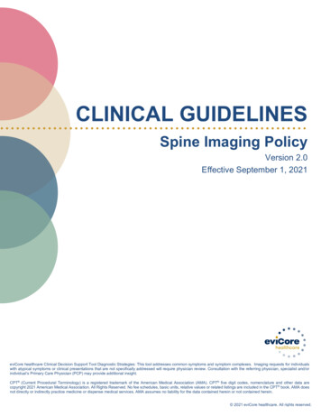
h) Describe the process of of embryonic gut rotationRotation of the Embryonic Gut2.1.4.3.5.11
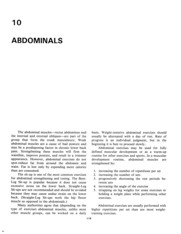
2) Gastrointestinal (GI) System Overviewa) Describe the general location, function, and relationships among the following structures: esophagus, stomach, small intestine, large intestine, spleen, pancreas, liver, and gall bladderb) Identify the principal arterial supply to and venous drainage from the foregut, midgut, and hindgut12
3) Foreguta) Describe the parts and anatomical relations of the following foregut organs: esophagus, stomach, duodenum, spleen, pancreas, liver, and gall bladder13
14
G09B: Arteries, Veins, and Lymphatics of the GI System(Dr. Albertine)At the end of this lecture, students should be able to master the following:1) Regional Vascularizationa) Arteries of the GI Systemii) Describe the specific arterial supply to the midgut (Superior mesenteric artery)15
iii) Describe the specific arterial supply to the hindgut (Inferior mesenteric artery)iv) Describe the inferior mesenteric arterial anastamoses with systemic arteries16
b) Veins of the GI Systemi) Describe the specific venous drainage of the foregut (Splenic vein and tributaries)ii) Describe the specific venous drainageof the midgut (Superior mesenteric vein)iii) Describe the specifc venous drainage of the hindgut (Inferior mesenteric vein)17
2) Portal Venous Systema) Describe the three veins that combine to form the hepatic portal vein (splenic, superior mesenteric, inferiormesenteric veins)b) Describe the main portal-caval anastamosesi) Distal end of the esophagusii) Rectum/Anusiii) Umbilicus18
3) Lymphatic Drainagea) Explain the rule in predicting lymph flow in the abdomenb) Trace the postnodal lymph flow from the cysterna chyli to its return to the venous blood19
G10A: Midgut and Hindgut (Dr. Albertine)At the end of this lecture, students should be able to master the following:1) Midguta) Describe the parts and anatomical relations of the following midgut organs: duodenum, jejunum, ileum,cecum, vermiform appendix, and colon (to the splenic flexure)b) Describe the structural differences between the jejunum and ileum20
2) Hindguta) Describe the parts and anatomical relations of the following hindgut organs: colon (splenic flexure andbeyond), rectum, and anal canal21
G10B: Autonomic Innervation of the GI Tract (Dr. Albertine)At the end of this lecture, students should be able to master the following:1) Functions of the autonomic nervous supply to the abdominal visceraa) Gastrointestinal tracti) Sympathetic: decreases motility and absorption; decreases contraction of the circular and longitudinalmuscle fibers; contraction of sphincter muscles; and decreased glandular secretionsii) Parasympathetic: increases motility and absorption; contraction and relaxation (peristalsis) of longitudinal and circular muscle fibers; relaxation in sphincter muscles; and increased glandular secretionsb) Pancreasi) Sympathetic: decreased secretion of both insulin and digestive enzymesii) Parasympathetic: increased secretion of insulin and digestive enzymesc) Liveri) Sympathetic: increased glycogenolysis/gluconeogenesisii) Parasympathetic: increased metabolism and secretionsd) Spleeni) Sympathetic: contraction of capsule2) Overview of the autonomic innervation of the GItracta) CNS origini) Sympathetics - T5-L2ii) Parasympathetics – CN X and S2-S4b) Splanchnics – transport autonomic fibers from thesympathetic trunk (sympathetics) or spinal nerves (parasympathetics) to the prevertebral plexusi) Sympathetic splanchnic nerves(1) Greater splanchnic – carry sympathetics fromthe T5-T9 level of the spinal cord to the prevertebralplexus (celiac and superior mesenteric)(2) Lesser splanchnic – carry sympathetics from theT10-T11 level of the spinal cord to the pervertebralplexus (celiac and superior mesentric)(3) Least splanchnic- carry sympathetics from theT12 level of the spinal cord to the prevertbral plexus(aorticorenal and inferior mesenteric)(4) Lumbar splanchnic- carry sympathetics from L1-2level of the spinal cord to the prevertebral plexus (inferior mesenteric and inferior hypogastric)ii) Parasympathetic splanchnic nerves(1) Sacral splanchnic – transports parasympatheticfibers from the S2-S4 spinal nerves to the prevertebralplexus (inferior hypogastric plexus)22
c) Prevertebral plexus- serves as a common pathway for both sympathetics and parasympathetics to travel tothe end-organi) Celiac plexus and celiac ganglia(1) Hepatic plexus to liver and gall bladder(2) Gastric plexus to stomach(3) Splenic plexus to spleen(4) Pancreatic plexus to pancreas and proximal portion of the duodenumii) Superior mesenteric plexus and superior mesenteric ganglia(1) To head of the pancreas, distal duodenum, jejunum, ileum, cecum, ascending colon, and transversecoloniii) Aorticorenal plexus and aorticorenal ganglia(1) Ureteral plexus to the suprarenal gland, kidney, and proximal ureteriv) Inferior mesenteric plexus and inferior mesenteric ganglia(1) Superior rectal plexus to the descending colon, sigmoid colon, and upper portion of rectumv) Inferior hypogastric plexus and pelvic ganglia(1) Middle rectal plexus to middle portion of the rectum(2) Inferior rectal plexus to the23
d) Trace the sympathetic and parasympathetic pathways to the following regions of the GI tract, include specific levels of origin, location of motor-motor synapse, and all preganglionic and postganglionic nerves traveledi) Foregut (symp. T5-T9 via greater splanchnic n.) (para. CN X)ii) Pancreas (symp. T6-T9 via greater splanchnic n.) (para. CN X)iii) Liver (symp. T6-T9 via greater splanchnic n.)iv) Spleen (symp. T6-T8 via greater splanchnic n.)v) Midgut (symp. T9-T11 via lesser splanchnic n.) (para. CN X)vi) Hindgut (symp. L1-L2 via lumbar splanchnic nn.) (para. S2-S4 via sacral splanchnics)24
3)Enteric Systema) Describe the relationship between the enteric system and the sympathetic and parasympathetic systemsb) Describe the role of the enteric system in the following functionsi) Gastric secretionsii) Gastrointestinal blood flowiii) Peristalsisc) Describe the location and nerve fiber contents of the following components of the enteric system:i) Myenteric plexusii) Submucosal plexus25
G11: Posterior Abdominal Wall (Dr. Albertine)At the end of this lecture, students should be able to master the following:1) Posterior Abdominal Musclesa) Describe the specific attachments and actions of the following muscles: iliacus, quadratus lumborum, psoasmajor, and psoas minor musclesb) Describe the pathway through the diaphragm of the following structures: esophagus, aorta, and inferiorvena cavac) Describe the median, medial, and lateral ligaments of the diaphragm26
2) Nervesa) Describe the pathway and general distribution of the following nerves: subcostal, iliohypogastric, ilioinguinal, lateral femoral cutaneous, femoral, genitofemoral, and lumbosacral trunk nervesb)Describe the origin and synapse site of the lower thoracic and lumbar sympathetic splanchnic nerves27
c)Describe the origin and synapse site of the vagus and sacral parasympathetic nerves28
3) Vasculature and Lymphaticsa) Describe the course of the aorta through the abdomenb) Describe the course, distribution, and vertebral level of the branches of the abdominal aorta in descendingorderc) Describe the course of the inferior vena cava through the abdomend) Describe the differences in the branches of the abdominal aorta and inferior vena cava29
e) Trace lymph flow from the aortic and preaortic lymph nodes to its return to venous blood30
4) Suprarenal Glandsa) Describe the location of the suprarenal glandsb) Describe the differences in embryonic origin and function of the cortex and medullac) Identify the three arterial sources to and two venous routes from the suprarenal glandsd) Describe the sympathetic innervation of the suprarenal glands (greater, lesser, and least splanchnic nerves)5) Kidneysa) Describe the locations of the right and left kidneysb) Identify the right and left surface projections of the kidneysc) Describe the relations of the right kidney to the following structures: suprarenal gland, liver, 2nd part of theduodenum, right colic flexure, 12th rib, diaphragm, psoas, quadratus lumborum, and transversus abdominisd) Describe the relations of the left kidney to the following structures: suprarenal gland, stomach, spleen, pancreas, jejunum, descending colon, 11th and 12th ribs, diaphragm, psoas, quadratus lumborum, and transversusabdominis31
e) Describe the autonomic innervation of the kidneyi) Sympathetic(1) Preganlionic neurons(a) Greater, lesser, and least splanchnic nerves(b) Aorticorenal ganglia (motor-motor synapse)(2) Postganglionic neurons(a) Renal plexus to kidneyii) Parasympathetic(1) Preganglionic neurons(a) Posterior vagal trunk(b) Pelvic splanchnic nerves (S2-4)(c) Renal plexus(d) Wall of organ (motor-motor synapse)32
6) Uretersa) Describe the pathway of the ureters through the abdomen and pelvisb) Describe the differences in the relations of the right and left uretersc) Describe the autonomic innervation of the uretersi) Sympathetic(1) Preganlionic neurons(a) Greater, lesser, and least splanchnic nerves(b) Aorticorenal ganglia (motor-motor synapse)(2) Postganglionic neurons(a) Renal plexus to ureterii) Parasympathetic(1) Preganglionic neurons(a) Posterior vagal trunk(b) Pelvic splanchnic nerves (S2-4)(c) Renal plexus (variable)(d) Wall of organ (motor-motor synapse)7) Bladdera) Describe the features of the four regions of the bladder (apex, base, neck, and inferior surface)b) Contrast the differences between the male and female bladderc) Describe the vascularization of the bladder33
d) Describe the source and function of sympathetic innervation of the bladderi) Preganglionic neurons(1) Lumbar splanchnic nerves(2) Inferior mesenteric ganglion (motor-motor synapse)ii) Postganglionic neurons(1) Superior hypogastric plexus(2) Inferior hypogastric plexus(3) Vesicle plexus to bladdere) Describe the source and function of parasympathetic innervation of the bladderi) Preganglionic neurons(1) Pelvic splanchnic nerves(2) Inferior hypogastric plexus34
G12: Gluteal Region, Perineum Related to the Ischioanal Fossa(Dr. Albertine)At the end of this lecture, students should be able to master the following:1) Gluteal Region2) Perineuma) Describe the location of the perineum35
3) Ischioanal Trianglea) Identify the boundaries of the ischioanal triangle (lateral, posterior, anterior, superior, and inferior)b) Describe the contents of the ischioanal triangle (levator ani, ischioanal fat pad, pudenal canal, pudenl neurovascular bundle, anococcygeal body, rectum, and anus)36
c) Describe the vascular supply of the ischioanal fossa (Internal pudendal artery and its branches)d) Describe the differences in the male and female perineal bodies37
G13: Urogenital Triangle (Dr. Albertine)At the end of this lecture, students should be able to master the following:1) Overviewa) Identify the boundaries of the urogenital triangleb) Compare and contrast the vascularization between the male and female urogenital triangles38
c) Describe the innervation of the urogenital triangle2) Deep Perineal Spacea) Identify the boundaries of the deep perineal spaceb) Compare and contrast contents between the male and female deep perineal spaces39
3) Superficial Perineal Spacea) Identify the boundaries of the superficial perineal spaceb) Compare and contrast contents between the male and female superficial perineal spaces40
G14A: Female Reproductive System (Dr. Albertine)At the end of this lecture, students should be able to master the following:1) Generala) Describe the general location and function of the ovaries, uterine tubes, uterus, vagina, clitoris, bulb of thevestibule, greater vestibular glands, paraurethral glands, labia minora, and labia majora2) Ovariesa) Describe the location and anatomical contents of the following ovarian and uterine ligamentsi) Mesovariumii) Broad ligament(1) Mesosalpinx(2) Suspensory ligament of the ovary(3) Ligament of the ovaryb) Describe the vascularization of the ovary (ovarian artery, ovarian venous plexus, ovarian vein)c) Describe the autonomic innervation of the ovaryi) Sympathetic innervation(1) Preganglionic neurons(a) Lesser, and least splanchnic nerves(b) Superior mesenteric ganglion or renal ganglia (motor-motor synapse)(2) Postganglionic neurons(a) Superior mesenteric plexus or renal plexus(b) Ovarian plexus to ovary41
3) Uterine Tubes and Uterusa) Describe the following parts of the uterine tubei) Isthmusii) Ampullaiii) Infundibulumiv) Ostiumv) Fimbriaeb) Describe the relation of the uterine tubes to the ovaries and broad ligamentc) Describe the relation of the uterus to the bladder, rectum, small intestines, ovaries, and vaginad) Describe the following components of the uterusi) Fundusii) Body (uterine cavity)iii) Cervix (internal os, external os, cervical canal)42
e) Describe the muscular (pubococcygeus muscle) and ligamentous (perineal body, pubocervical ligament,cardinal ligament, uterosacral ligament, and round ligament) support of the uterusf) Describe what a physician sees when looking through a speculum during a pelvic exam (cervical exam)g) Describe the vascularization of the uterus and uterine tubes (internal iliac artery, uterine artery to uterus,tubal branch of uterine artery to uterine tubes, and corresponding veins)i) Describe the relation between the uterine artery and the ureter43
h) Describe the autonomic innervation of the uterusi) Sympathetic(1) Preganglionics neurons(a) Lesser, least, and lumbar splanchnic nerves (T10-L2)(b) Superior mesenteric ganglion (motor-motor synapse)(2) Postganglionic neurons(a) Superior mesenteric plexus(b) Inferior mesenteric plexus(c) Superior hypogastric plexus(d) Inferior hypogastric plexus(e) Uterovaginal plexusii) Parasympathetic(1) Preganglionic neurons (S2-4)(a) Pelvic splanchnic nerves(b) Inferior hypogastric plexus(c) Uterovaginal plexus(d) Organ wall (motor-motor synapse)4) Vaginaa) Describe the relation of the vagina to the cervix, urethra, pelvic floor, and vestibuleb) Describe the following parts of the vagina: introitus, vaginal vault, vaginal rugae, vaginal fornixc) Describe the differences in the upper and lower parts of the vaginal walld) Describe the vascularization of the vagina (internal iliac artery, uterine artery, vaginal branches, and corresponding veins)i) Describe the anastamoses among the ovarian, uterine, and vaginal arteriese) Describe the innervation to the upper and lower parts of the vaginai) Upper part- parasympathetic from S2-4(1) S2-4 spinal nerves(2) Pelvic splanchnic nerves(3) Inferior hypogastric plexus(4) Uterovaginal plexusii) Lower part- somatic(1) Pudendal nerve44
5) Clitorisa) Describe the structure of the clitorisi) Corpora cavernosa(1) Root(a) Ischiocavernosus muscle(2) Bodyii) Corpus spongiosum(1) Glans clitorisiii) Prepuce of the clitorisb) Describe the suspensory ligament of the clitorisc) Describe the autonomic innervation of the clitorisi) Sympathetic(1) Preganglionic neurons (from L1-2)(a) Sacral splanchnic nerves(b) Inferior hypogastric plexus (motor-motor synapse)(2)Postganglionic neurons(a) Uterovaginal plexus(b) Cavernous nerves to clitorisii) Parasympathetic(1) Preganglionic neurons (S2-4)(a) Pelvic splanchnic nerves(b) Inferior hypogastric plexus(c) Uterovaginal plexus(d) Cavernous nerves(e) Organ wall (motor-motor synapse)45
b) Describe the vascularization of the clitoris (internal iliac artery, internal pudendal artery, deep and dorsalclitora arteries, and corresponding veins)6) Bulb of the Vestibule, Greater Vestibular Glands, and Paraurethral Glandsa) Describe the location and composition of the bulb of the vestibulei) Corpus spongiosum(1) Bulbospongiosus muscleb) Describe the location and function of the greater vestibular glandsc) Describe the location of the paraurethral glands46
7) Vulvaa) Describe the location of the labia majora and their relation to the labia minora and mons pubisb) Describe the location labia minora and their relation to the clitoris, vestibule, and bulb of the vestibulec) Describe the following components of the vestibulei) Urethral opening(1) Opening of ducts of paraurethral glandsii) Vaginal opening(1) Opening of duct of greater vestibular glandiii) Hymend) Describe the cutaneous innervation of the vulva (pudendal nerve, S2-4)8) Ovulationa) Describe the pathway of an ovum from ovulation to implantationi) Ovaryii) Uterine tube (Ostium, infundibulum, ampulla, isthmus)iii) Uterus (endometrium of the uterine body)47
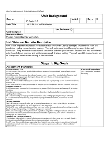
G14B: Male Reproductive System (Dr. Albertine)At the end of this lecture, students should be able to master the following:1) Generala) Describe the general location and function of the testis, epididymis, ductus deferens, seminal vesicle, ejaculatory duct, prostate gland, urethra and penis2) Scrotuma) Identify the layers of the scrotum and their correlation to the layers of the abdominal wallb) Describe the cutaneous innervation of the scrotum (genitofemoral n., L1-L2)c) Explain how an inguinal hernia may affect scrotal nerves (genital branch of the genitofemoral nerve)Layers of Abdominal Wall1. SkinSuperficial fascia2. Fatty (Camper’s)3. Membranous (Scarpa’s)4. External oblique5. Internal oblique6. Transversus abdominis7. Transversalis fascia8. Extraperitoneal fatty tissue9. PeritoneumCorresponding Layers in the Scrotum1. Skin2. Disappears - no fat in scrotal wall3. Dartos muscle and fascia4. External spermatic fascia5. Cremasteric muscle and fascia6. No contribution7. Internal spermatic fascia8. Areolar connective tissue9. Tunica vaginalis48
3) Testis and Epididymisa) Describe the site of production of spermatozoab) Describe the vascularization of the testis (testicular artery and vein, pampiniform plexus of veins)c) Trace the sympathetic innervation of the testisi) Preganglionic neurons (Celiac ganglion (motor-motor synapse))ii) Postganglionic neurons (Testicular plexus to testis)d) Describe the function and regions of the epididymis (head, body, tail)4) Spermatic Corda) Describe the contents and the pathway of the following anatomical structures within the spermatic cordi) Ductus deferens (and the artery of the ductus)ii) Testicular artery and Pampinform plexus of veinsiii) Cremasteric muscleiv) Genital branch of the genitofemoral nerve (L1-L2)v) Autonomic fibers for testis and epididymisvi) Lymphatic vessels49
5) Ductus Deferensa) Describe the pathway of the ductus deferens relative to the inguinal canal, inferior epigastric artery, external iliac artery, ureters, seminal vesicle, and bladderb) Describe the vascularization of the ductus deferens (internal iliac artery, umbilical artery, artery of theductus, pampiniform plexus of veins, testicular vein)c) Describe the sympathetic innervation of the ductus deferensi) Preganglionic neurons (L1-2)(1) Lumbar splanchnic nerves(2) Inferior hypogastric plexus and pelvic ganglia (motor-motor synapse)ii) Postganglionic neurons(1) Deferential plexus to ductus deferens6) Seminal Vesiclesa) Describe the location seminal vesicles and the function of their secretionsb) Describe the relationship among the ductus deferens, seminal vesicles, and the ejaculatory ducts7) Prostate Gland and Ejaculatory ducta) Describe the prostate’s relationship to the ejaculatory ducts, bladder, urethra, and rectumb) Identify the five lobes of the prostatec) Describe the function of the prostatic secretionsd) Describe the vascularization of the prostate (internal iliac artery, inferior vesicle artery, prostatic branchesand venous plexus, vesicle venous plexus and veins)e) Describe the autonomic innervation of the prostatei) Sympathetic innervation(1) Same as seminal vesiclesii) Preganglionic neurons (S2-4)(1) Pelvic splanchnic nerves(2) Inferior hypogastric plexus(3) Prostatic plexus(4) Wall of organ (motor-motor synapse)f) Describe the process of a digital prostate examg) Explain why men will commonly experience impotence after a prostatectomy50
51
8) Urethra and Bulbourethral Glandsa) Describe the three parts and pathway of the male urethrai) Prostaticii) Membranousiii) Spongyb) Describe the location, innervation, and function of the urethral sphincters in urination and ejaculationi) Internal urethral sphincterii) External urethral sphincterc) Describe the location and function of the bulbourethral glands9) Penisa) Describe the structure of the penisi) Corpora cavernosa (Deep arteries of the penis)ii) Corpus spongiosum (Spongy urethra)iii) Root of the penis (Ischiocavernosus muscles)iv) Bulb of the penis (Bulbospongiosus muscle)v) Glans penisvi) Prepuce (foreskin)b) Describe the facial layers of the penisi) Superficial fasciaii) Deep (“Buck’s”) fasciaiii) Tunica albuginiac) Describe the ligaments of the penisi) Suspensory and Fundiform ligaments of the penisd) Describe the vascularization of the penise) Describe the sympathetic innervation of the penis from L1-2i) L1-2 spinal nerves - white rami communicantes - sympathetic trunk - sacral splanchnic nerves - inferiorhypogastric plexus - prostatic plexus - cavernous nervesf) Describe the parasympathetic innervation of the penis from S2-4i) S2-4 spinal nerves - pelvic splanchnic nerves - inferior hypogastric plexus - prostatic plexus - cavernousnervesg) Explain the role of the parasympathetic nervous system (S2-S4) in erectioni) Dilation of arterial supply to erectile tissuesh) Explain the role of the sympathetic nervous system (L1-L2) in ejaculation and detumescencei) Contraction of smooth muscle within ductus deferens and accessory glandsii) Contraction of internal urethral sphincteriii) Contraction of smooth muscle within arteries, arresting blood flow to erectile tissuesi) Explain the role of the pudendal nerve and the penis52
53
10) Fertilization PathwayTrace the ejaculation pathway: Seminiferous tubules - rete testis - straight tubules - efferent ductules of the testis- epididymis (head, body, tail) - ductus deferens - ejaculatory duct - prostatic urethra - membranous urethral spongy urethra – urethral orificeHomologsMALETestesCorpora cavernosa of penisCorpus spongiosum of penisBulbourethral glandsProstate glandScrotumSpongy urethraProstatic utricleFEMALEOvariesCorpora cavernosa of clitorisCorpora spongiosum of clitoris and bulb of the vestibuleGreater vestibular glandsParaurethral glandsLabia majoraLabia minoraUterus54
55
Gray’s Anatomy for Students (GAFS), Chapters 4-5 Gray’s Dissection Guide for Human Anatomy (GDGHA), Labs 10-17 Unit #2- Abdomen, Pelvis, and Perineum G08- Overview of the Abdomen and Anterior Abdominal Wall (Dr. Albertine) G09A- Peritoneum, GI