Transcription
[Recent Advances in Chest Medicine]BLUE-Protocol and FALLS-ProtocolTwo Applications of Lung Ultrasound in the Critically IllDaniel A. Lichtenstein, MD, FCCPThis review article describes two protocols adapted from lung ultrasound: the bedside lungultrasound in emergency (BLUE)-protocol for the immediate diagnosis of acute respiratoryfailure and the fluid administration limited by lung sonography (FALLS)-protocol for the management of acute circulatory failure. These applications require the mastery of 10 signs indicating normal lung surface (bat sign, lung sliding, A-lines), pleural effusions (quad andsinusoid sign), lung consolidations (fractal and tissue-like sign), interstitial syndrome (lungrockets), and pneumothorax (stratosphere sign and the lung point). These signs have beenassessed in adults, with diagnostic accuracies ranging from 90% to 100%, allowing consideration of ultrasound as a reasonable bedside gold standard. In the BLUE-protocol, profileshave been designed for the main diseases (pneumonia, congestive heart failure, COPD,asthma, pulmonary embolism, pneumothorax), with an accuracy . 90%. In the FALLS-protocol,the change from A-lines to lung rockets appears at a threshold of 18 mm Hg of pulmonaryartery occlusion pressure, providing a direct biomarker of clinical volemia. The FALLS-protocolsequentially rules out obstructive, then cardiogenic, then hypovolemic shock for expediting thediagnosis of distributive (usually septic) shock. These applications can be done using simplegrayscale machines and one microconvex probe suitable for the whole body. Lung ultrasoundis a multifaceted tool also useful for decreasing radiation doses (of interest in neonates wherethe lung signatures are similar to those in adults), from ARDS to trauma management, andfrom ICUs to points of care. If done in suitable centers, training is the least of the limitationsfor making use of this kind of visual medicine.CHEST 2015; 147(6):1659-1670BLUE 5 bedside lung ultrasound in emergency; FALLS 5 fluid administration limitedby lung sonography; LUCI 5 lung ultrasound in the critically ill; LUCIFLR 5 Lung Ultrasound in theCritically Ill Favoring Limitation of Radiation; PLAPS 5 posterolateral alveolar and/or pleural syndromeABBREVIATIONS:Daily concerns of the intensivist are acuterespiratory and circulatory failure. The needfor fast and accurate management calls for avisual approach, which is what ultrasoundprovides. Portable machines suitable for useat the bedside have been available sinceManuscript received June 1, 2014; revision accepted November 22, 2014.AFFILIATIONS: From the Service de Réanimation Médicale, HôpitalAmbroise-Paré, Boulogne-Billancourt, France.CORRESPONDENCE TO: Daniel A. Lichtenstein, MD, FCCP, Servicede Réanimation Médicale, Hôpital Ambroise-Paré, 9 Ave Charlesde-Gaulle, 92100 Boulogne-Billancourt, France; e-mail: 82. This article, therefore, could havebeen written 33 years ago. Echocardiography has been used for a long time in theICU and now is currently used inside thethorax through the transesophageal route.1,2Echocardiography is an elegant way to solve 2015 AMERICAN COLLEGE OF CHEST PHYSICIANS. Reproduction ofthis article is prohibited without written permission from the AmericanCollege of Chest Physicians. See online for more details.DOI: 10.1378/chest.14-13131659
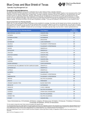
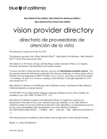
these respiratory and circulatory concerns. This articledescribes a complementary tool: lung ultrasound.HistoryLung ultrasound originally was not meant to be usedin emergent care. Except for echocardiography usedin cardiology and sonography used in obstetrics,ultrasound in general was a tool for radiologists, andthe lung in particular was not considered suitable forthis imaging technology.3 Since 1989, François Jardin’sICU explored, applied, and made lung ultrasound witha portable unit a standard of care in critically ill patients.Based on our 25 years of experience using lung ultrasound in the critically ill (LUCI), the American Collegeof Chest Physicians and La Société de Réanimation deLangue Française jointly proposed LUCI as a standardof care.4Since 1991, intensivists have been using whole-bodyultrasound, including vascular access, search for freeblood, and so forth, and lung ultrasound.5 Gradually, theICU community understood the relevance of lung ultrasound in critical care. Publications began to emerge andare now quite prevalent in the literature; thus, only a fewregarding the lung are quoted in the present article.6-42Tools Used for the BLUE-ProtocolOne application of lung ultrasound is the onsite exploration of acute respiratory failure: the bedside lungultrasound in emergency (BLUE)-protocol. Althoughthe new generation of intensivists benefits from a varietyof excellent machines, we keep using our 1992 technology (last updated in 2008) for several reasons: Welike its resolution; 32-cm width in settings where eachlateral centimeter counts; 7-s start-up time; flat, easyto-clean, fluid-proof design; 5-MHz microconvex probeallowing whole-body analysis from 0.6 to 17 cm; simpletechnology based on three clearly identified buttons,instant response, respect of artifacts, and ease of maintenance; intelligent narrow cart (preventing any drop);and low cost. These reasons make up the first of sevenprinciples: The simplest equipment is suitable for lungimaging.43 Any modern machine can be used, however.We believe that redesigns of the same machine for usein modern facilities (using wireless transmission, Dopplerand transesophageal echocardiography, etc) are appropriate as long as these do not interfere with the criticalproperties of small size, cost-effectiveness, and immediatestart-up time. The alternative, which we have beenusing for 25 years, is to have one simple, cost-effectiveunit and one comprehensive echocardiographic uniton hand.The second principle is to use gravity rules (gas towardthe sky, fluids toward the earth) to locate disorders. Thethird principle is to define standardized thoracic points,called BLUE-points, to allow for reproducible analyses44(Fig 1), and the fourth is to precisely define the pleuralline, the first of 10 basic signatures. The fifth principle inlung ultrasound focuses specifically on artifacts. TheA-line is a repetition of the pleural line, indicating gas(Fig 2). The sixth principle analyzes lung sliding, whichis a twinkling visible at the pleural line that spreadshomogeneously below it (generating the seashore signin M-mode). Lung sliding and A-lines define a normalFigure 1 – A, B, The bedside lung ultrasound in emergency (BLUE)-points. This figure shows the standardized points used in the BLUE-protocol. Twohands (from roughly the patient’s size) are applied as follows: upper little finger just below clavicle, fingertips at middle line, and lower hand just belowupper hand (thumbs excluded). The point coined “upper BLUE-point” is at the middle of the upper hand. The “lower BLUE-point” is at the middle of thelower palm. These four points roughly follow the anatomy of the lung, and avoid the heart as much as possible. The posterolateral alveolar and/or pleuralsyndrome (PLAPS)-point is built from the horizontal line continuing the lower BLUE-point and the vertical line continuing the posterior axillary line.The intersection, rigorously, is the PLAPS-point, but the user can move the probe in two directions: (1) as posteriorly as possible in order to get moreposterior information in these supine, sedated patients or (2) downward when no PLAPS is detected at first sight. Usually, after two intercostal spaces,the probe scans the abdomen. Like the six spots of ECG, the six BLUE-points help in reproducible analysis. They were sufficient for providing the 90.5%accuracy of the BLUE-protocol. (Adapted with permission from Lichtenstein.45)1660 Recent Advances in Chest Medicine[147#6 CHEST JUNE 2015]
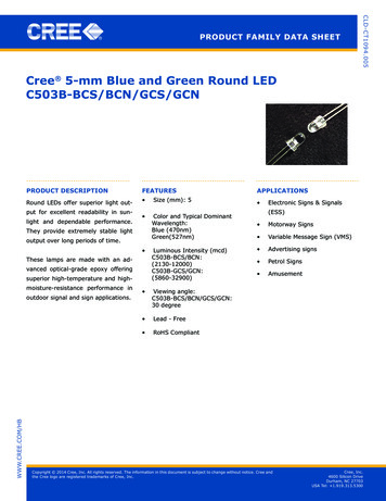
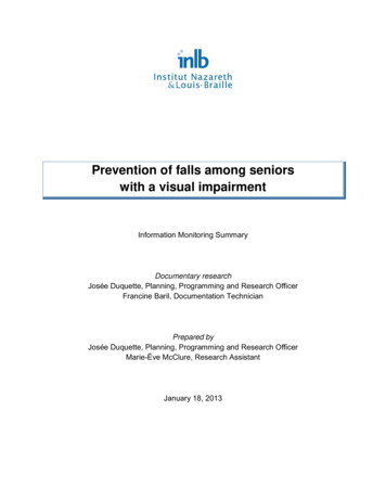
Figure 2 – Pleural line and A-line. The bat sign: The ribs (vertical arrows)and the pleural line (upper horizontal arrows) outline a silhouettereminiscent of a bat. This allows confident recognition of the pleural linein all circumstances, even in challenging examinations (patients withdyspnea, patients who are agitated, bariatric patients). The pleural linealways corresponds to the parietal pleura (and to the visceral pleuralonly if joined). The Merlin space encompasses the pleural line, the shadowof both ribs, and the lower border of the image. The A-line: Inside theMerlin space, a repetition of the pleural line is seen (lower horizontalarrows), occurring at a standardized distance from skin to pleural line. Itindicates gas below the pleural line. A-lines can be complete, as here, orpartial (as in Figs 3 and 8).lung surface46 (Figs 2, 3). The seventh principle is basedon the fact that all acute, life-threatening disordersare superficial. This allows to standardize the field(Table 1).Diagnosis of pleural effusion is an old application oflung ultrasound51,52 and basically yields two standardsigns: the quad sign and the sinusoid sign (Figs 4, 5).47Alveolar syndrome (lung consolidation) is also an olddiagnostic application of lung ultrasound.53 This fluiddisorder usually is superficial48 and, thus, accessible toultrasound, particularly in the diagnosis of nontranslobarconsolidations, which yield the fractal (or shred) sign(Fig 5), and translobar forms, which yield the tissue-likesign (Fig 6).48 Interstitial syndrome generates lung rocketson lung ultrasound (Fig 7).49 This application is a mainpoint of discussion in this article.Pneumothorax was first approached in lung ultrasonography using the sole abolition of lung sliding.54-56This sign had a poor specificity until it was associatedwith the A-line sign (Fig 8).46,57 Abolished lung slidinggenerates the stratosphere sign in M-mode. Lungsliding (or its equivalent, the lung pulse)58 or B-linesrule out pneumothorax. The lung point (Fig 9) is apathognomonic sign.59 Pneumothorax occurring inpatients with severe dyspnea or adherences is beyondthe scope of this article.journal.publications.chestnet.orgFigure 3 – Lung sliding. The left image shows the bat sign (arrowheadsindicate the pleural line). In the Merlin space, there is a partial A-line(arrows), meaning that there is gas below the pleural line. A partialA-line (or even not visible, a pattern then coined “O-line,” for non-Anon-B) is a common finding, with unchanged pathophysiologic meaning.Note that a comet-tail artifact is seen arising from the pleural line (*); itis ill defined, not erasing A-lines (arrows); less echoic than the pleuralline; short; and standstill (in real time), allowing easy distinction withthe B-line (see Fig 7). This artifact, devoid of meaning, is coined “Z-line.”The right image shows the seashore sign of lung sliding. Lung sliding isvisible in real-time but not in a frozen image (left image). It can, however, be demonstrated using the M-mode. Above the pleural line, thepattern is stratified (motionless soft tissues). Exactly from and below thepleural line (arrowheads), the pattern is sandy, hence, the seashore sign.This indicates that the gas pattern indicated by the A-line in the leftimage is alveolar gas (ruling out pneumothorax).Patients, Diseases, and Profiles in theBLUE-ProtocolThe BLUE-protocol was developed based on the studyof 300 consecutive adults with acute respiratory failurewho were admitted to our ICU and given a diagnosis.The most frequent cause of respiratory failure waspneumonia (32%) followed by acute hemodynamicpulmonary edema (24%); exacerbated COPD (18%);severe asthma (13%); pulmonary embolism (8%);pneumothorax (4%); and countless rare causes, includingeasy-to-diagnose ones, such as massive pleural effusion(3%). We excluded rare, unknown, and multiple diagnosesbecause they generate methodologic issues. TheTABLE 1] Published Performances of Lung Ultrasoundin Critically Ill Patients Compared WithCT ScanningUltrasoundPleural effusion47Sensitivity, %Specificity, %94979098Interstitial syndrome49100100Complete pneumothorax461009679100Alveolar consolidation48Occult pneumothorax501661
Figure 4 – PLAPS and pleural effusion. This image at the PLAPS-pointshows the quad sign, which is an image outlined by the pleural line(upper horizontal arrows), the rib shadows (vertical arrows), and aclearly defined line (lower horizontal arrows) called the lung line, regularand roughly parallel to the pleural line ( 15 -20 ). The lung line indicates the visceral pleura. Note the long comet-tail artifact (*) arising fromthe lung line but not from the pleural line, hence coined “sub-B-line.” Thissub-B-line indicates that the lung in contact is aerated, not consolidated(ie, one more piece of information). This effusion is roughly anechoic.Even if echoic, the quad sign is a universal sign of pleural effusion regardless of its echogenicity. In free pleural effusions, the lung line movestoward the pleural line on inspiration, shaping the sinusoid sign, a basicsign that indicates unloculated pleural effusion, and mostly low viscosityof the fluid, indicating that a small needle is suitable if thoracentesis isenvisioned. The distance between the pleural and lung lines is roughly10 mm (the present view taken on expiration). We consider an effusionvolume of roughly 75 to 150 mL in adults. In the BLUE-protocol, thispattern of isolated, minute pleural effusion is, sensu stricto, a PLAPS.See Figure 1 legend for expansion of abbreviations.BLUE-protocol is integrated within the control of acuterespiratory failure, which requires an understanding ofanatomy, physiology, pathophysiology, clinical signs,traditional imaging, and the biology of dyspnea. TheBLUE-protocol is fully based on pathophysiology.One feature of the BLUE-protocol is the establishedprofiles, that is, signs associated with locations. Theseprofiles are labeled simply to indicate abridgedconcepts. The A-profile is shorthand for “anterior lungsliding with A-lines profile.” The remaining eightprofiles follow the same labeling convention: B-profile(hemodynamic pulmonary edema), B9-profile, A/B-profile,C-profile, A-profile without DVT but with posterolateralalveolar and/or pleural syndrome (A-no-V-PLAPSprofile) (pneumonia), A-profile plus DVT (pulmonaryembolism), A9-profile (pneumothorax), and nude profile(COPD/asthma).At the anterior chest wall, lung sliding with predominantA-lines define the A-profile. Lung sliding is explained bythe respiratory movements of the visceral pleura againstthe parietal pleura. The A-line is displayed when normalsubpleural interlobular septa are too thin for disturbing1662 Recent Advances in Chest MedicineFigure 5 – PLAPS and lung consolidation: nontranslobar consolidation.The shred sign: At the PLAPS-point, a disorder can be described betweena lung line (because there is an associated anechoic pleural effusion)(lower horizontal arrows) and a shredded deep border, where thispattern is replaced by gas barriers (all vertical arrows). This shreddedline instantaneously demonstrates a lung consolidation. The whole makesanother example of PLAPS, here combining free pleural fluid with alveolarfluid, an extremely frequent pattern in critically ill patients. At the step aPLAPS is found (ie, after detection of an A-profile, then of a free venousnetwork), pneumonia is a likely cause of the respiratory failure. Thedistance between the pleural and lung lines in this standardized viewtaken on expiration is 25 mm. One can assume that there is more fluidthan shown in Figure 4 (between roughly 350 and 700 mL or slightlymore, according to our index). The upper horizontal arrows indicate thepleural line. See Figure 1 legend for expansion of abbreviation.the reverberation of the pleural line. The A-profileindicates a normal anterior lung surface. Associatedwith a DVT, it is connected with pulmonary embolism.The venous step is the longest part of the BLUE-protocol.The anterior lung analysis takes 0.5 min, negativevenous scanning 2 min, and posterior lung step 0.5 minby an experienced operator using the standardizedBLUE-points.Lung sliding with lung rockets define the B-profileand usually indicate hemodynamic pulmonary edema.When interlobular septa are enlarged by edema, theultrasound flow penetrates the lung, but the majorimpedance gradient between gas and fluids traps theultrasound flow, hence showing a persistent to-and-frodynamic, generating the B-line. Three B-lines betweentwo ribs, a pattern called lung rockets, correspond tothe anatomic number of subpleural interlobular septa.Hemodynamic pulmonary edema creates a transudative,pressurized pulmonary edema, therefore associatinglung sliding with a culminant (anterior) location of lungrockets. Anterior lung rockets associated with abolishedlung sliding define the B9-profile. In inflammatoryinterstitial syndrome (ie, pneumonia), each subpleuralinterlobular septum should exudate fibrin, behaving likeglue, resulting in abolishing lung sliding. Unilateral lung[147#6 CHEST JUNE 2015]
hemodynamic pulmonary edema or pulmonary embolismare posterior.60Anterior A-lines associated with abolished lungsliding define the A9-profile. The A9-profile suggestspneumothorax—the lung point is mandatory. In pneumothorax, the abolished lung sliding is explained by theabsence of visceral pleura and the A-line by the absenceof any fluid structure abutting the parietal pleura. Thelung point is explained by the slight inspiratory increaseof volume of the collapsed lung and, therefore, an increasedparietal contact making an abrupt ultrasound change.Figure 6 – PLAPS and lung consolidation: translobar consolidation. Thetissue-like sign: This huge tissue-like image (similar to the spleen [S]) atthe PLAPS-point touches the pleural line (arrowheads); there is no (oralmost no) pleural effusion. The deep limit is the mediastinal line, whichis not sharply defined in this image, but another sign can be used here:The interpleural distance in this image is 10 to 11 cm, a size not compatible with a pleural effusion. This image definitely indicates a consolidation. Note the other features. The location is the left lower lobe, totallyhepatized. The tissue-like sign is homogeneous; necrotic areas areunlikely (they would appear as hypoechoic, rounded areas). There is noair bronchogram and no loss of volume (as an atelectasis would cause).There are some drops of subpulmonary effusion. Real time would haveshown an abolished lung sliding (this patient had a pneumonia). SeeFigure 1 legend for expansion of abbreviation.rockets define the A/B-profile. This asymmetry of interstitial signs is also linked to pneumonia.Anterior lung consolidation, regardless of number andsize (up to simply a thick, irregular pleural line), definesthe C-profile. In the BLUE-protocol, the C-profile isassociated with pneumonia. Following the secondprinciple of lung ultrasound, consolidations seen inAt the posterior chest wall, lung consolidations andpleural effusions are assessed together for simplicitybecause both disorders usually come together, hence thepractical term “PLAPS” (Fig 5). The A-no-V-PLAPSprofile is connected with pneumonia. The A-profile withno DVT and no PLAPS (ie, nude profile) is linked withasthma and COPD (two bronchial diseases with similartherapy combined for simplification).In developing the BLUE-protocol, all study patients,including the excluded ones, benefited from receiving aprofile. The A-profile was seen in 53.8%, the B-profile in27.3%, the A9-profile in 3.4%, the B9-profile in 3.4%, theC-profile in 7.6%, and the A/B-profile in 4.6%.The BLUE-Protocol: When and HowIs it Used, What Occurs Practically,With How Much Accuracy?The BLUE-protocol is done each time the physicianhas clinical doubts after the physical examination. Themachine is brought to the bedside, the probe applied atFigure 7 – A, B, Lung rockets. An elementary signature of interstitial syndrome, the B-line, can be described by seven criteria. This is always a comet-tailartifact and always arises from the pleural line. It always moves in concert with lung sliding. Almost always (roughly 95% for each of these last fourfeatures), it is long and up to the edge of the screen (in the left image, some do not reach the 17-cm depth); is well defined, like a laser; obliterates theA-lines; and is hyperechoic like the pleural line. Usually, all seven criteria are present (always the first three ones) and define the B-lines with a precisionthat avoids confusion with other comet-tail artifacts, such as Z-lines and E-lines. (E-lines are comet-tail artifacts with most criteria of B-lines but arisingabove the pleural line, indicating subcutaneous emphysema.) Lung rockets, defined as three B-lines or more between two ribs, demonstrate interstitialsyndrome. The left image shows septal rockets. Three or four B-lines can be counted between two ribs (ie, roughly 6 or 7 mm apart, the anatomicdistance between two subpleural interlobular septa in adults). Septal rockets indicate the thickening (usually edematous) of these septa. The right imageshows ground glass rockets. One can count here twice as many B-lines than on the left image. Ground glass rockets are correlated with scanographicground glass areas. Lung rockets are used in daily routine to assess acute respiratory or circulatory failure, among other uses.journal.publications.chestnet.org1663
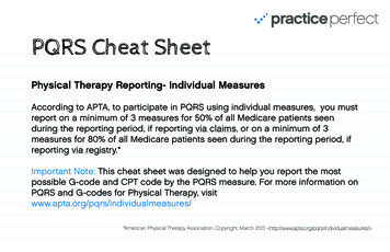
Figure 8 – Pneumothorax and A9-profile. The diagnosis of pneumothorax requires a two-step approach. The first step is to detect theA9-profile, associating the A-line sign with the abolition of lung sliding.The left image shows the A-line sign. The Merlin space always displaysA-lines (arrowheads), meaning gas below the pleural line (arrows). TheA-line shown here is ill defined, again more like an O-line, but there isdefinitely no B-line. The dots delineate the M-mode shooting line. Theright image shows the stratosphere sign. This homogeneous, stratifiedpattern demonstrates what is seen on real-time imaging (ie, the constantand complete abolition of lung sliding). The arrows indicate the pleuralline. The A9-profile is very sensitive but not specific to pneumothorax.Note that the two images are not only side by side but also exactly side byside (take a ruler at the pleural line) without any lag that may generate,in acute conditions, one element of confusion. This case is of a eupneicpneumothorax and represents a first step for learning (the signs of dyspneic pneumothorax are standardized too but add one more degree ofcomplexity because they obey the rules of shooting at a mobile target).the anterior standardized points, and the BLUE-protocolbegun (Fig 10), first searching for lung sliding. If lungsliding is present, the association with predominantA-lines defines the A-profile, and a venous scan isdone following a sequential order.62 An A-profileassociated with a DVT is 99% specific to pulmonaryembolism. For this reason, the veins are analyzedbefore the rest of the lung. PLAPS are common toseveral causes, the veins must preferably be assessedbefore searching for PLAPS. Finding a DVT afterdetecting an A-profile makes sense. If no DVT isfound, PLAPS are sought for the PLAPS point. If present,the A-no-V-PLAPS-profile is defined, suggestingpneumonia. If absent, the nude profile (all items normal)is defined, suggesting COPD or asthma. The A9-profilerequires the search for a lung point, which if positive,rules in pneumothorax. The B-profile makes hemodynamic pulmonary edema the priority diagnosis. TheB9-profile, A/B-profile, C-profile (anterior consolidations) highly suggest pneumonia. Eighty-six percent ofARDS cases have one of the four profiles of pneumonia.61All in all, the BLUE-protocol provides a 90.5% accuracy,61with detailed accuracies ranging from 81% to 100%(Table 2).1664 Recent Advances in Chest MedicineFigure 9 – Pneumothorax and lung point. The lung point: Each time anA9-profile is detected (at the anterior wall, by definition), the search forthe lung point is the second mandatory step, time permitting. At acertain location of the thorax (lateral, posterior), probe standstill, lungpatterns such as lung sliding and lung rockets replace the A9-profile. Thechange in rhythm with respiration is abrupt (arrow in right image). Thelung point is pathognomonic for pneumothorax, indicating its volume.(In this patient, the lung point was roughly located at the PLAPS point,corresponding to a radiovisible pneumothorax.) It indicates that theequipment is suitable (real-time instant-response acquisition, suitableresolution, mastery of filters). Arrows in the left image indicate thepleural line. See Figure 1 legend for expansion of abbreviation.Frequently asked questions are answered in Lichtenstein.45For example, a frequent question is, “Why is the heartnot included?” Looking at the heart to solve a pulmonaryfailure is a legitimate, yet indirect approach. Thesuffering organ is the lung, so lung ultrasound providesa direct approach. Echocardiography is associated butnot included (searching for left-sided heart anomaliesin the absence of lung rockets makes less sense becausepulmonary edema has been ruled out as a cause ofrespiratory failure). Small anterior lung consolidations(C-lines) suggest pneumonia 18 times more frequentlythan pulmonary embolism.61Of importance, the BLUE-protocol is only designedto be piloted by the physician’s common sense andintegrated with other basic data. A nude profile willsometimes require confident elimination of pulmonaryembolism (CT scan, scintigraphy); an anterior smallconsolidation will, rarely, be one sign of pulmonaryembolism. Used this way, the BLUE-protocol showsmaximal efficiency.A Development of the BLUE-Protocol:Lung Ultrasound for Diagnosing AcuteCirculatory Failure—the FALLS-ProtocolFor this major concern, successive tools have beenused, with echocardiography currently being one ofthe most popular.1,2 Many others are competing,[147#6 CHEST JUNE 2015]
Figure 10 – BLUE-protocol decisiontree showing the practical steps of theBLUE-protocol. See Figure 1 legendfor expansion of abbreviations.(Adapted from Lichtenstein andMezière.61)providing an impressive list of parameters whencombined, suggesting that no gold standard is currently available. The fluid administration limited bylung sonography (FALLS)-protocol is not yet supported by clinical studies but should be considered asa potential source of help in difficult situations. It isbased on sequential concepts: Pulmonary edema generates a thickening of the interlobular septa of whichtheir subpleural end is accessible using lung ultrasound63,64; A-lines transform into B-lines at a pulmonary artery occlusion pressure threshold of 18 mm Hgat the anterior chest wall in critically ill patients65;and no artifact has ever been described betweenTABLE 2A-lines and B-lines, indicating that B-lines appear(and vanish) all of a sudden, making septal thickening an on-off parameter. The use of lung artifactsallows for a direct assessment of lung water, morespecifically interstitial lung water (what no bedsidetool can do). The FALLS-protocol assumes that pulmonary edema is the most harmful consequence offluid overload in an extreme emergency (see limitations presented later in this section).The FALLS-protocol follows the Weil classification ofshock.66 The best of simple cardiac sonography and someBLUE-protocol are used. With the same unit and thesame probe, we first search for a substantial pericardial] Accuracy of the BLUE-ProtocolSpecificity, %PPV, %Acute hemodynamicpulmonary edemaMechanism of DyspneaB-profileProfiles of BLUE-ProtocolSensitivity, %979587NPV, %99Exacerbated COPD orsevere acute asthmaNude profile (A-profile with no DVTand no PLAPS)89979395Pulmonary embolismA-profile with DVT81999498PneumothoraxA9-profile (with lung point)8810010099PneumoniaThe four profiles89948895B9-profile1110010070A/B LAPS-profile42968378BLUE 5 bedside lung ultrasound in emergency; NPV 5 negative predictive value; PLAPS 5 posterolateral alveolar and/or pleural syndrome; PPV 5 positivepredictive value. (Adapted from Lichtenstein and Mezière.61)journal.publications.chestnet.org1665
effusion (assimilated to tamponade), then an enlargedright ventricle (assimilated to pulmonary embolism)(if poor cardiac windows, the BLUE-protocol can be usedinstead), and then an A9-profile (suggesting a tensionpneumothorax). At this step, obstructive shock canreasonably be ruled out.The B-profile is sought next. In its absence, a cardiogenicshock from left origin (ie, the far majority) can be ruledout by definition.The next step is performed in patients with neither theA9-profile nor the B-profile. The A-profile or equivalent(A/B-profile mainly) usually is seen, indicating that thepatient is a FALLS-responder. Only hypovolemic anddistributive shock are remaining causes, and the therapeutic part begins, which is fluid resuscitation. TheA-profile shows that fluids can be administrated, anotion of interest for intensivists who use volume resuscitation in distributive shock. Intensivists who wouldrather use vasopressors may appreciate that the FALLSprotocol allows them to avoid giving these drugs inunderestimated hypovolemia (ie, a safety factor useful atthe initial step). The improvement of clinical/biologicsigns of circulatory failure with an unchanged A-profileunder fluid therapy reasonably defines hypovolemicshock. The FALLS-protocol as a new tool for diagnosinghypovolemia should be appreciated in these complexsettings (prolonged surgery, prolonged intensive care)occurring in complex, challenging, and bariatricpatients.If no clinical improvement occurs, fluid therapy continues.The apparition of anterior B-lines (one can search morelaterally) means that an iatrogenic interstitial syndromelikely has been generated by the fluid administration.Interstitial edema is an early step, preceding alveolaredema.67,68 This step is clinically and biologically silent23and is the FALLS-end point (ie, the time to discontinuefluid therapy). Schematically, the
journal.publications.chestnet.org 1661 lung surface 46 ( Figs 2, 3 ). Th e seventh principle is based on the fact that all acute, life-threatening disorders are superfi cial. Th is allows to standardize the fi eld ( Tbale 1 ). Diagnosis of pleural eff usion is an old application o