Transcription
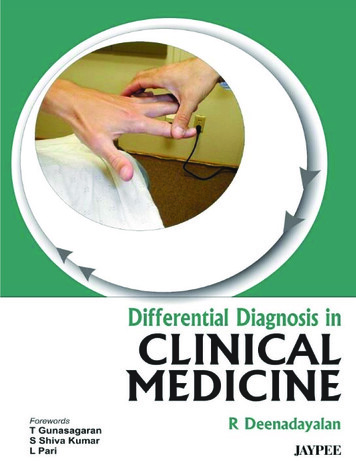
Differential Diagnosis inClinical Medicine
Differential Diagnosis inClinical MedicineR Deenadayalan MD (Gen Med)FormerlyProfessor of MedicineStanley Medical College and HospitalChennai, Tamil Nadu, IndiaMeenakshi Medical College Hospital and Research InstituteEnathur, Kancheepuram, Tamil Nadu, IndiaForewordsT GunasagaranS Shiva KumarL Pari JAYPEE BROTHERS MEDICAL PUBLISHERS (P) LTDNew Delhi Panama City London
Jaypee Brothers Medical Publishers (P) Ltd.HeadquarterJaypee Brothers Medical Publishers (P) Ltd4838/24, Ansari Road, DaryaganjNew Delhi 110 002, IndiaPhone: 91-11-43574357Fax: 91-11-43574314Email: jaypee@jaypeebrothers.comOverseas OfficesJ.P. Medical Ltd.83 Victoria Street, LondonSW1H 0HW (UK)Phone: 44-2031708910Fax: 02-03-0086180Email: info@jpmedpub.comJaypee-Highlights Medical Publishers Inc.City of Knowledge, Bld. 237, ClaytonPanama City, PanamaPhone: 507-301-0496Fax: 507-301-0499Email: cservice@jphmedical.comWebsite: www.jaypeebrothers.comWebsite: www.jaypeedigital.com 2012, Jaypee Brothers Medical PublishersAll rights reserved. No part of this book may be reproduced in any form orby any means without the prior permission of the publisher.Inquiries for bulk sales may be solicited at:jaypee@jaypeebrothers.comThis book has been published in good faith that the contents provided bythe author contained herein are original, and is intended for educationalpurposes only. While every effort is made to ensure accuracy of information,the publisher and the author specifically disclaim any damage, liability, orloss incurred, directly or indirectly, from the use or application of any of thecontents of this work. If not specifically stated, all figures and tables arecourtesy of the author. Where appropriate, the readers should consult witha specialist or contact the manufacturer of the drug or device.Differential Diagnosis in Clinical MedicineFirst Edition: 2012ISBN: 978-93-5025-768-5Printed at
Dedicated toMy Children and GrandsonWho are a GreatSource of Inspiration
Dr T GunasagaranVice ChancellorMeenakshi UniversityEnathur, Kancheepuram 631 552ForewordEvery book is meant to bring concept to those who care to readthem. Books on medical subjects are vast in number and everyauthor strives to fill a need that he himself has felt. Some authorsachieve this objective, but others, their intentions though aregenuine meander into a dreary desert of words.Prof R Deenadayalan MD (Gen Med) has made a seriousattempt in trying to help a hard pressed medical student bypresenting him with a work based on two decades of experiencesuperimposed on those of his teachers. Thus, a simple but avery useful glossary of differential diagnosis of clinical signs andentities has been created. Though this cannot replace a formaltextbook, it will serve as a ready reckoner to the beleagueredmedical student who labors under an ever increasing load ofinformation and changing priorities.The faculty of the Meenakshi University, I am sure, will find thiscontribution very useful and I am definite that they will recommendit to their students and colleagues. This book will be a bedsidecompanion to students and staff alike.Dr T GunasagaranVice ChancellorMeenakshi UniversityEnathur, KancheepuramTamil Nadu, India
Dr S Shiva Kumar MDProfessor of Medicine (Retd.)Stanley Medical College and HospitalChennai-600 001Res: 3362/O, AE, 8th Street,Anna Nagar, Chennai 600040Phone: 26212522Mobile: 9840224019ForewordDr R Deenadayalan has a reputation as an excellent teacher ofclinical medicine to both undergraduate and postgraduatemedical students. In this era, where modern diagnostic facilitiesare easily available, clinical medicine has unfortunately taken aback seat. But medicine is not an exact science, as even with allthe modern diagnostic facilities, diagnosis can be difficult. In manyrural areas diagnostic facilities are not available and clinicianhas to rely only on clinical methods for diagnosis. It is, therefore,apt that Dr R Deenadayalan has written this book on DifferentialDiagnosis in Clinical Medicine which would be of immense benefitto undergraduates, postgraduates and clinicians to refer to aparticular system and look for differential diagnosis. It should beremembered that clinical medicine is an art as well as scienceand one cannot replace clinical medicine even in this presentera.I congratulate Dr R Deenadayalan for the immense efforts tobring this book and wish him good luck in his endeavors.Dr S Shiva KumarProfessor of Medicine (Retd.)Stanley Medical College and HospitalChennai, Tamil Nadu, India
Dr L Pari MDProfessor of MedicineMeenakshi Medical CollegeMeenakshi General HospitalSenior Civil Surgeon, Govt. General Hospital16, 1st Main Road,Srinivasa NagarChennai-600 099Professor of Medicine, Madras Medical College (Formerly)Authorised Medical AttendantReg. No. 20216ForewordIt is with great pride and pleasure that I write the foreword to thisbook on clinical medicine by Prof Dr R Deenadayalan. He issincere teacher, very popular among students. They had parentalrespect and great regard towards him.I know Prof Dr R Deenadayalan for several years. He has putin a lot of efforts to bring out this concised book. The book is anexpression of his teaching career and service to patients over along period in various hospitals.Medicine is both a science and an art, continuously changingand challenging. Obviously, it is too far vast a field to eversummarize in a textbook of any size. The tremendousdevelopments in the field of medicine have increased the bulk oftextbooks of medicine. A sincere attempt has been made toincorporate both the clinical methods and the critical aspects.Thus, this book is a handy one with adequate information. Despitethe enormous information available in a number of textbooks orat the push of a key on computer, it is less frequently that thestudents or house officers are benefited by these. Hence, a readyreckoner like a book of this kind, will be of immense use to them.
xiiDifferential Diagnosis in Clinical MedicineThis book has been designed to provide a rapid and thoughtfulinitial approach to medical problems seen by students andinternees with greater frequency. Questions that frequently comefrom faculty to the house staff on rounds, have been anticipitatedand important ways of arriving at diagnosis are presented. Thisapproach will facilitate the evidence-based medicine discussionsthat will follow the work up of the patient.This well-conceived book should enhance the ability of everymedical student to properly evaluate a patient in a precise timelyfashion and to be stimulated to work the various possibilities indiagnosis.I am sure that this book will prove to be a worthy addition tomedical education, aiding in proper diagnosis and hence timelymanagement. It will be useful throughout the arduous but incrediblyrewarding journey of learning medicine for students.Dr L PariProfessor of MedicineMeenakshi Medical CollegeMeenakshi General HospitalSenior Civil Surgeon, Govt. General HospitalProfessor of Medicine, Madras Medical College (Formerly)Authorised Medical AttendantChennai, Tamil Nadu, India
PrefaceMedicine is science. Practicing medicine is an art. When a doctorcan make a proper history and physical examination, the correctdiagnosis can be made. Because only after making a correctdiagnosis, the physician can give a correct treatment. At time,patients may not bother about the diagnosis. The patients aremainly worried about relief of symptoms. To make a correctdiagnosis, this book may be of some use.In the book, the clinical usefulness is discussed. I have takencare, so that the book will be of some help for the undergraduatesas well as postgraduates.I have been teaching medicine nearly for two decades. So, Ithink that I can to some extent fulfill the needs of the students.There are so many books on clinical medicine, but, still this bookalso will fulfill the needs of a practicing doctor.Suggestions to improve the book are welcome and it will bevery much appreciated.R Deenadayalan
AcknowledgmentsIn preparing this book, I have taken the help of my AssistantProfessors and other colleagues.Prof S Shiva Kumar had been helping me in preparation ofthis book. I must thank him for his advice in preparing this book.I must thank the stenographer Mr S Thanthoney for preparingthe book.
Contents1. General Examination . 1 Fever 1 Hypothermia 6 Delirium 7Medical Causes of Itching 7 Skin Pigmentation 8Nails 17 Cancer 23 Smell 24 Sweating 25Joints 25 Lymph Node 31 Testes 32Endocrine 33 Pain 36 Blood Pressure 40Body Development 41 Parasite 41Cigarette Smoking 42 Skeleton 43Examination of Head 44 Chest 45 Alcohol 45Blood 46 Lungs 46 Infection 46 Ear 47Gait 47 Eye 47 Gastrointestinal (GI) Tract 48Diabetes 48 Biochemistry 48 Vomiting 49Miscellaneous 502. Neurology . 58 Differential Diagnosis in Neurology 58Pain Sensitive Structures in Nervous System 58Headache 58 Cerebral Blood Flow 59Nonmyelinated Nerve Fibers 60CNS Infections through Olfactory Nerve 60Syndromes Connected with Olfactory Nerve 60Syndromes Associated with Optic Nerve 60Testing of Vision 61Argyll Robertson Pupil and Holmes Adie Pupil 62Horner's Syndrome 63 Spinal Cord-testing 63Ascending Tracts 64 Descending Tracts 65Functions of Dominant Hemisphere 65Mononeuropathy Multiplex 66Functions of Neurons 67 Types of Glial Cells 67Frontal Lobe-testing 68 Parietal Lobe 69
xviii Differential Diagnosis in Clinical MedicineOccipital Lobe-testing 70 Temporal Lobe-testing 70Neurological Causes for Syncope 70Etiology for Neurological Disorders 70Causes of Muscle Weakness 71Causes of Postural Hypotension 71Causes of Temporary Ophthalmoplegia 71Correctable Causes of Dementia 72Uncorrectable Causes of Dementia 72Clinical Testing for Dementia 72 Intelligence 73Intelligence Quotient 73 Facial Expression 73Paresthesia 74 Dermatoma 74 Topognosia 74Two Point Discrimination 74 Graphesthesia 74Causes of Fasciculation 75Causes of Muscle Cramps 75Nocturnal Muscle Pain 76 Motor System 76Anterior Horn Cells Diseases 77Diseases Affecting the Myoneural Junction 78Myotome 78 Spinal Nerve Root Lesions 78Functions of Extrapyramidal System 79Structures of Extrapyramidal System 79Gross Abnormalities of Extrapyramidal System 80Causes of Chorea 80 Causes of Athetosis 81Reflexes Associated with Extrapyramidal System 81UMN 81 Features of UMN Lesion 81Disorders of Cerebellum 82Pathways for Cerebellum 84Peripheral Nerve Palpation 86 Plantar Reflex 87Causes of Mutism 87 Signs of Papilledema 88Abnormal Dopamine Metabolism 88Causes of Trismus 88 Pyramidal Tract Function 89Pyramidal Tract Lesion 90 Myotonia 91Reflexes 92 Superficial Reflexes and Deep Reflexes 93Radial Inversion 95Myopathy and Anterior Horn Cell Disease 96
Contents xixMuscle Wasting 98 Palpation of Muscle 99Ataxia 100 Neuralgia 102 Foot Drop 102Gait 102 Brainstem Reflexes 103Mononeuritis Multiplex 103 Management of Pain 103Wasting of Small Muscles 105 Flaccid Paraplegia 105Uses of Alcohol (in Neurology) 106Parasites Producing Muscle Disorders 106Causes of Toe Walking 106 Gower's Sign 106Neuropathic Joint 107 Flexor Spasm 107Optic Atrophy 107 Myotonia 108Bilateral LMN Palsy 110 Bilateral UMN Palsy 111Abdominal Reflexes 111 Proptosis 112Clonus 113 Recurrent Stroke 115Recurrent Diplopia 116 Causes of Facial Pain 117Facial Nerve Complete Discussion 1183. Abdomen . 125 Differential Diagnosis in Neurology 125Causes of Anorexia 125Causes of Dysgeusia 125Causes of Heart Burn 125Causes of Gain in Weight 126Causes of Loss of Weight 126Causes of Visceral Pain 126Causes of Peritoneal Pain 127Causes of Acute Abdomen 127Causes of Acute Diarrhea 127Causes of Chronic Diarrhea 128Causes of Black Stools 128GIT Disorders in Families 129Causes of Hairy Tongue 130Causes of Dry Tongue 130Causes of Macroglossia 131Causes of Microglossia 131
xx Differential Diagnosis in Clinical MedicineCauses of Strawberry Tongue 131Causes of Magenta Tongue 131Causes of Tremor of Tongue 132Functions of Tongue 132 Atrophic Glossitis 133Hepatomegaly 135 Splenomegaly 136Arterial Bruit 140 Venous Hum 141Causes of Increased and Absent Bowel Sounds 142Mass in the Abdomen 142 Causes of Constipation 144Causes of Bleeding Gum 144 Liver Biopsy 145Hepatic Failure 146 Causes of Dysphagia 148Medical Causes of Constipation 149Small Bowel Diarrhea 150 Large Bowel Diarrhea 150Portal Circulation 151Non-cirrhotic Portal Hypertension 153 Viral Hepatitis 154Causes of Tenesmus 155 Causes of Renal Colic 1554. Cardiology . 158 General Considerations in Cardiology 158Conducting System of the Heart 159Ischemic Chest Pain 160 Angina 161Hematogenous Pericardial Effusion 163Complications of Infective Endocarditis 163Jaundice in CVS 164 Fever in CVS 164Tricuspid Incompetence 166Causes of Refractive Cardiac Failure 167Atrial Fibrillation 168Presentation of Ischemic Heart Disease 169Aortic Regurgitation 169 Pericardial Effusion 171Pericarditis 171 Myocardial Infarction 172Pulmonary Infarction 172 Dissecting Aneurysm 173Congenital AV Fistula 173 Acquired AV Fistula 174Cardiomyopathy 176 Sudden Death 176Causes of PND 177 Cardiac Failure 177Cardiotoxic Drugs 178 Resistant Arrhythmias 178
Contents xxiAortic Stenosis 179 Aortic Sclerosis 179Contraindications for Stress Testing 179Complications of Inferior Wall Infarction 180Clinical Features of Shock 180Usefulness of Carotid Sinus 181Diabetes and Heart 181CVS Disorders due to Alcohol 182Effects of Nicotine on Heart 182 Mitral Stenosis 184Complications of Prosthetic Valves 185Features in Acute MR 185Innocent Murmur and Organic Murmur 186Osler Nodes 187 Shoulder Hand Syndrome 187Congenital Heart Disease 188Rare Causes of Syncope 188Central Cyanosis and Peripheral Cyanosis 189Arterial Pulsation and Venous Pulsation 189Raised Neck Vein 1905. Respiration . 191 Respiration 191 Pneumonia 233Pleural Effusion 238 Mediastine Lesion 248Diaphragmatic Paralysis 252 Respiratory Function 252Sputum Examination 253 Miscellaneous 253Index . 255
1GeneralExaminationFEVER1. How fever is beneficial?1. Leukocytes show maximum phagocytic activity between38-40 C2. During fever the circulating iron level goes down. Iron ishelpful for the growth and reproduction of bacteria. So,when circulating iron level goes down, bacterial growth isprevented3. Fever produces direct inhibiting effect on certain viruseslike polio and coxsackie viruses.2. How fever causes weight reduction?1. BMR2. protein breakdown (catabolism)3. Water loss4. Anorexia (loss of appetite)3. Fever blisters (Herpes simplex) seen in1. Pneumonia2. Malaria3. Streptococcal infections4. Meningococcal infections5. Rickettsial fever
Differential Diagnosis in Clinical Examination2Rare in1.2.3.4.TyphoidTBSmallpoxMycoplasma pneumoniae4. Beneficial effects of fever1. Neurosyphilis2. Some forms of chronic arthritis3. Gonorrhea4. Malignancy—In fever there is release of endogenouspyrogens which activate T cells and this enhances hostdefense mechanism.5. Ill effects of fever1. Epileptiform fits2. Weight loss3. Sweating causes salt and water depletion4. Depletion – Dehydration – Delirium6. Fever without infection1. Pontine hemorrhage2. Factitious fever3. Habitual hyperthermia4. Drugs – Atropine, etc.5. Malignancy – Leukemia, Hodgkin, etc.6. Rheumatological disorders, e.g. SLE, rheumatoid arthritis,etc.7. Fever in cardiovascular system disorders1. Rheumatic fever2. Infective endocarditis3. Atrial myxoma4. (TB) pericarditis (effusion)5. Polyarteritis nodosa
General Examination6.7.8.9.Myocardial infarctionPulmonary thromboembolismCCFTemporal arteritis8. Fever in respiratory system disorders1. Pyogenic infectious of lung – suppurative2. Bronchitis3. Pneumoconiosis4. Pneumonia5. Pleurasy9. Fever in gastrointestinal tract disorders (abdominal)1. TB peritonitis and TB abdomen2. Crohn’s disease3. Acute appendicitis4. Subdiaphragmatic abscess5. Perinephric abscess10. Fever in liver disorders1. Infective hepatitis – preicteric stage2. Amoebic liver abscess3. Malignancy4. Cholecystitis11. Fever with malignant disorders1. Hypernephroma2. Ca pancreas3. Ca lung4. Ca bone5. Hepatoma6. Hodgkin7. Non-Hodgkin’s lymphoma8. Acute leukemias3
4Differential Diagnosis in Clinical Examination12. Fever in hematological disorders1. Hodgkin’s disease2. Infections mononucleosis3. Blood transfusion reactions (mismatched)4. Hemorrhage into body cavities5. Septicemic conditions13. Fever in neurological disorder1. Meningitis2. Encephalitis3. Cerebral abscess4. Poliomyelitis – early stage5. Acute polyneuritis6. Head injury7. Pontine hemorrhage8. Cerebral malaria14. Causes of fever with sweating1. Malaria2. TB3. RS: Lung abscessBranchiectasis – acute stagePneumonia4. CVS: Infective endocarditisMyocardial infarctionAterial myxoma5. Renal : Pyelonephritis6. Others : FilariasisAny pyogenic infection15. Special forms of fevera. Charcot’s feverIn acute cholecystitis with inflammation of the cystic duct,the patient is afebrile in daytime. But, evening temperature
General Examinationb.c.d.e.f.g.h.5shoots up to 105 F accompanied by chills. This is calledCharcot’s fever.Pel-Ebstein fever – Fever lasting for 7–10 days andafebrile for about a week.Pretibial fever– LeptospirosisFactitious fever – Patient develops fever voluntarily byinfecting contaminated materialHabitual– Fever with normal sedimentation rate.hyperthermiaUsually occurs in young femaleBlack water fever – MalariaBlack fever– Kala-azarBrake bone fever – Dengue1. 1 F rise of temperature raises the BMR by 7%2. 1 F rise of temperature the heart rate by 10 beats3. Heart rate is increased by a maximum of 15 beats perminute during pregnancy.16. Fever in muscle disorders1. Polymyositis2. Born holm disease3. Crush injury to muscles17. Fever in bone and joint involvements1. Osteomyelitis – acute2. Arthritisa. Rheumatoidb. Rheumaticc. Pyogenic3. Malignancy – Osteosarcoma18. Fever in renal disorders1. Acute glomerulonephritis2. Pyelonephritis3. Cystitis4. UTI
6Differential Diagnosis in Clinical ExaminationInfection without fever1. Immunosuppressed patients.19. Types of fever1. Continuous2. Remittent3. Intermittent– Fever is present continuousl y.Fluctuation of temperature is 1 F– Fever present. Fluctuation 2 F– Fever present intermittently.20. Causes of remittent fever1. Typhoid2. Infective endocarditis3. Kala-azar4. Plague (Remiteant or continuous)5. Infectious mononucleosis6. TB21. Causes of continuous fever (sustained fever)1. Pneumonia22. Causes of intermittent fever1. Malaria23. Causes of hyperthermia1. Heat stroke2. Hyperthyroidism3. Sepsis4. Pontine hemorrhage5. Anticholinergic drug overdoseHYPOTHERMIA24. Causes of hypothermia1. Pituitary insufficiency2. Addison’s disease3. Hypoglycemia
General Examination4.5.6.7.8.9.10.11.12.7Cerebrovascular diseaseMyocardial infarctionCirrhosisPancreatitisAlcoholDrugs – Barbiturates, alcohol, phenothiazinesExposure to coldHypothyroidismWernicke’s encephalopathyDELIRIUM25. Causes of delirium1. Fever2. Head injury3. Uremia4. Liver cell failure5. Hypoxia6. Hyponatremia7. Postictal state8. Drugs – Alcohol (withdrawal), Atropine9. SenilityMEDICAL CAUSES OF ITCHING26. Medical causes of itching (pruritus)1. Cholestatic jaundice (more at nights)2. Primary biliary cirrhosis3. Hodgkin’s disease4. Uremia – chronic renal failure5. Diabetes mellitus6. Hyperthyroidism and hypothyroidism7. Polycythemia rubra vera – especially after a hot bath8. Advanced stages of pregnancy due to intrahepatic cholestatis
Differential Diagnosis in Clinical Examination89.10.11.12.13.Sleeping sicknessMalignancy – leukemia – mylomaPsychogenicSenile pruritusDrugs – Aspirin, opium derivatives, quinine, penicillin, sulfagroup of drugs14. Onchocercus volvulus15. Carcinoid syndrome16. Iron deficiency27. If there is rubella infection in the antenatal period the childwill have:1. Mental deficiency2. Nerve deafness3. Cataract4. Patent ductus arteriosus5. Pulmonary stenosis6. Pulmonary artery branch stenosisSKIN PIGMENTATION28. Causes of palmar erythema1. Chronic liver disease2. Long standing cases of rheumatoid arthritis3. Thyrotoxicosis4. During pregnancy – disappears after delivery5. Chronic leukemia6. Chronic fever7. Alcoholism8. SLE9. Polycythemia29. Skin pigmentations seen in (including oral cavity)1. Chronic renal disease2. Chronic liver disease
General Examination93. Malabsorption syndrome4. Addison’s disease5. Peutz-Zager syndrome30. Causes of spider naevi1. Liver disorders – Hepatic encephalopathy2. Rheumatoid arthritis3. Pregnancy appears between 2-5th month – disappearsafter delivery4. Sometimes in normal individuals (Here it is less than 5 innumber)31. Causes of flapping tremor1. Hepatic failure2. Chronic renal disease3. Cardiac failure4. Respiratory failure32. Causes of malar flush1. Mitral stenosis2. Lupus erythematosis33. Striae of skin of abdominal wall(Due to rupture of elastic fibers of skin)– Multiparus women– In multiparus women if there is sudden in abdominal sizelike– Abdominal tumor– Obesity– Cushing syndrome34. Causes of yellowish discoloration of skin1. Jaundice2. Carotene pigments3. Quinacrine
10Differential Diagnosis in Clinical Examination4. AtabrineSlight degrees of bilurubin is first seen near the franulumof the tongue.35. Causes of graying of hair (Loss of formation of melanocytes)1. Aging2. Hereditary3. Albinism4. Pernicious anemia5. Over vitiligo6. Chloroquine toxicity7. Associated with B12 deficiency (Megaloblastic anemias)Macule: Up to 1 cm, circumscribed flat discoloration not palpablee.g. Flat naeviPetechiaePurpuraDrug eruptionRubella, rubeola, typhoid, rheumatic fever, etc.Papules: Up to 1 cm, circumscribed elevated superficial solidlesione.g. Elevated naeviWartsSecondary syphilisChickenpox, Smallpox, etc.Nodules: Up to 1 cm, may be in level with or above or beneaththe skin surface.e.g. XanthomaSecondary syphilisEpitheliomaErythema nodosumVesicles: Up to 1 cm, circumscribed elevated, contain serousfluide.g. Chickenpox – Smallpox – Herpes zoster
General Examination11Bullae: Larger than 1 cm. Circumscribed, elevated containserous fluide.g. Burns and ScaldsPurpuraPetechiaeUp to 2 cm in size 1 – 2 mm size36. Causes of hyperpigmentation of skin1. Scleroderma2. Addison’s disease3. Cirrhosis liver4. Hemochromotasis5. Fetty’s syndrome6. Pernicious anemia7. Folic acid deficiency8. Malnutrition9. Starvation10. Porphyria lutea11. Drugs: BusulphanArsenical12. Pellagra13. Malignancy (internal)14. Irradiation15. Alkaptonuria16. Arsenic poisoning17. Post dermal kala-azar18. Onchocerca volvulus37. Causes of hypopigmented patches1. Tenia versicolor leprosy2. Vitiligo3. Leprosy38. Causes of blushing of skin1. Emotional2. FeverEcchymoses2 – 5 mm size
Differential Diagnosis in Clinical Examination123.4.5.6.HyperthyroidismCarcinoid tumorDrugs allergyHypercapnia (Exessive CO2 in blood)39. Causes of ichthyosis1. Lepromatous leprosy2. Hodgkin’s disease3. Malabsorption4. Vitamin A deficiency5. Hypothyroid state6. Pellagra7. Refsum’s disease – rare8. Congenital40. Some of the autoimmune disorders1. Hashimoto’s thyroiditis2. Addison’s disease3. Thyrotoxicosis4. Primary atrophic hypothyroidism5. Idiopathic hypoparathyroidism6. Insulin dependent diabetes7. Pernicious anemia8. Rheumatic fever41. Raynaud phenomena seen in (Collagen vascular disorders)1. Scleroderma2. Disseminated lupus erythematosis3. Rheumatoid arthritis4. Dermatomyositis5. Primary pulmonary hypertension (in early stages)6. Myeloma7. Atrial myxoma8. Thoracic outlet syndrome9. Shoulder hand syndrome
General Examination10.11.12.13.14.13Drugs: Reserpine and methyldopa, guanethidinePrimary systemic sclerosisCREST syndromePolymyositiesSjögren’s syndromeColor changes – Palar, cyanosis, erythemiaSymptoms are: 1.Numbness, 2.Tingling, 3. Burning sensation42. Cafe au lait spots seen in1. Multiple neurofibroma2. Albright’s syndrome43. Pallor without anemia1. Shock2. Myocardial infarction44. Causes of intermittent jaundice1. Drugs like–Methyldopa, oral contraceptives, salicylates, INH,Chloramphenicol, carbon tetrachloride, trichloroethylene2. Acute intermittent porphyria3. Migrating worms obstructing the ampulla of Vater4. Inflammatory edema of ampulla of Vater5. Gallstones intermittently obstructing the bile duct6. Spasm of bile duct7. Fever like malaria – RBC destruction8. III trimester of pregnancy9. Transient formation in pulmonary thromboembolism10. Lobar pneumonia45. Causes of bigger teeth1. Maternal diabetes mellitus2. Maternal hypothyroidism3. Big babySmall teeth1. Darwin syndrome
14Differential Diagnosis in Clinical Examination46. Fear of swallowing (Odynophasia)1. Rabies2. Tetanus3. Hysteria4. Pharyngeal paralysis due to fear of aspiration5. Painful esophagitis47. Causes of erythema nodosum1. Leprosy2. Rheumatic fever3. Tuberculosis4. Sarcoidosis5. Fungal infections– coccidioidomycosis– histoplasmosis6. Infection with hemolytic Streptococcus7. Cat scratch fever8. Drugs like – Sulfathiazole48. Diseases spread by dogs1. Rabies2. Hydatid disease3. Tetanus4. Asthma by allergy5. Leptospirosis6. Brucellosis7. Blastomycosis49. Diseases spread by cats1. Toxoplasmosis2. Rabies3. Asthma by allergy4. Cat scratch fever5. Tularemia
General Examination1550. Diseases spread by rats1. Plague2. Leptospirosis3. Tetanus4. Asthma by allergy5. Weil’s disease51. Diseases spread by laboratory animals1. Asthma2. Toxoplasmosis3. Rabies52. Diseases spread by fish1. Food allergy2. Food poisoning3. Asthma4. Allergic skin lesions5. Diphyllobothrium latum—fish tapeworm53. Diseases spread by pigs1. Cysticercosis – Tapeworm2. Encephalitis3. Brucellosis4. Leptospirosis54. Parasites producing eye lesions1. Toxoplasma2. Onchocercus volvulus3. Loa Loa55. The hormones (only two) which are not controlled by otherhormones (Other endocrine glands)1. Parathyroid hormone2. Insulin56. Blue sclera1. Osteogenesis imperfecta2. Marfan syndrome
16Differential Diagnosis in Clinical Examination3. Iron deficiency anemia4. Pseudohypoparathyroidism5. Newborn and young children (Normal)57. Spider naevi seen in1. Hepatic failure2. Rheumatoid arthritis3. Normally (Particularly in children) – Rare4. Pregnancy (Appear between 2nd and 5th month anddisappear within 2 months after delivery)58. NAILS (Transverse ridges)1. Beau’s lines Trauma, systemic stress2. Terry’s nail Cirrhosis (Tips-pink proximatewhite)3. Mee’s lines Hypoalbuminuria parallelwhite transverse lines4. Lindsay’s nail Renal failure, distal red proximalwhite5. Onycholysis Fungal infection/psoriasis/hyperthyroidism6. Spoon nails Iron deficiency anemiaLichen planusHypothyroidismSyphilisCoronary arterial diseaseRheumatic fever7. Subungual splinter Infective endocarditishemorrhage8. Clubbing Parrot-beak appearance9. Wider nail Acromegaly10. Long narrow nail Hypopituitarism11. Yellow nail syndrome Nail plates yellow12. Hypoplastic nail Turner’s syndrome13. Eggshell nail Syphilis
General Examination14. Hippocratic nails15. Brittle nail(Onychorrhexis)17 Respiratory and circulatorydisease/cirrhosis The free end of nail islaminated and irregular seen inhypocalcemic malnutrition59. Nails1. Rate of growth of nail is 0.5 mm per week (0.1 mm per day)2. Nail growth is faster in summer than in winter3. Nails in hands grow about 4 times faster than nails in toes4. Nails of long fingers grow more rapid than in small fingers5. It is an analog of clear in the lower arrivals60. Causes of Dupuytren’s Contracture: (One or both sides maybe affected) (palmar fibrosis)1. Alcoholic liver disease2. Trauma3. Epilepsy4. Old age5. Diabetes mellitus6. May be hereditary61. Causes of generalized lymphadenopathy1. Lymphatic leukemia2. Lymphoreticular malignancy3. Secondary syphilis4. Measles5. Infectious mononucleosis6. Sarcoidosis7. Toxoplasmosis62. Causes of bilateral exophthalmos1. Thyrotoxicosis2. Craniostenosis3. Acromegaly
Differential Diagnosis in Clinical Examination184.5.6.7.8.MyxedemaCavernous sinus thrombosisHyperpituitarismLymphomasLeukemia63. Causes of unilateral exophthalmos1. Cavernous sinus thrombosis2. Primary tumors within the orbit3. Retro-orbital intracranial tumors4. Diseases of nasal air sinuses (mucococle, carcinoma)5. Arteriovenous aneurysms6. Thyrotoxicosis7. Myxedema64. Polydactyly (supernumerary fingers)CongenitalFamilialAssociated with certain syndromes– Laurence-Moon-Biedl syndrome– Juvenile obesity– Retinal degeneration– Genital hypoplasia– Mental retardation65. Koilonychia causes (spoon nails)1. Iron deficiency anemia2. Syphilis3. Lichen planus4. Rheumatic fever5. Hypothyroidism6. Fungal dermatosis66. Causes of big lips1. Cretinism
General Examination192. Myxedema3. Acromegaly67. Hutchinson’s triard1. Hutchinson’s teeth2. Interstitial keratitis3. Labyrinthine deafness68. Causes of saddle nose1. Syphilis2. Leprosy3. Wegener’s granuloma4. Achondroplasia5. Fracture nasal bone6. Hurler’s syndrome7. Down syndrome69.S.No. KF ring1.2.3.4.5.6.Cornea between ring and limbusis normalMay be interruptedGolden brown in colorSeen in the desmous membraneAlways pathologicalBetter seen in slit-lampsexamination70. Causes of pescavus1. Fredrick’s ataxia2. Peroneal muscular atrophy3. Spina bifida occulta4. Hereditary spastic paraplegia5. Roussy-Lévy syndromeArcus senilisCornea seen betweenring and limbusContinuousGrayish white—PhysiologicalCan be seen bynaked eye
20Di
Diagnosis in Clinical Medicine which would be of immense benefit to undergraduates, postgraduates and clinicians to refer to a particular system and look for differential diagnosis. It should be remembered that clinical medicine is an art as well as science and one cannot replace
