Transcription
Med Oral Patol Oral Cir Bucal. 2008 Apr1;13(4):E261-5.Caries diagnosis assinting programDiagnostic performance of radiovisiography in combination with a diagnosisassisting program versus conventional radiography and radiovisiographyin basic mode and with magnificationLeopoldo Forner Navarro 1, Mª Carmen Llena Puy 2, Franklin García Godoy 3(1) Professor of Operative Dentistry and Endodontics, Department of Stomatology. University of Valencia (Spain)(2) Professor of Preventive Dentistry, Faculty of Experimental and Health Sciences, CEU Cardenal Herrera University. Moncada(Spain). Health Area 8, Valencian Public Health Service, Valencia (Spain)(3) Assistant Dean of Clinical Sciences. College of Dental Medicine. Nova Southeastern University. Fort Lauderdale. USACorrespondence:Prof. Dr. Forner-Navarro.Clínica Odontològica. Universitat de València.C. Gascó Oliag, 1.46010 Valencia. SpainE-mail: forner@uv.esReceived: 06/07/2007Accepted: 07/09/2007Indexed in:-Index Medicus / MEDLINE / PubMed-EMBASE, Excerpta Medica-SCOPUS-Indice Médico Español-IBECSForner-Navarro L, Llena-Puy MC, García-Godoy F. Diagnostic performance of radiovisiography in combination with a diagnosis assistingprogram versus conventional radiography and radiovisiography in basicmode and with magnification. Med Oral Patol Oral Cir Bucal. 2008Apr1;13(4):E261-5. Medicina Oral S. L. C.I.F. B 96689336 - ISSN /v13i4/medoralv13i4p261.pdfAbstractObjective. To evaluate and compare the diagnostic efficiency of a digital radiographic system (RadioVisioGraphy–RVG-) in combination with a diagnostic assisting program (Logicon) with RVG in basic mode, magnification andconventional radiography (Kodak).Study design. A study was made of 384 proximal surfaces corresponding to 192 posterior teeth. The diagnostictechniques employed comprised Kodak DF-58 Ultraspeed X-rays and a Trophy RVG system used in normal mode,magnification, combination with a caries diagnosis assisting program by Logicon; Kodak DF-58 Ultraspeed radiographs were used. The same X-ray source was used in all cases. The images obtained were compared to histologicalsections of the teeth used. These were brewed by abrasion and observed using a light microscope. A single operatormade all observations. Sensitivity, specificity, the positive and negative predictive values, and the probability ratiowere calculated.Results. Application of the Logicon program increased sensitivity, specially in lesions with caries extending into thedentin. In contrast, conventional radiography yielded a higher specificity and positive predictive value. The negativepredictive values were similar for both tecniques.Conclusions. The RVG digital radiography and the Logicon assistance program was effective in diagnosing caries.Key words: Digital radiology, caries diagnosis.IntroductionRadiography is considered essential in the diagnosis ofapproximal dental caries, though its diagnostic efficacyis dependent upon good image quality. Radiographs havelimitations and are unable to reveal the earliest stages ofdental caries, and moreover underestimate the extent ofdemineralization (1), an important consideration whendeveloping a treatment plan. Radiographs have additionallimitations, with risk of false positive and false negativeArticle Number: 10489696 Medicina Oral S. L. C.I.F. B 96689336 - ISSN 1698-6946eMail: medicina@medicinaoral.comdiagnoses with diminishing lesion size. Inter-observervariations in the interpretation of the same image alsoposes a limitation; consequently, adequate training andcalibration measures are required when conducting experimental or epidemiological studies involving a number ofobservers. Another major difficulty arises when attemptingto diagnose occlusal caries - though in contrast the earlydiagnosis of proximal superficial caries is relatively simple.The morphological and structural features of the occlusalE261
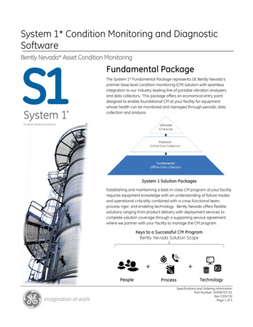
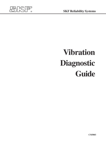
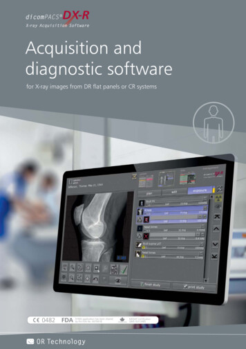
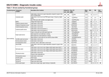
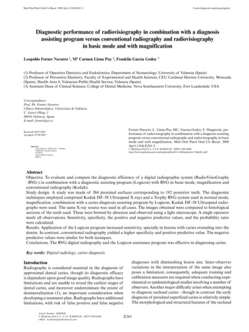
Med Oral Patol Oral Cir Bucal. 2008 Apr1;13(4):E261-5.surface make it more difficult to detect incipient cariouslesions - of which only a third are correctly diagnosed inpractice. Even lesions with minimal dentinal advance areonly detected in two-thirds of cases (2).Digital radiology involving image processing techniquescommenced with the introduction of RadioVisioGraphy(RVG) -Trophy Radiologie. Marne-La Vallée, France(3). Computed radiology with scanning laser stimulatedluminescence was introduced in the early eighties (4), andin future, light-induced fluorescence may prove useful forthe diagnosis of occlusal caries. The specificity of thistechnique is less than that of direct visualization, as confirmed from sections of theeth, though the sensitivity ofthe technique is higher than that of clinical examination/inspection in determining lesion depth (5). In this studyvalidation was performed with confocal microscopy.Computing procedures have been added to all these innovations to assist the decision taking process, with theaim of optimizing the use of modern imaging diagnosticsystems (6). Digital radiology has been found to be usefulin diagnosing incipient carious lesions in vitro (7).The present study compares the diagnostic efficacy ofconventional bitewing radiographs, RVG together with adiagnosis assistance program (Logicon Caries DetectorTM;Logicon, Inc., Los Angeles, CA, USA), RVG in basicmode and with magnification, and conventional radiographs - the findings being contrasted with the histologicobservations of the corresponding dental sections.Material and MethodsThe study sample comprised 192 teeth removed for orthodontic or periodontal reasons with or without cariouslesions, and with the crown intact. The exclusion criteriawere: dental fractures, consumptive processes (wear orabrasion), teeth with developmental alterations causingmorphological and/or structural anomalies, teeth exhibitingextensive carious destruction of the pulp chamber or canalinvasion, and cases of severe coronal morphological andstructural alterations. In the latter case selection was madevisually in some instances and radiographically in others.For the radiographic study, the teeth were mounted ingroups of 8 teeth (4 maxillary and 4 mandibular, of whichtwo each were premolars and molars). Articulation ofthe two blocks of teeth was achieved with plastic miniarticulators, to reproduce the anatomical relations amongthe neighboring and opposing teeth. Once articulated,the teeth were kept stable on a support with the occlusalplane parallel to the floor and the long axis of the toothperpendicular to this plane. A soft tissue simulator waspositioned between these models and the X-ray source,to simulate a realistic reproduction of the actual oralconditions.An ORIX-AET 65 kV intraoral radiodiagnostic apparatuswas used. The selected exposure time was 0.8 seconds.Paralleling was achieved with a plastic Klauser paralleli-Caries diagnosis assinting programzer - the Hawe-Kwik-Bite (Hawe Neos Dental, Bioggio,Switzerland) - which both fixed the radiographic film andmarked the distance between it and the end of the X-raytube. Conventional radiography was in turn conductedwith the Kodak DF-58 Periapical Ultraspeed (size 2) (Kodak, USA). Processing of the exposed film was performedwith a Periomat developer (Dürr Dental, Germany). Visualization was carried out with a viewer under conditionsof environmental illumination, without opacification ormagnification. Digital recording of the same models withteeth was also performed using the same X-ray generatingtube, with an exposure time of 0.4 seconds, employing aTrophy RadioVisioGraphy system. The latter consisted ofa scintillation sensor that transmits the absorbed radiation via optic fibers to the CCD or couple transfer devicethat register the image. The associated electronics in turnprocess the image obtained. Te image was equalized tooptimize resolution and enhance diagnostic performance.The image producing unit in turn allows variations inimage presentation, offering a series of options such asimage equalization and zoom amplification - which mighthelp the observer viewing the image.Once digitally recorded, the images were analyzed usingspecific software for the diagnosis of caries (Logicon).In a selected area for diagnosis, this program providesenamel and dentinal densitometric readings that canbe correlated to the possible presence of carious lesionsthrough an information based in previous studies madeby the manufacturer (fig. 1).The approximal caries observed with both conventionalradiography and standard and magnified RVG wereclassified according to the following criteria: 0 noradiotransparency or visible restoration; 1 enhancedradiotransparent zone in the external half of the enamel;2 radiotransparent zone occupying the full thickness ofthe enamel, though without reaching the amelodentinaljunction; 3 increased radiotransparency involving theenamel, amelodentinal junction and external half of thedentine; 4 increased radiotransparency penetrating tothe internal half of the dentine, either with or withoutapparent involvement of the pulp (8).Histopathological verification of the carious lesions wascarried out by specimen abrasion and observation undera Nikon SMZ-10A photomacroscope (x32 magnification)equipped with a Nikon FDX-35 camera and its corresponding adapter (Nikon H-III) (Nikon, Japan) with a lightsource. The histopathological diagnostic criteria were: 0 no evidence of caries, 1 presence of a carious lesion (orlesions) in the external half of the enamel only; 2 presence of a carious lesion (or lesions) in the inner half of theenamel, including the amelodentinal junction but withoutspread to the dentine; 3 presence of a carious lesion (orlesions) through the enamel to the external half of thedentine (fig.2); 4 presence of a carious lesion (or lesions)through the enamel to the inner half of the dentine (9).E262
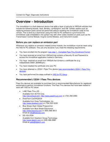
Med Oral Patol Oral Cir Bucal. 2008 Apr1;13(4):E261-5.Caries diagnosis assinting programFig. 1. Logicon densitometric analysis display from an approximal caries lesion.All observations were made by a single operator, with therepetition of 10% for intra-observer validation purposesbased on percentage concordance and the Cohen Kappatest. In order to analyze the results of the three diagnosticmodalities tested versus the histopathological findings,measurements were made of the corresponding sensitivity,specificity, positive and negative predictive values (PPVand NPV), and likelihood ratio.ResultsThe results corresponding to intra-observer concordance are shown in Table 1 (range, 84-95%), along with theCohen Kappa values (over 0.9 in all cases). As suggestedby the data analysis afforded by both tests, intra-observerreliability was found to be very high - thus contributing toensure consistency of the results obtained.Comparison of the sensitivity, specificity, PPV and NPVand likelihood ratio for each of the diagnostic proceduresstudied (i.e., conventional radiology, standard RVG, RVGwith magnification and diagnosis assisted by the Logiconprogram) with a standard (in our case, the associatedhistopathological findings) showed the highest sensitivityin enamel to correspond to the Logicon program on themesial surfaces. In the case of the distal surfaces, maximumsensitivity was afforded by RVG with magnification. Asregards dentine, the Logicon program likewise yieldedthe highest sensitivity values. In contrast, the highestspecificity was afforded by conventional radiography forall surfaces studied. In the case of PPV, the best resultswere obtained with conventional radiography, while NPVshowed coincident values for mesial enamel assessmentbetween radiography and the Logicon program - with verysimilar values among all the diagnostic modalities and forall surfaces. The likelihood ratios for a positive reading ofthe diagnostic techniques showed values equal to unityfor standard RVG in relation to mesial dentine and distalFig. 2. Example of a histopathologic image from a caries lesion(score 3).E263
Med Oral Patol Oral Cir Bucal. 2008 Apr1;13(4):E261-5.Caries diagnosis assinting programTable 1. Intra-observer concordance values.Percentageconcordance9589899284Conventional X-raysStandard RVGRVG with magnificationLogicon softwareHistopathologyKappa test0.980.980.980.970.98RVG: radiovisiographyTable 2. Performance parameters for each of the diagnostic methods lEDED14518317520719 14 32 1939 20 26 34SpecificityMesialDistalEDED98 100 91 9685 95 80 9079 94 71 8768 81 61 76PPVMesialDistalEDED81 100 45 2536 22 28 1130 35 29 1738 18 20 18NPVMesialDistalEDED70 83 74 8667 82 72 8666 83 73 8770 82 68 88Probability ratioMesialDistalEDED7 2 0.71.1 11 0.70.9 2.3 1.1 1.41.2 1 0.6 1.4Rx: conventional radiography, RVGst: standard radiovisiography, RVGmag: radiovisiography with magnification, PPV: positive predictivevalue, NPV: negative predictive value, E: enamel, D: dentineenamel, and for the Logicon program in mesial dentine.This indicates that positive readings with these explorations do not increase the probability of caries affecting thesurfaces studied. In the rest of cases, the likelihood ratiosranged from 0.6 to 7, reflecting the different incrementsin the probability that a given carious tooth will yield apositive reading with the corresponding diagnostic technique (Table 2).DiscussionDigital radiology detects 70% of deep dentinal lesions,versus only 45% when using other radiological techniques(including conventional X-rays and xeroradiography),without incrementing the proportion of false positivereadings (10).In the initial version of radiovisiography (RVG), theauthors acknowledged that the resolution afforded wasslightly inferior to that of a conventional radiograph though images could be magnified. Additional advantagesover conventional X-rays are the possibilities of improving brigthness and contrast (3). The radiographic imagedepends on a number of factors; these include exposuretime, geometric parameters such as the distance betweenthe X-ray source and film, or the direction of the tube, theamount and extent of mineral loss in the tooth, the typeof film used, the conditions of development, and the conditions of observation. As a result, the apparently simpleprocess of "seeing" and X-ray can lead to important diagnostic errors if the above mentioned influencing factorsare not adequately controlled. In this sense, Cederberg &others (11), in relation to the diagnosis of dental lesions,showed background illumination to have no effect uponthe diagnostic capacity of the observer. On the other hand,these authors dealt with images processed both digitally(Digora) and conventionally (Ultraspeed and EktaspeedPlus) - thus suggesting that the processing approach employed is also unlikely to greatly influence the diagnosticdecision of an experienced observer in situations of genuine carious pathology.In the present study a single X-ray source was used withknown and stable characteristics, and exposure wascontrolled for time, focal distance and position. The filmand digital detector features likewise remained constantthroughout the study. In order to avoid one of the mainsources of error in the production of radiographic images,we at all times used an automatic processor to ensuresimilar conditions for all the X-ray films obtained. Onthe other hand, and although digital imaging obviatessuch steps, variability may nevertheless result from thepossibility of modifying and improving the images; as aresult, the digital images obtained were equalized underconstant parameters to ensure the best possible imageunder known and normalized conditions. In the case ofthe radiographs, we conducted visualization as uniformlyE264
Med Oral Patol Oral Cir Bucal. 2008 Apr1;13(4):E261-5.and simply as possible, i.e., using a viewer without environmental illumination (the only way to control for thisfactor), without magnification.Digital radiological systems allow image modificationand analysis in an attempt to improve their diagnosticcapacity. In this sense, a number of experimental studieshave tried to precisely assess the true capacity of these technical options. While modifications in the basic functionsof the global digital radiological systems do not seemto afford much improvement over direct digital imaging(12), magnification of the initial image - a function alsooffered from the early digital radiological techniques - doesin effect increase the diagnostic precision of proximalcarious lesions versus non-magnified images (13). In ourstudy, slightly higher sensitivity values were obtained formagnified RVG versus the standard RVG technique, whilefor the rest of the methods the values were either similaror slightly inferior.A study comparing bitewing radiographs with RVGin basic mode in the analysis of occlusal and proximalsurfaces reported results similar to our own in terms ofspecificity and PPV in application to proximal surfaces(9). The sensitivity results of conventional radiographyversus RVG in application to the proximal surfaces wereinferior to our own, however.Heaven & others (14), using a software supported diagnostic system (CariesFinder), reported significant differences between the latter procedure and conventionalradiography using D-Speed film. In our study, the highestsensitivity values were obtained with the Logicon program,while maximum specificity was afforded by conventionalradiography. Wenzel (15) stablished the consistency ofLogicon studying proximal surfaces in patients whenthe analysis is repeated on the same surface; this workshowed that inter-observer agreement caries diagnosisdid not improved using the program. A recent study (16)compared clinical and Logicon-assisted RVG diagnosis,showing that dentists were able to detect 20 percent morecases of caries penetrating in dentin with Logicon versusthose founded by clinical criteria. When comparing witha similar study (17) we obtained less sensibility and higherspecificity.The results obtained suggest digital radiology to be effective in the diagnosis of dental caries. On the other hand, weconsider that although diagnosis assisting software offers agood diagnostic performance, further research is requiredin this field since such systems furthermore contributeto improve communication with the patient and afforddocumental support of the diagnosis made.Caries diagnosis assinting programevaluation of RadioVisioGraphy. Oral Surg Oral Med Oral Pathol. 1989Aug;68(2):238-42.4. Sonoda M, Takano M, Miyahara J, Kato H. Computed radiography utilizing scanning laser stimulated luminescence. Radiology. 1983Sep;148(3):833-8.5. Ferreira AG, Ando M, Lagerweij MD, Isaacs RL, Analoui M, StookeyGK. Light-induced fluorescence detection of occlusal caries: a pilotstudy. J Dent Res. 1988;77(special issue, AADR abstracts):211.6. Duncan RC, Heaven T, Weems RA, Firestone AR, Greer DF, Patel JR.Using computers to diagnose and plan treatment of approximal caries.Detected in radiographs. J Am Dent Assoc. 1995 Jul;126(7):873-82.7. Forner L, Llena MC, Almerich JM, García-Godoy F. Digital radiologyand image analysis for approximal caries diagnosis. Oper Dent. 1999Sep-Oct;24(5):312-5.8. Pitts NB. Systems for grading approximal carious lesions and overlapsdiagnosed from bitewing radiographs. Proposals for future standardization. Community Dent Oral Epidemiol. 1984 Apr;12(2):114-22.9. Russell M, Pitts NB. Radiovisiographic diagnosis of dental caries:initial comparison of basic mode videoprints with bitewing radiography.Caries Res. 1993;27(1):65-70.10. Wenzel A, Larsen MJ, Fejerskov O. Detection of occlusal caries without cavitation by visual inspection, film radiographs, xeroradiographs,and digitized radiographs. Caries Res. 1991;25(5):365-71.11. Cederberg RA, Frederiksen NL, Benson BW, Shulman JD. Comparison of digital and film images using different background lightingconditions. J Dent Res. 1998;77(special issue, AADR abstracts):227.12. Kullendorff B, Nilsso
Caries diagnosis assinting program Med Oral Patol Oral Cir Bucal. 2008 Apr1;13(4):E261-5. Caries diagnosis assinting program Diagnostic performance of radiovisiography in combination with a diagnosis assisting program versus conve