Transcription
Removable PartialDenture ManualRobert W. Loney, DMD, MS2011
Removable PartialDenture ManualRobert W. Loney, DMD, MS2011
Table of Contents - iTable of ContentsIntroduction to Removable Partial Dentures . 1Alternatives to RPD's. 1Indications for RPD's . 1Treatment Objectives . 1Components of a Partial Denture . 3Classification . 4Kennedy Classification . 4Applegate's Rules for Applying the Kennedy Classification . 5Anatomy Tour for Complete and Partial Dentures. 6Irreversible Hydrocolloid Preliminary Impressions . 11Making the impression . 11Evaluating Irreversible Hydrocolloid Impressions . 12Pouring a Model . 12Two-Pour Technique . 12Trimming . 13Surveying, Path of Insertion, Guiding Planes . 14Parts of a Surveyor: . 15Selecting the Path of Insertion of a Removable Partial Denture . 16The Optimal Path of Insertion . 17Selection and Preparation of Guiding Planes: . 20The Effects of Guiding Planes on Retention and Stability . 21Alteration of Other Axial Contours . 22Summary of Removable Partial Denture Abutment Modifications . 23Rests and Rest Seats . 24Functions of Rests. 24Preparation of Rest Seats . 25Rest Seat Form . 25Occlusal Rest Seats . 25Lingual Rest Seats . 27Composite Buildups for Cingulum Rests: . 30Round Lingual Rest Seat Form. 31Incisal Rests . 31
Table of Contents - iiMajor Connectors . 32Functions of a Major Connector . 32Requirements of a Major Connector. 32I. Mandibular Major Connectors . 33Lingual Bar . 33Lingual Plate (Linguoplate) . 34Continuous Bar Retainer (Kennedy Bar, Double Lingual Bar) . 35Labial Bar . 35II. Maxillary Major Connectors . 35Anterior-Posterior Palatal Strap . 35Full Palatal Plate . 36Single Palatal Strap . 36Anterior Palatal Plate . 37Minor Connectors . 38Functions. 38Basic Types of Minor Connectors: . 38Gridwork Design . 41Direct and Indirect Retainers . 43Extra-Coronal Direct Retainers . 43Requirements of Direct Retainers . 44Factors affecting the magnitude of retention . 44Direct Retainers For Tooth-Borne RPD’s . 461. Circumferential (Circle or Akers) clasp. 462. Ring clasp . 473. Embrasure (Double Akers) Clasp . 474. "C" clasp (Hair-pin or Reverse action) . 48Direct Retainers For Tooth and Tissue Borne RPD's . 491. Bar Clasps . 492. The R-P-I Clasp . 503. Combination Clasp . 524. RPA Clasp . 52The Distal Rest Concept . 52Direct Retainer Selection . 55Indirect Retainers . 56Denture Bases and Replacement Denture Teeth . 59Functions. 59Materials . 59Flange Extension . 60Acrylic Resin Finish Lines . 61Partial Denture Replacement Teeth . 61
Table of Contents -iiiPrinciples of Partial Denture Design. 63Denture Bases . 64Direct Retainers . 64Number of Direct Retainers . 66Indirect Retainers . 66Major Connectors . 66Distal Extension Case Considerations . 67General Considerations . 68Drawing the Design . 66Design Session . 71Clinical Protocol for Removable Partial Dentures . 79Diagnosis & Treatment Planning. 79Final Impressions for Partial Dentures . 80Framework Impressions. 80Evaluating the Impression . 82Removable Partial Denture Framework Fabrication . 84Partial Denture Framework Adjustment . 84Pre-clinical Inspection and Adjustment . 84Clinical Adjustment . 85Occlusal Adjustments to the Framework . 87Special Adjustments for Distal Extension Cases . 88Finishing and Polishing of Adjusted Surfaces . 88Maxillo-Mandibular Relations, Altered Cast Impressions and Wax Try-in Procedures . 89Altered Cast Impressions . 91Interim Removable Partial Dentures . 93Indications. 93Fabrication . 94Interim Partial Denture Design . 94Coordinating Fixed and Removable Prosthodontics . 95Coordinating Complete and Removable Partial Dentures . 96RPD Checklist . 97Summary of RPD Design . 103
Introduction to Removable Partial Dentures - 1Introduction to Removable Partial DenturesA.Definitions1. Partial denture: A prosthesis that replaces one or more, but not all of the natural teethand supporting structures. It is supported by the teeth and/or the mucosa. It may be fixed(i.e. a bridge) or removable.2. Removable partial denture (RPD): A partial denture that can be removed and replacedin the mouth by the patient.3. Interim denture (provisional; temporary): A denture used for a short interval of time toprovide:a. esthetics, mastication, occlusal support and convenience.b. conditioning of the patient to accept the final prosthesis.4. Retention: Resistance to removal from the tissues or teeth5. Stability: Resistance to movement in a horizontal direction (anterior-posteriorly ormedio-laterally6. Support: Resistance to movement towards the tissues or teeth7. Abutment: A tooth that supports a partial denture.8. Retainer: A component of a partial denture that provides both retention and support forthe partial dentureB.Treatment Objectives1.preserve remaining teeth and supporting structures2.restore esthetics and phonetics3.restore and/or improve mastication4.restore health, comfort and quality of lifeC.Alternatives to RPD's (Treatment Options - Important for Informed Consent)1. No Treatment (Shortened Dental Arch)- Most patients can function with a shortened dental arch (SDA)- Requires anterior teeth 4 occlusal units (symmetric loss) or 6 occlusal units(asymmetric loss) for acceptable function (opposing PM 1 unit, opposing molars 2 units)- RPD doesn’t usually improve function if minimal occlusal units present2. Fixed partial denture – requires abutments at opposite ends of edentulous space, moreexpensive than RPD, must grind down abutments, flexes and can fail if too long.3. Implant supported prosthesis – most costly, closest replacement to natural dentition,less costly over long term4. Complete denture (if few teeth left, with poor prognosis); if replacement of missingteeth is very complex or costlyD.Indications for RPD's1.lengthy edentulous span (too long for a fixed prosthesis)2.no posterior abutment for a fixed prosthesis3.excessive alveolar bone loss (esthetic problem)
Table of Contents - 24.5.6.7.8.poor prognosis for complete dentures due to residual ridge morphologyreduced periodontal support of remaining teeth (won't support a fixed prosthesis)cross-arch stabilization of teethneed for immediate replacement of extracted teethcost/patient desire considerationsTreatment Sequence for Partial DenturesIf an RPD is part of planned Treatment:PLAN THE RPD BEFORE BEGINNING ANY OTHER TREATMENT Survey, tripod, heights of contourDraw design on surveyed castDesign approved before any treatment started:o Affects direct restorationso Can influence need for/preparations for crownso Insures RPD can be completed successfullyo NO EXCEPTIONSCLINICAL STEPS1.2.3.4.5.6.7.8.9.Diagnosis, Treatment Plan, HygieneDiagnostic CastsDraw design & list abutment modifications on Prosthesis Design pageInstructor ApprovalComplete Phase 1 treatmentAbutment modificationsPreliminary impression to check abutment modificationsCrown or Fixed partial denture’s for removable partial denture abutments (if necessary)Final Framework Impression (must include hamular notches/retromolar pads for distalextension removable partial dentures10. Make two casts11. Draw design on 2nd cast12. Instructor approval/corrections13. Complete RPD Framework Prescription (instructor signature required)a. Second poured cast with design sent to Lab with 1st pour14. Inspect wax-up15. Framework Adjustment16. Altered Cast impression, if needed17. Try-in with teeth in wax18. Process, deliver to patient
Introduction to Removable Partial Dentures - 3E.Components of a Partial Denturea.Major Connector: The unit of a removable partial denture that connects the parts of oneside of the dental arch to those of the other side. It's principal functions are to provideunification and rigidity to the denture.b.Minor Connector: A unit of a partial denture that connects other components (i.e. directretainer, indirect retainer, denture base, etc.) to the major connector. The principlefunctions of minor connectors are to provide unification and rigidity to the denture.c.Direct Retainer: A unit of a partial denture that provides retention against dislodgingforces. A direct retainer is commonly called a 'clasp' or 'clasp unit' and is composed offour elements, a rest, a retentive arm, a reciprocal arm and a minor connector.d.Indirect Retainer: A unit of a Class I or II partial denture that prevents or resistsmovement or rotation of the base(s) away from the residual ridge. The indirect retaineris usually composed of one component, a rest.e.Denture Base: The unit of a partial denture that covers the residual ridges and supportsthe denture teeth.
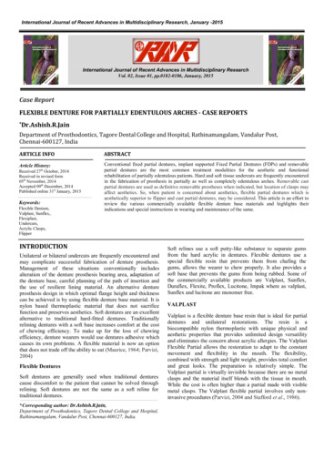
Table of Contents - 4F.ClassificationThe Need for ClassificationThere may be over 65,000 possible combinations of teeth and edentulous spaces. Aclassification system facilitates communication between dentists. Since there are severalmethods of classifying partial dentures, the use of non-standard classifications could lead toconfusion. Therefore, the Kennedy system has been adopted by most dentists.Kennedy ClassificationIn 1923, Kennedy devised a system that became popular due to its simplicity and ease ofapplication. A tremendous number of possible combinations can be reduced to four simplegroups.Class I - bilateral edentulous areaslocated posterior to all remaining teeth.Class III - unilateral edentulous areabounded by anterior andposterior natural teeth.Class II - unilateral edentulous arealocated posterior to all remaining teeth.Class IV - a single, but bilateral(crossing the midline) edentulousarea located anterior to remaining teeth.
Introduction to Removable Partial Dentures - 5Applegate's Rules for Applying the Kennedy ClassificationRule 1:Classification should follow rather than precede extraction.Rule 2:If the 3rd molar is missing and not to be replaced, it is not considered in theclassification.Rule 3:If the 3rd molar is present and to be used as an abutment, it is considered in theclassification.Rule 4:If the second molar is missing and not be replaced, it is not considered in theclassification.Rule 5:The most posterior edentulous area determines the classification.Rule 6:Edentulous areas other than those determining classification are called modificationspaces.Rule 7:The extent of the modification is not considered, only the number.Rule 8:There is no modification space in Class IV.
Anatomy Tour - 6Anatomy Tour for Complete and Partial DenturesIdentify the following structures, and answer the corresponding questions regarding anatomy thatis important in the fabrication of complete and partial dentures. The tour does not provide acomprehensive overview of all critical anatomy, but a sample of structures that are easily visiblein the dentate mouth and on casts. Use your instructor as a resource.LIPS:! Vermilion Border-When a denture provides insufficient lip support (teeth set too far palatally), thevermilion border becomes narrow, or disappears, adversely affecting appearance.- Note the width and fullness of the vermilion border of your partner(s), so you havesome idea of the normal shape and contour of these tissues.! Height, maxillary mm, mandibular mm! Philtrum-If denture teeth are set too far facially, the maxillary lip is stretched so that thedepression of the philtrum is lost. This looks unnatural. Note the normal appearance ofthe philtrum on several of your colleagues. The appearance varies.! Nasolabial Angle- The angle measured between the columella of the nose and the philtrum of the lip.- Normally, approximately 90 as viewed in profile.! Estimate your partner’s nasolabial angle ! Tissue of the Upper Lip-Gather the loose tissue of the upper lip between your thumb and index finger, so youhave some idea of the tension of the tissue or support of the lip on a dentate individual.If a denture is too “full”, no tissue can be gathered without struggling.If there is insufficient denture support, a large portion of the lip can be gatheredCHEEKS:! Masseter Muscle- Have a partner clench while you palpate this muscle externally and internally! What happens internally, when the muscles are active?! What aspect of a denture base will be affected by this structure?! Will this muscle be active when you make an open mouth impression?! How will you record its contours for your denture base?! Who has the most pronounced masseter in your group?
Anatomy Tour - 7RESIDUAL RIDGES (Identify on casts, and corresponding portions of dentures)If ridges are severely resorbed, inform the patient, so they know denture fabrication and retentionwill be difficult.! Maxillary! MandibularVESTIBULES (Identify on casts and corresponding portions of dentures)If vestibules are short, inform the patient, so they know denture fabrication and retention will bedifficult.! Maxillary Buccal! Mandibular Buccal! Mandibular LingualMAXILLA:! Maxillary TuberositiesThese will be more easily observed in edentulous patients (identify, then see casts)What influence will these structures have on dentures:a) if they are oversized?b) if they are severely resorbed?c) if they are severely undercut? i.ii.! Incisive PapillaImportant landmark for setting of teeth (midline of papilla to labial of incisor !10mm)On a partner, note the position and size of the papilla in relation to the teeth:a. How far anterior to the incisive papilla are the central incisors? mmb. What is the mediolateral relation of the incisive papilla to the midline of the maxillaryteeth! “Hamular” NotchThe posterior border of a complete denture and some partial dentures must pass throughthis notch, between the bony tuberosity and hamulus. Denture border must terminateon “soft displaceable tissue”, to provide comfort and retention. In some patients thenotch is posterior to where the depression in the soft tissue appears.! Use the head of your mirror to palpate the notch & mark with an indelible marker.! Posterior Border of the Hard Palate- What significance is this border for maxillary complete and partial dentures?
Anatomy Tour - 8! Soft Palate! Vibrating LineCritical posterior border of complete dentures and some partial dentures. The junctionof movable and immovable portions of the soft palate. If the denture terminatesposterior to this, movements of the soft palate cause it to dislodge and drop. If thedenture terminates anterior to this, on the hard palate, no seal is created, the denture isunretentive and often uncomfortable.!Within a group of six students:!!!Have an instructor demonstrate how to find and mark the vibratingline on one student, using an indelible stickHave an instructor draw an indelible line at an incorrect position onone of the two students and a correct position on the other, and guesswhich one is correct.Locate and draw the vibrating line on the remaining students, checkwith instructor.! Fovea PalatiniBilateral indentations near the midline of the soft palate. Close to the vibrating line inmany individuals, but not accurate enough for using as a landmark for the posteriorborder of the denture! Median Palatine Raphe (midline palatine suture)a. A bony midline structure, that can be raised and may require relief when coveredby a denture. Covered with thin mucosa and can ulcerate easily if impinged upon.b. Occasionally a torus palatinus may be present, creating similar, often morepronounced problems.! Identify who has the most prominent raphe/torusMANDIBLE:! Pear Shaped PadA soft pad containing glandular tissue, with an inverted pear shape, which is theposterior terminus of the mandibular complete dentures and Class I & II partialdentures. The pear shaped pad is usually created from scarring after extractions, and istherefore more easily seen in edentulous casts.! Identify the retromolar area or pad on a partner, and have an instructor check.! Identify the pear shaped pad on the distributed cast! Buccal ShelfThe primary denture bearing area of a mandibular denture.Between extraction sites of molars and the external oblique ridge. Does not tend toresorb much.! Identify on a partner.
Anatomy Tour - 9! Anterior Border of the Ramus- Do not extend dentures posterior to the pear shaped pad onto this structure, ordiscomfort will result! External Oblique Ridge- Do not extend dentures laterally from the buccal shelf onto this structure or discomfortwill result! Mylohyoid Ridge (Internal Oblique Ridge)- Origin of the mylohyoid muscle, largest muscle in the floor of the mouth, whichinfluences the length of the flange of the lingual flange of a denture.- Can be quite prominent, and/or sharp, requiring relief in the final denture, particularlytoward the posterior.! Identify, palpate and indicate the colleague with the most prominent mylohyoid ridge! Lingual ToriRaised bony structures that may require relief when covered by a denture. Covered withthin mucosa and can ulcerate easily if impinged upon! Genial Tubercles (no demo)- Attachment for the genioglossus muscle in the midline of the lingual vestibule.- When the residual ridge is highly resorbed the tubercles may be higher than the ridgeand present problems for comfort.FRENA (singular frenum):- Frena must be relieved in a denture to allow for movement, without impingement.- If frena are very prominent, adequate relief can weaken a denture.- If too much relief is provided, retention is lost.- Frena can be displaced easily with viscous impression materials such as alginate, so itis important to check their prominence intraorally.! Pull the cheeks and lips of a colleague to see how frena change shape.! Note the shape, size and direction of the following and look on the distributed denturefor corresponding areas to these frena:! maxillary labial! mandibular labial! lingual- these are narrow in shape! maxillary buccal! mandibular buccal- these are broader in shape! Who has the most prominent frena in your group?
Anatomy Tour - 10OTHER STRUCTURES:! Pterygo-Mandibular Raphe- Connects from the hamulus to the mylohyoid ridge.- Rarely significant, but when prominent, it can cause pain, or loosening of the denture,requiring a relief “groove ” by the hamular notch! Have partner open wide as possible -identify raphe.! Is it prominent?! Retrozygomal Fossae (Space)- Palpate the zygomatic process in the buccal vestibule just buccal to the first maxillarymolar.- The vestibular space posteriorly is the retrozygomal fossae.- It is commonly incompletely captured in preliminary impressions without a syringetechnique - this leads to a short flange and non-retentive denture.! Coronoid Process- Place mirror head lateral to tuberosity, have partner move mandible to opposite side.- Note binding or pain during this excursion.- This gives some indication of the width of the space available for a denture flange.- If the flange is made too thick, it will cause pain and sometimes dislodgment.
Preliminary Impressions - 11Irreversible Hydrocolloid Preliminary ImpressionsSelection of a stock tray:1. A space of 5 -7 mm should exist between the tray and the tissues to provide bulk for strengthand accuracy of the material.2. The tray should be just short of the labial vestibule and slightly beyond the vibrating line3. Compound may be placed on peripheries of stock tray to extend borders if needed.Extension should be made only to provide coverage of critical anatomy, not for the purposeof displacing or distorting the vestibular tissues, which should be registered accurately toobtain a peripheral seal on a denture.Handling the Material:1. Pre-measure material – do not take containers to your operatory. Do not handle containerswith contaminated gloves/hands. This makes infection control easier.2. Do not leave containers open in a humid environment - humidity and high temperatures cancause deterioration of the powder.3. Do not mix in a bowl contaminated with dental stone - gypsum can cause acceleration of thealginate. Conversely alginate contamination of a bowl used to mix stone can diminish thestrength of the cast or model produced. Keep separate bowls and spatulas for alginate andstone.Measuring and Mixing alginate1. Fluff the powder before
8. Crown or Fixed partial denture’s for removable partial denture abutments (if necessary) 9. Final Framework Impression (must include hamular notches/retromolar pads for distal extension removable partial dentures 10. Make two casts 11. Draw design on 2nd cast 12. Instructor approval/corrections 13.File Size: 1MBPage Count: 108Explore furtherRemovable Partial Dentures - A System of Design .www.dentalnotebook.comImplant compatibility chartwww.tncdental.comDesigning a Removable Partial Denture (Kennedy's .www.slideshare.netDentures vs. Partials vs. Bridges vs. Implants: Everything .chinookdentureclinic.comRPD Design PDF Dentures Mouthwww.scribd.comRecommended to you b