Transcription
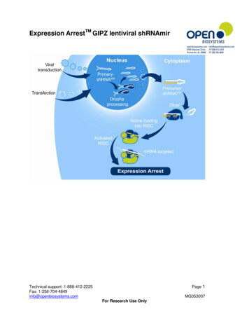
APPLICATION NOTEDharmacon Lentiviral shRNAmir forstable and regulatable RNAiProduct descriptionRNA interference (RNAi) has revolutionized the study of biologyand offers numerous applications in basic biology as well as in drugdiscovery research. Since the discovery of RNAi, several tools have beendeveloped to enable loss-of-function studies in mammalian systems.Vector-based short hairpin RNA (shRNA) expression and syntheticshort interfering RNA (siRNA) are currently the basis for all mammalianRNAi experiments (Figure 1). Each technology has specific advantages.shRNA can be used to perform many of the experiments similar to thosedone with siRNA. Additionally, when combined with viral delivery andintegration into the genome, shRNA has the added benefit of long-termknockdown from a single treatment and the incorporation of a genetictag (or barcode) allowing highly parallel pooled screening.We have further refined this technology with the creation of genome-scalemicroRNA-adapted shRNA libraries. shRNAmir are expressed as primary-miRNA(pri-miRNA) transcripts. These constructs were created by redesigning the wellstudied human miRNA, miR-30, to express an artificial siRNA/miRNA. The stemof the primary miR-30 transcript was replaced with gene-specific duplexes fordifferent target genes (Figure 2). This design does not affect normal miRNAmaturation and allows endogenous miRNA processing to produce maturesiRNAs. The shRNAmir design harnesses endogenous enzymatic processingby the RNase III Drosha, which increases subsequent Dicer recognition andspecificity. shRNAmir triggers enter the RNAi pathway ahead of either shRNAor siRNA, and are processed by both Drosha and Dicer, leading to more siRNAsproduced in the cell that are available for incorporation into the RISC complex fortarget mRNA degradation (Figure 3). [Silva et al. 2005, Boden et al. 2004, Cleary etal. 2004, Gregory et al. 2005].Additionally, the lentiviral vector format cantransduce hard-to-transfect cell lines like primaryand non-dividing. These lentiviral vectors incombination with shRNAmir design offer a powerfultool for RNAi studies.A.B.Figure 1. Evolution of RNAi triggers broadens RNAi applications.dharmacon.horizondiscovery.comWhole genome shRNAmir lentiviral libraries havebeen created using both the Dharmacon GIPZ lentiviral vector (Figure 4) that incorporates greenfluorescent protein (GFP) to track shRNAmirexpression, and the Dharmacon TRIPZ lentiviralvector (Figure 5), which shares many of the benefitsof GIPZ and includes a tetracycline (doxycycline)inducible promoter that produces tightlyregulatable RNAi.
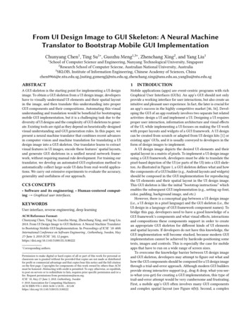
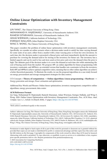
Revised versionsFrohCMVPuroRAmpRSV40 WPRERshRNA5ʹ ΨLTRΨhCMV5'ΨLTRRRERRE5ʹ LTR5' LTRFigure 4. The pGIPZ vector expresses turboGFP (tGFP) as a marker of shRNAmir expression.pGIPZRAmppUCoriSV40 orishRNAmir is constitutively expressedA. Vector map highlightingthecomponentsof pGIPZ.as a polycistronic vector expressing tGFP, puromycin resistance marker (Puror) and theshRNAmir. B. HEK293T cells transducedby GIPZ at a low MOI. Green fluorescence is anpUC oriSV40 oriAmpRindicator that the shRNAmir-containing transcript is being expressed.hCMVtGFPPuroRPuroRWPREpTRIPZSV40 oripUC oriSV40 orihPGKU6RREshRNAΨRREPuroRB.C.Ψ5' LTRpUC oriRSV/5'Ψ LTRpLKO.1IREStGFP5' LTRR Dox 48 hr500ng/mlAmpE.pSMART 2.0AmpRpUC orihCMVRREIRESWPRE500 ng/ml Dox 72 hrRREcPPSV40 oriPuroRmicroRNAWPRERREΨΨ3.tGFPshRNA3' SIN LTRNon Silencing shRNAmirNo Doxycycline (Dox) 48 hrpUC oriPuroR3' SIN LTRhCMVRRE5ʹ LTR RSV/5ʹΨ LTRΨshRNAAmpRAmpRD.cPRREΨ5ʹ LTRIRES5ʹ LTRpSMARTrtTA32.0tRFP3' SIN LTRUBCTRERRE2.A.RREWPREshRNA3' SIN LTR5' LTRΨIRESΨRRE1.Post Dox 72 hrU6pSMART 2.0hPGK3' SIN LTRPost Dox 24 hrRSV/5ʹ LTR 5ʹΨ LTRshRNAΨRSV/5' LTRΨMulti TaghCMV SiteCloningtGFPIRESPuropLOCR5'ΨLTRmicroRNAWPRE3' SIN LTR 3' SIN LTRRREGene expression knockdown5ʹΨLTRΨ5' LTRpSMPuroRRREFigure 5. A. The TRIPZ vector incorporates many of the same elements of the GIPZlentiviral backbone including a fluorescent marker (tRFP) of shRNAmir expression.RSV40 oriAmporiAdditionally, it containsallof thepUCcomponentspLKO.1 necessary for inducible expression of theshRNAmir. B-D. tRFP expression is shown as a marker of controlled, inducible expressionfrom this vector. Transduced cells were fed media containing 500 ng/mL doxycycline (Dox)and maximal inductionwithin72 hours. E. Subsequently, The cells were washedRpUCwasori seenAmphCMVand fed medium without doxycycline.Seventy-two hours later tRFP reflected a minimalRREtGFPnuc -2a- Blast R WPRElevel of expression.ORFIRESRRE3' SIN LTRFigure 3. shRNAmir is processed via the endogenous microRNA pathway. 1) shRNAmirtriggers modeled on primary miRNA are processed by Drosha and Dicer to producemature siRNA targeting a complementary mRNA. 2) shRNA triggers are approximatelymodeled on precursor miRNA and are processed by Dicer to produce mature siRNA.3) Chemically synthesized siRNA enter the RNAi pathway post-Dicer cleavage andincorporate into RISC to target complementary mRNA. All three RNAi triggers use theendogenous RNAi pathway but have distinct entry points.RREWPREΨRREpUC ori5ʹ LTR5' LTRpGIPZ3' SIN LTR 3' SIN LTRThis tech note summarizes data provided by Rigel Therapeutics from theirpilot experiments conducted to evaluate the Dharmacon GIPZ and TRIPZLentiviral shRNA Genome Scale Libraries. Seventy-eight GIPZ shRNAmirconstructs designed against 19 target genes were transduced into multiplecell lines at low multiplicity of infection (MOI). The GIPZ shRNAmir exertedknockdown effects at the both RNA and protein level. Knockdown of thetargeted genes with GIPZ shRNAmir mimicked known cellular phenotypesinduced by small molecule inhibitors. The GIPZ shRNAmir were also employedto dissect the components in a signaling pathway using a SMAD reportersystem. Their findings support previous data showing that the shRNAmirconstructs produced effective knockdown at low copy.shRNA3' SIN LTRFigure 2. shRNAmir design is based on the primary microRNA-30 (mir-30) transcript.A. Endogenous mir-30 primary transcript. B. shRNAmir expressed from a miR-30 context.The mature mir-30 sequence has been replaced with a gene specific duplex. This designallows specific processing by Drosha and Dicer and active loading into the RISC complex.RREWPREΨIRESΨtGFPRRERREΨRREAmpR5ʹ moiceershCMVmCMVhEF1αIRESPuroRWPRESV40 oriSMARTvector universal scaffold3' SIN LTR5ʹ LTR5' LTR3' SIN LTR5' LTRΨSMARTchoicepromoters5' LTRSeventy-eight shRNAmir in either the GIPZ or the TRIPZ lentiviral vectors were2.0SV40 oriAmpRpUCpSMARToripSMevaluated and percent remaining mRNA expression was measured by qPCR.H1299 human lung carcinoma cells were transduced at an MOI of 1-2, selectedSV40 oriori gene expressionwith puromycin forAmp48R hourspUCandwas measured by qPCR.Three independent experiments were performed for each shRNAmir. Previouswork done in concert withhCMVthe National Cancer Institute showed that themCMVGIPZ library was capableofachieving 70% knockdown in approximatelyhCMVhEF1αRRRE-2a-In H1299,WPREORFmEF1BlastshRNA2 of 3 hairpins. The findingshereweretGFPsimilar.72% of the shRNAnucα IRESSMARTchoiceCAGMultiTag expression by 70% (Figure 6). Overall thiswere shown to knockdowngeneCloning SitePGKvalidated the previous expectationsof the library for consistency and level ofUBCpLOCknockdown.5ʹ LTR
A.100%B.Dharmacon %54%50%50%45%40%40%30%23%20%16%10%Dharmacon TRIPZ100%6% 7%28%25%22%20%14%14%22%22%30%20%9% 10%8%35%35%23%25%22%24%17%24% 26%10%1%0%0%Figure 6. Demonstration of knockdown efficiency. In H1299, a human lung carcinoma cell line, 72% of the shRNA were shown to knock down gene expression by 70%. The red linedenotes this level of knockdown. Multiple hairpins target each gene either in A. GIPZ or B. TRIPZ. All samples were measured using qPCR. The value shown is an average of threeindependent experiments.Constitutive and inducible knockdown at theprotein levelmRNA knockdown is a quick and useful validation of shRNAmir function.However, the resulting decrease in protein is usually what is thoughtto alter phenotypes. The cell surface marker AXL was targeted in HeLacells and changes in protein level were measured by a fluorescentimmunocytochemistry assay using FACS analysis to determine the ability ofGIPZ and TRIPZ to knockdown protein levels. Every shRNAmir in both vectorsprovided 70% knockdown of the RNA in HeLa cells (data not shown). Theoption of inducible shRNAmir expression provides experimental flexibility,and the uninduced state is an ideal control for many experiments. However,one concern about inducible expression systems is the possibility of a basalexpression level in the absence of the inducing agent (tetracycline in this case)which is commonly referred to as ‘leaky’ expression.There are two main components on the pTRIPZ vector enabling induction: thetetracycline response element (TRE) and the transactivator. The transactivatorexpressed from pTRIPZ is rtTA3, a modified form optimized for increasedresponse to tetracycline without increasing background. TRIPZ demonstratedtightly controlled expression. The level of protein expression is comparedbetween untransduced cells and cells that are transduced but no tetracycline(‘OFF’) has been added to induce expression of the shRNAmir. These proteinlevels should be the same if there is no unwanted expression of the shRNAmir,and this is what was found by using a FACS assay and measuring AXL (Figure 7).Untransduced - ‘OFF’ (-DOX) No Leak‘OFF’ (-DOX) - ‘ON’ ( DOX) Inducible Knockdown% of Max.10080‘ON’ ( DOX)‘OFF’ (-DOX)60UntransducedUnstained40200100101102FL 8 Log: APCFigure 7. Decrease inprotein level without‘leaky’ expressionshown by FACS analysis.Immunocytochemistrywas used to detect the AXLprotein by FACS. The leftshift of the peaks denotes adecrease in the AXL proteinlevel after knockdown withTRIPZ shRNA targetingAXL. The untransducedand ‘OFF’ sample primarilyoverlap suggesting verylittle uninduced or leakyexpression. The left-shiftedred peak compared to theblue shows the knockdown.Phenotypic validationA high content screen ultimately relies on phenotypic changes. In thisexample, H1299 cells were transduced with constructs from the GIPZ librarytargeting genes associated with cell cycle progression and cancer. Manyof the phenotypes presented as polyploidy including endoreduplication,a cellular state in which the genome of the cell is duplicated but it doesnot under go mitosis. The polyploidy phenotype can present as one verylarge nucleus or multinucleated phenotypes and matched very well withprevious data using siRNA or small molecule described in the literature(Figure 8, see page 4).The library was able to mimic known cellular phenotypes induced by smallmolecule inhibitors for every target gene chosen, thereby validating its useas a screening tool with a phenotypic assay. Any of these results could beincorporated as a positive control into a high content screen for additionalnovel genes inducing the phenotype.DharmaconSmall moleculeGIPZ shRNAmir inhibitorUntreatedAURKBEg5/KIF11ACTA1Figure 8. Knockdown of the targeted genes produces phenotypes known to result fromdisrupting the cell cycle. AURKB, and PLK1 knockdown produced an endoreduplicationphenotype. EG5 knockdown produced the expected preprometaphase ‘stellate’ phenotypeand endoreduplication. Knockdown of ACTA1 produced a multinucleated phenotype.
Figure 9. The cell line HeLa/SMAD-gRL was engineeredto express luciferase whenstimulated with TGF-β. Thecell line allowed luciferaseto be used as a readout forsignaling through this pathway.Unknown genes’ contributionto this pathway can now becorrelated to luciferase activity.Genes without redundancyin the pathway will result in astronger disruption in luciferaseactivity when knocked down.The converse is also true. Thisis reflected in the luciferasereadout of TGFbR1 which isrequired for signaling and theSMADS that have redundantpartners with alternatedownstream signaling. (Adaptedfrom Rigel’s data presentation.)A.B.C.D.Figure 10. Luciferase as a read out for SMAD signaling using the cell line HeLa/SMAD-gRL. A. Control dose response curve was generated from the HeLa/SMADgRLcell line without disrupting the pathway. SMAD genes known to participate in thissignaling pathway were knocked down using multiple hairpins to each gene (theirclone IDs are shown in the legend). A decrease in the luciferase output suggests adisruption in the SMAD signaling pathway. The response curves were compared tothe control. Multiple hairpins to three of the target genes are shown here B. TGFbR1,the TGF-β receptor, C. SMAD2 and D. SMAD4.Pathway analysisOne of the more sophisticated experiments that can be done with RNAiis to dissect the components in a signaling pathway. Changes in manygenes’ expression can be difficult to detect directly. Using a reportersystem can alleviate this by linking expression of a reporter protein (forexample, luciferase) to a promoter that is known to be activated when thepathway of interest is involved. The well characterized TGF-β pathway wasused as a model. TGF-β, a cell signaling molecule involved in proliferationand differentiation in many cell types regulates gene expression throughSMAD pathway. Signaling through this pathway initiates with the TGF-βreceptor binding TGF-β and involves many elements of the SMAD signalingpathway. SMADs represent a broad family of genes that can overlap in theircontribution to signaling [reviewed in Derynk, R. et al. 2003]. A cell line(HeLa/SMAD-gRL) was designed to expresses luciferase from a SMADresponse element-based promoter (Figure 9) coupling TGF-β signalingactivity to luciferase activity, a functional readout more amenable to a highthroughput assay. Dose response curve was first generated by stimulationof this pathway without disrupting gene expression (Figure 10a). After eachgene suspected to be in the pathway was knocked down the resulting doseresponse curve was compared back to the control (Figures 10 b-d). Changesin luciferase activity were used to determine the relative contribution ofmany genes in the TGF-β/SMAD signaling pathway. Knockdown of the TGF-βreceptor, TGFbR1, significantly disrupted luciferase expression (Figure 10b).Knockdown of SMAD2 and SMAD4 (Figures 10c and 10d respectively) resultedin a significant but incomplete disruption of the reporter. This reflects theredundant nature of multiple SMADs with overlapping signaling roles.This data provides benchmarks for luciferase activity while disrupting genesknown to be involved in the pathway. Going forward, using the current datato gauge participation in TGF-β signaling pathway, this assay is amenable toa high throughput screen of the entire genome. Knockdown of the TGF-βreceptor, TGFbR1, significantly disrupted luciferase expression (Figure 10b).Knockdown of SMAD2 and SMAD4 (Figures 10c and 10d respectively) resultedin a significant but incomplete disruption of the reporter. This reflects theredundant nature of multiple SMADs with overlapping signaling roles. Thisdata provides benchmarks for luciferase activity while disrupting genes knownto be involved in the pathway. Going forward, using the current data to gaugeparticipation in TGF-β signaling pathway, this assay is amenable to a highthroughput screen of the entire genome.ConclusionThe ability of the GIPZ and TRIPZ lentiviral shRNAmir constructs to produceeffective knockdown has been evaluated at the mRNA and protein level aswell as in functional analysis. qPCR results confirmed not only that a majorityof the constructs in this library are capable of inducing sufficient knockdown( 2 out of 3), but that this can be achieved at low MOIs in most cases. This hassignificant advantages for the customer who is interested in knockdown of asingle gene or the customer who is interested in a larger scale screening tool:1. One of the main concerns of an RNAi experiment is the possibility foroff-target effects. The ability to achieve significant knockdown at low MOIswill reduce the possibility of concentration dependent off-target effects.2. Fewer resources can be used to perform more experiments.3. Any downstream event dependant on knockdown will be more robust. Forexample, this was shown for the TGF-β pathway (Figures 9 and 10). Knownphenotypes caused by small molecule inhibitors were mimicked and theessential components of the pathway were able to be distinguished fromthe redundant components.dharmacon.horizondiscovery.com
References1. Silva, J.M. et al. (2005) Second-generation shRNA libraries covering themouse and human genomes. Nat. Genet. 37, 1281–1288.2. Boden, D. et al. (2004) Enhanced gene silencing of HIV-1 specific siRNAusing microRNA designed hairpins. Nucleic Acids Res. 32, 1154–1158.3. Cleary, M.A. et al. (2004) Production of complex nucleic acid libraries usinghighly parallel in situ oligonucleotide synthesis. Nat Methods 1, 241–248.4. Gregory, R.I. et al. (2005) Human RISC couples microRNA biogenesis andposttranscriptional gene silencing. Cell 123, 631–640.5. Derynk, R. et al. (2003) Smad-dependent and Smad-independent pathwaysin TGF-beta family signalling. Nature 425, 577-84.t 44 (0) 1223 976 000 (UK) or 1 800 235 9880 (USA); 1 303 604 9499 (USA)f 44 (0)1223 655 581w horizondiscovery.com/contact-us or Horizon Discovery, 8100 Cambridge Research Park, Waterbeach, Cambridge, CB25 9TL, United KingdomShuttle is a trademark of Lonza. All trademarks are the property of Horizon Discovery Company unless otherwise specified. 2018 HorizonDiscovery Group Company—All rights reserved. First published January 2015. UK Registered Head Office: Building 8100, Cambridge ResearchPark, Cambridge, CB25 9TL, United Kingdom.V2-1018If you have any questions, contact
The pGIPZ vector expresses turboGFP (tGFP) as a marker of shRNAmir expression. A. Vector map highlighting the components of pGIPZ. shRNAmir is constitutively expressed as a polycistronic vector expressing tGFP, puromycin resistance marker (Puror) and the shRNAmir. B. HEK293T cells transduced by