Transcription
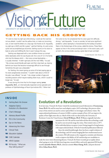
University of RochesterFlaum Eye InstituteAUTUMN 2011 NewslEtterGetting Back His Groove“If I were to close my right eye while driving, I could see the road butfirst came to me, he complained that his vision gave him difficultynot any details. I couldn’t see the yellow lines, or make out oncomingdriving,” said Aquavella. “It was no wonder, he had some significantcars, or even read the letters on a stop sign. But thanks to Dr. Aquavella,problems. Lattice dystrophy is characterized by the buildup of proteinmy right eye is 20/25 and life is great,” said Karl Sterling. An avid runner,fibers in the thickest layer of the cornea, called the stroma. These fiberscyclist and accomplished jazz drummer, Sterling counts on his vision toappear as lines in the cornea and disrupt vision. In the worst cases, suchlead an active and fulfilling life. But it wasn’t always this way.as Karl’s, the cornea erodes causing a great deal of pain and lightSterling was diagnosed with a rare condition known as corneallattice dystrophy when he was 8 years old. He is one of 12 in his family( c o n ti n ued o n page 5 )who has this disease which — like many corneal dystrophies —is usually inherited. “It didn’t get bad until the mid-1990s,” he said.“My corneas would literally split open and then close back up, leavingbehind scar tissue that became increasingly difficult to see through.Sometimes the pain was unbearable.”For Sterling, this meant giving up some of his best paying gigs ashis vision progressively worsened. “I couldn’t take about a third ofthe jobs I was offered,” he said. “I do a large number of gigs andrecording sessions which require reading sheet music, which I could nolonger see,” he said.It was during this time that he first began seeing JamesAquavella, M.D., an internationally respected cornea specialist andprofessor of Ophthalmology at Flaum Eye Institute (FEI.) “When KarlINSIDE1Getting Back His Groove1Adaptive Optics:Evolution of a Revolution2Chair’s Message3Advisory Board Profile3 FEI in the Community4 Eye on the News6Advancing the Vision9 Clinical Trials10 Education Update10 CME Calendar11 ARVO Update12 FEI Faculty: “What arethey talking about?”Evolution of a RevolutionIn its last issue, Vision for the Future visited the translational research laboratories of GeunyoungYoon, Ph.D. He and his team have used adaptive optics (AO) technology (the process wherearrays of tiny mirrors are used to correct optical flaws) to investigate and enhance vision. Theunprecedented performance of custom LASIK is a prime example of how AO can improve thequality of how light enters the eye. Much of this work was directed by the University ofRochester Center for Visual Science’s David Williams, Ph.D. Williams originally had otherintentions for AO. He and his team believed that this technology couldbe used to non-invasively image the retina. Their initial work, in 1977,led to individual photo-receptors (cones) being imaged in living humanswith unprecedented detail. A flood of watershed publications followed.Since the initial breakthrough, there has been a rapid evolution ofthe technology, and AO retinal imaging labs have mushroomed acrossthe country. FEI still remains at the vanguard as scientists and clinicianswork to improve the fidelity of AO and develop new modalities tostudy the most vision threatening diseases. Vision for the Future recently got an update aboutwhat’s going on in the AO labs from long-time research fellow and newly appointed FEI facultymember Jennifer Hunter, Ph.D., clinician scientist Mina Chung, M.D., and post-doctoralfellow Ethan Rossi, Ph.D.( c o n ti n ued o n page 8 )
C h a i r ’ sM e s s a g escientific impact of the Flaum Eye Institute,out a new on-line publication “Insight”built upon a decade of incredibleto make sure everyone within ourindividual and collective efforts.organization of almost 200 individualsIn this issue you’ll learn about the questof Richard Libby, Ph.D., assistantprofessor of Ophthalmology, HumanGenetics, and Visual Science, to underSteven E. Feldon,M.D., M.B.A.stand the molecular triggers of retinalneuron death in glaucoma (page 4).You’ll also discover how our team ofscientists and clinicians, including one ofAnother initiative involves providing moremanagerial and technical support forour faculty and staff within a refinedorganizational structure that has thecapacity to be efficient, responsive, andsupportive of even more rapid growthover the next five years.Reaching for the Topour newest faculty members, JenniferAs you can tell, my enthusiasm for theHunter, Ph.D., is using adaptive opticspotential of the Flaum Eye Institute toI came to the University of Rochesterimaging of the retina to find better wayshave regional, national, and internationalnearly 10 years ago to take on theto treat blinding diseases like macularimpact on vision health is greater thanchallenge of building a nationallydegeneration. We also welcome Ryanever. There are already plans to add moreacclaimed eye institute. Since that time,Vida, O.D. who is working in conjunctionclinical and research faculty and to expandI have been privileged to work withwith Scott MacRae, M.D. at the FEIour space to meet the increased demand.dedicated faculty, staff, and communityRefractive Surgery Center.Our readership encompasses professionalleaders. Their dedication and effort haveled the Flaum Eye Institute along a steepgrowth trajectory addressing its missionsof research, education, patient care, andtechnology transfer. Of course, theultimate goal of preventing blindness andrestoring and enhancing vision guides allof these missions. Our sustaining donors,Bausch Lomb, Glover-Crask Foundation, Research to Prevent BlindnessWe continue to reach out into thecommunity with more screenings andpatient support and education programs.We welcome your feedback on theseefforts. If you can think of new ways wecould provide additional outreach, pleasetalk to us. The entire faculty and staff lookforward to your thoughts on how we canserve outside the walls of FEI.and academic colleagues, staff, community leaders, donors, and patients. You arevery much a part of what the Flaum EyeInstitute is today and what it aspires tobecome. With your support, our goals asan eye institute can be met and surpassed.And I conclude with my thanks to all ofyou that are part of this journey.Sincerely,Foundation, David and Ilene Flaum,Aggressive pursuit of our academicand the estate of Lynn and Jack Lutzmissions requires that the organizationalhave been pivotal by contributing morestructure and culture evolve. In the lastthan 20 million in support of ouredition of this newsletter, I mentioned ourprogrammatic and infrastructure needs.interest in developing as a learningHundreds of additional major and annualorganization as envisioned by Peter SengeDirector, David and Ilene Flaum Eye Institutegifts add to this outpouring of generosity.in his classic book The Fifth Discipline.Chair, Department of OphthalmologyIn addition to supporting our philanthropy,One of our first steps involves a revisionUniversity of Rochester School ofour Advisory Board continues to provideof our mission statement to include inputMedicine & Dentistryoutstanding guidance in our strategic andfrom the board, faculty, and staff. I lookoperational planning, as exemplified byforward to sharing our efforts in theClayton Osborne, who is profiled in thisnear future. Another step has been anissue. Also recognized in this Vision for theexpansion of our quality improvementFuture is the important clinical andteams with increased participation fromall levels of the organization. To improveinternal communications, we have rolled2is updated on what’s happening at FEI.Steven E. Feldon, M.D., M.B.A.
ADVISORY Board PROFILEThe Flaum Eye Institute Advisory Board is integral to our mission to serve the community throughpatient care and education while advancing the treatment of eye disease through translationalresearch. Consisting of prominent leaders and philanthropists, their wise counsel is helping todevelop FEI as a national leader in ophthalmology and as a sustainable engine of economic growthat the University of Rochester and throughout the region. This is the second in a series of profilesto introduce you to these dedicated men and women who are helping FEI to chart its course.C l ay t o n Osb o r n eStaying on BoardClayton “Clay” Osborne has a passion for coaching leaders andwas going on at the Eye Institute. By then I had really developed abuilding learning organizations. After 25 years in the corporate,passion for the type of work that occurs here. The technology and theeducation and government sectors, he is now president of Trueinnovative models for treating and researching eye disease wereInsights Consulting. Osborne is recognized for his acumen in talentcompelling; I just had to stay.”management, human resource development, organizational effec-As an active member of the Board, Osborne has served on severaltiveness and diversity, and is an adjunct professor at the St. Johnsub-committees including community awareness. Currently he isFisher College Executive Leadership Program. He is also Minnetthelping FEI to evolve as a learning organization. No one appreciates hisProfessor Emeritus at Rochester Institute of Technology, where heexpertise here more than Feldon.consults for the RIT Leadership Institute, and has been invited to“We are so fortunate to have Clay Osborne on our advisory board,”serve on several boards including ESL Federal Credit Union andsaid Feldon. “His vast experience in building relationships betweenRochester’s Center for Governmental Research. He holds a bachelor’sthe public and private sectors as well as his expertise in developingdegree and master’s degrees in social work from SUNY Albany.organizations has been of tremendous value to me in directing theOsborne originally came to sit on the Flaum Eye Institute’s AdvisoryBoard as a liaison from Bausch Lomb. The former Advisory Boardliaison Barbara Kelley had just retired from the company. B L continuedFlaum Eye Institute through its rapid growth. As FEI strives to evolve asa learning organization, Clay’s guidance has been inspirational.”As a strong advocate for improving health care strategies, Osborneto need someone to maintain its commitment to help the University ofsees the learning organization concept as one of the catalysts for futureRochester establish a world class center for vision research and patientgrowth at FEI.care and Osborne was the perfect fit. As Vice President of Human“FEI’s strongest asset is the faculty, staff and leadership whoResources, Talent Management, Learning and Diversity at B L, he wasDr. Feldon has been able to attract and retain,” he said. “I believe thatconnected to virtually all of the company’s far-flung enterprises.with further cultivation of the talent and diversity that exists at FEI —Within a year and a half of joining the FEI Advisory Board, B Las well access to private and public funding — the opportunity for morewas taken private and some of its operations moved out of Rochester.growth and innovation is incredible,” he said. “If even a small percent-After an eighteen-year tenure, Osborne decided to retire from B L andage of what is planned comes to fruition, I see tremendous potential forestablish True Insights Consulting LLC. With his responsibilities to B LFEI to provide eye care not only throughout the region, but beyond.fulfilled, he decided to remain a member of the FEI Advisory BoardTen years from now, FEI could be the model for how to build an eyewhere he is still a key figure.institute in the U.S. and perhaps internationally. Under Dr. Feldon’s“When Steve Feldon asked me to stay on, it was a pretty easydecision,” he said. “It didn’t take long to become captivated by whatLeadership, FEI has captured my imagination. I am glad that I made thechoice to stay on board.”FEI in theCommunityThe new academic year always brings a busy schedule of FEI extending its reach into the region to bring healthcare and education through communitybased lectures and screenings. FEI extends its appreciation to all the staff students and volunteers who helped out at during the following events:FEI residents and medical studentvolunteers provided free glaucoma screenings at the Rochester PublicMarket as part of the University of Rochester Medical Center’s women’shealth screening. The screenings are made possible through a grant fromthe Friends of the Congressional Glaucoma Caucus Foundation.September 10th :The Foundation for Fighting Blindness(FFB) invited Mina Chung, M.D., to deliver a presentation to itslocal members titled “Adaptive Optics Imaging for Inherited RetinalDegenerations”. FFB is dedicated to eradicating the entire spectrum ofretinal degenerative diseases through the research it supports.September 22nd :Scott MacRae, M.D., gave a lectureabout options for people facing cataract surgery at Men’s Health Day.The program sponsored by the URMC was held at the downtownHyatt Regency Hotel.September 23rd :marked the second meeting of a patient runsupport group for Graves’ disease. The meeting was chaired by patientPatricia Marino, Ph.D., and James Aquavella, M.D., discussed dryeye coping strategies for those suffering from thyroid eye disease.October 6th :3
EYE ON THE NEWSNew FacultyFEI has promoted Jennifer Hunter, Ph.D.,to assistant professor of Ophthalmology onthe research faculty. Hunter oversees manyof the operations of FEI’s adaptive opticsscanning laser ophthalmoscope laboratoriesin addition to pursuing her research intoimaging the living retina. Hunter completedher Ph.D. in Physics and Vision Scienceat the University of Waterloo in Ontario,Canada. She first came to the Universityof Rochester as a postdoctoral researchJenniferHunter, Ph.D.associate working in the laboratories ofDavid Williams, Ph.D., and became aresearch assistant professor at FEI in 2010. She has an extensive recordof publications and presentations and is the associate chair of theVision and Color Division of the Optical Society of America .to occur by a similar molecular pathway. As with development, themolecular process regulating RGC death in glaucoma is unknown.Understanding the molecular process of RGC death is an importantstep to developing a neuroprotective treatment for glaucoma and willprovide fundamental insight into how the nervous system develops.Several years ago, Libby and colleagues showed that the moleculeBAX was required for RGC death both in development and glaucoma.BAX is a molecule that is known to be critical for neuronal apoptosis;in fact, BAX activation is the point of no return in this process.Unfortunately, inhibiting BAX activation is not a good therapeuticoption. The reason for this is that BAX is too far down the degeneration pathway. Even after BAX is turned off, the damage has alreadybeen done and RCGs are still too sick to function properly. However,BAX activation provides us with a known entry point into thedegeneration pathway of RGCs. Using this knowledge, the team hasbegun to unlock the cascade of upstream biochemical events stepby-step, hoping ultimately to understand the entire cell death signalingRyan Vida, O.D., joins FEI as a seniorinstructor. Vida works with the FEIrefractive surgery team at its Brighton, NYlocation. Originally from Pittsburgh, Penn.,he graduated from the Indiana UniversitySchool of Optometry where he completedhis clinical training at the cornea/contactlens clinic in Bloomington, Ind. and atseveral Department of Veterans AffairsRYA N VI D A , O . D .Medical Centers along the east coast. In July2011, he completed an Indiana Universityresidency in refractive and ocular surgery at Wang Vision Institute inNashville, Tenn. Vida recently authored chapters in two upcominginternationally released ophthalmology textbooks and is a memberof a national corneal topography consult team. He is a member of theAmerican Academy of Optometry and the Optometric Council onRefractive Technology.FEI Researchers Make HeadwayUnderstanding the Root Cause of Glaucomacan activate BAX.Through experimentation it was determined which of thesemolecules is required for BAX activation in developing RGCs and inRGCs after a glaucomatous-like injury. Surprisingly, it was found that amolecule BBC3 is required for BAX-dependent cell death in thedeveloping retina, but not for BAX-dependent cell death in developedadults. This result is surprising because BBC3 had not been previouslyimplicated in retinal development, and it clearly shows that RGCdevelopmental cell death is molecularly different from glaucoma.Overall, the work illustrates the selectivity of physiological (normal) andpathological (disease) RGC death and defines a branching point in thecell death pathways. This work has also helped define the potentialsignaling pathways that could kill RGCs in glaucoma, putting us onestep closer to developing a neuroprotective treatment for the disease.FEI Researcher Receives 1.4 Million NEI Grantto Study Training-Induced VisualRecovery After Brain DamageBrain damage, most commonly resulting from stroke, can haveRichard Libby, Ph.D., and Jeffrey Harder recently published in thedevastating effects on vision. Many times, these insults to neuraljournal Molecular Neurodegeneration, findings that may ultimatelytissue in the primary visual cortex (V1) result in what is referred to aspoint toward better treatments for glaucoma. Retinal ganglion cells“cortical blindness”. In this condition, an individual’s eyes may be(RGCs) are the retinal neurons that connect the eye to the brain. RGCsperfectly healthy, but the brain can no longer process the visual signalsare the neurons thought to die in glaucoma.it receives because of the damaged pathways. Recently, FEI researcherRetinal development is a complex process involving both theKrystel Huxlin, Ph.D., has proven that vision can be partially restoredgeneration and circuit formation of many different types of RGCs.in cortically blind regions of the visual field through a rigorous programBefore we are born about twice as many RGCs are made as arethat “retrains” the brain to see. She hypothesizes that training worksnecessary. To get a normal functioning retina, the extra neurons mustby stimulating an alternative brain pathway through visual area MTbe pruned. The pruning process occurs by a mechanism calledwhich can process visual motion; this pathway is upstream of V1.apoptosis — a molecular process of cell death that involves killing a cellInitial results from a series of experiments — where cortically blindin a controlled way. To date, numerous factors have been suggestedtest subjects were retrained and regained some of their lost sight —to play a role in determining the ultimate number of neurons that wehave been encouraging. This early success has resulted in a slew ofare born with, but at a molecular level it is unclear what triggers thejournal articles and patentable technologies. The National Eye Institute‘extra’ cells to die.wants to better understand how this process works and has awardedIn glaucoma, a blinding disease that affects more than 4 million4pathway in RGCs. There are only a small number of molecules thatHuxlin and her team of experts, including the University of Rochester’sAmericans, the ultimate cause of vision loss is the death of RGCs.Duje Tadin, Ph.D., and New York University professors David Heeger,Interestingly, RGC death in glaucoma and development are thoughtPh.D., and Marisa Carrasco, Ph.D., a four-year R01 grant to character-
Ab o u tLattice DystrophyLattice dystrophy gets its name from an accumulation of amyloid deposits, or abnormalize what happens in the brain during visualprotein fibers, throughout the middle and back of the thickest of the cornea’s five layers,retraining.called the stroma. During an eye examination, doctors see these deposits in the stroma asExperiments will determine why visualclear, comma-shaped overlapping dots and branching filaments, creating a lattice effect.retraining, using moving stimuli, recoversOver time, the lattice lines will grow opaque and involve more of the stroma. They alsonot only motion vision but also staticgradually become confluent, giving the cornea a cloudiness that may also reduce vision.vision (useful in tasks such as reading andIn some people, these abnormal protein fibers can accumulate under the cornea’s outerrecognizing objects and faces). Severallayer — the epithelium. This can cause erosion of the epithelium resulting in temporaryhypotheses will be tested to determine whatvision problems and severe pain. Even the involuntary act of blinking can be painful. To easechanges may be occurring to the rest of thethis pain, a doctor may prescribe eye drops and ointments to reduce the friction on thevisual pathways during retraining. Results oferoded cornea. In some cases, an eye patch may be used to immobilize the eyelids. Withthis testing may provide critical informationeffective care, these erosions usually heal within three days, although occasional sensationsabout brain pathways and signal processingof pain may occur for the next six to eight weeks.mechanisms stimulated by training to evokeBy about age 40, some people with lattice dystrophy will have scarring under thevisual relearning in cortically blind fields.epithelium, resulting in a haze on the cornea that can greatly obscure vision. In this case, aThis knowledge is essential to bettercorneal transplant may be needed. Although people with lattice dystrophy have an excellentunderstand the type and degree of recoverychance for a successful transplant, the disease may also arise in the donor cornea in as littlepossible in damaged visual systems andas three years. Although lattice dystrophy can occur at any time in life, the conditionmay improve treatment strategies for thoseusually arises in children between the ages of two and seven years old.suffering from cortical blindness.Getting Back His Groove( c o n ti n ued F R O M C O V E R )sensitivity while leaving scars on the surface of the eye making it moregiven day, vision in his right eye ranges between 20/20 and 20/30.difficult to see. Lattice dystrophy almost always affects both eyes and“It can vary a bit,” laughed Sterling. “Since it’s plastic, I have to keep itresults in surgical intervention 80 to 90 percent of the time.”clean. When it gets a little dirty, it’s like looking through an unwashedIt wasn’t long until Sterling received his first PhototherapeuticKeratectomy (PTK). In this procedure, an excimer laser is used toremove the scar tissue and smooth the cornea to provide better optics.windshield.” Sterling maintains a regimented routine to keep his K-Proclean and also uses an antibiotic eye drop daily.“It’s just like an artificial knee joint or heart valve,” said Aquavella.“Between both eyes, I had three PTKs,” said Sterling. “For awhile it“He’ll continue to use the antibiotic throughout his life to reduce theworked pretty well, but by 2003 the problems returned and my visionrisk of an infection developing.”eventually got worse than before. That’s when Dr. Aquavella and Iagreed that I needed a cornea transplant.”Although Sterling notices that his K-pro is there, cosmetically, andit infrequently causes him some mild discomfort (especially when he isUnfortunately for Sterling, nearly three years after a successfulin bright sunlight and not wearing sunglasses) he says the benefits farsurgery, the natural-tissue transplant failed. This is not uncommon.outweigh any minor inconveniences. “During the last four years I haveSterling’s father had 12 natural-tissue transplants since 1970. The goodalmost no complaints,” he said. “With the bands (he plays in twonews was that during this time, Aquavella had been working with hisregularly) and as a certified personal fitness trainer, I travel a lot andHarvard University colleague Klaus Dohlman, M.D., perfecting the latestI need good vision to drive. The K-pro gives me this. Even better, I canversion of an artificial cornea (Keratoprosthesis) for transplantation.take any gig because I can see the sheet music again.”“Natural-tissue cornea transplantation enjoys a high success rate,”“I’m so pleased with how the K-pro worked out for me,” hesaid Aquavella. “I’ve performed thousands of them. Given Karl’s familycontinued. “I wouldn’t go anywhere else for my eye care. The doctorshistory, however, we thought this would be the ideal situation to use anreally know their business and the staff has become like family to me.artificial cornea.” Aquavella explained that even when a natural-tissueSince lattice dystrophy runs in my family, I’ve referred many of mygraft is successful, lattice dystrophy can return. “With Keratoprosthesisrelatives to Dr. Aquavella. My cousin has two K-pros and is doing(K-pro) there is no chance of this happening.”During the first two months after surgery, there were a few touchreally well. I get along fine with theone, but if ever I wanted to doand go moments that had to be managed. But all the while, the K-prothe second, I know where to come.provided a clear path for light to enter Sterling’s eye; something heI couldn’t live the life I choose to ifhadn’t experienced for years. “There was some turmoil with my eye, butit weren’t for Dr. Aquavella and theI knew that I was in good hands,” said Sterling. “Within six months I wasEye Institute. It’s great.”seeing great. With glasses I could get down to 20/25. Before the K-pro,20/40 (legal driving vision) with correction was the best I could get.”. . . and the beat goes on . . .Now Sterling is seeing better than ever through his K-pro. On any5
ADVANCING THE VISIONA m o s t g r a t e f u l T H ANK y o u t o o u r d o n o r s f o rt h e i r g e n e r o u s g i f t s a n d o n g o i n g s u p p o r t.The David and Ilene Flaum Eye Institute is most grateful to its donors for their generous gifts and ongoing support. We are especially appreciative tothe friends, patients, alumni and faculty who contributed to our Eye Institute Annual Fund. The Annual Fund is an essential source of funding thatwill help continue our groundbreaking work in vision care and research.The following donors have contributed in various ways to FEI between April 1, 2011, and September 1, 2011. Gifts can be designated to theAnnual Fund and mailed to: Rachel Wicks, Director of Advancement, FEI, 210 Crittenden Blvd., Box 659, Rochester, NY 14642.Or make a gift online by going to eyeinstitute.urmc.edu and clicking on “Support the Eye Institute.”C o r p o r at i o n sBonadio & Co., LLPDiamond PackagingE. G. Sackett Co.King Graphic Communications LLCKovalsky-Carr Electric CoAdvanced OpticalClearVisionEast Tennessee Council of the BlindHolbrook Peterson Smith PLLCSafiloThe Irving ClubF o u n d at i o n sAlvin F. & Ruth K. Thiem FoundationFlorescue Family FoundationM & T Charitable FoundationFoundation for the Jewish CommunityRochester Area Community FoundationIndividualsNorman A. AborjailySusan AckerLaura AllimanThomas P. AzzolinoCharlotte H. Baker and Robert B. BakerLawrence BarlakDr. Donald A. Barrett 68 (Res) andGayle BarrettDora BartonPeter BattistiKelly BechardHenry Joseph Beetz 59 (MS)Jay B. BirnbaumRosemary BoeselGail W. BrasleyMarlys A. Braunlich andHarry R. BraunlichLouis V. BresciaJennifer BrooksJoan O. Brown 50 (BS)Patricia W. BrownHunter BrushDaniel J. Burns 11 (MBA)Geraldine Calder and Leslie H. CalderRita P. CarboneBetsy Carver and John P. CarverDr. Matilda Chan 06 (Res)Dr. Percival H. Y. Chee 62 (MD)Eugene ChernoyDaniel J. ChessinMargaret A CiesinskiMichael D. Clark 63 (EdM) andMary E. ClarkMartha B. ClelandDr. Ronald J. Cole 62 (MD)Willie E. ColeyProfessor Otis Stephen’s ConstitutionalLaw ClassJoseph Griffin CookJudy M. Cornett and Richard C. ParrottVirginia J. Covert and Brian E. CovertShelly L. Coville and David Coville6John D’Amico Jr.Paula J. DaileySusan M. Davies and Thomas Y. DaviesGail L. DeKramer and Don DeKramerMary K. EdwardsCaitlyn ElamCarol B. Eldredge and Francis EldredgeAsta Eldrup-JorgensenJoyce ErmerSusanne Esan 49 (BA), 52 (MA)Preston Kirk Faulkner 08 (MS)Diane A. Feldon and Dr. Steven E. FeldonJames E. FineganMichele S. Fisher and Walter D. Fisher Jr.Barry William Florescue 66 (BS)Clara Fox and Edward R. FoxLauren Dru FrankSaeed M. GaseEleanor A. GaussDr. Matthew D. GearingerLudmila Fayn Girsh and Boris Fayn GirshGail M. GriersonLida GriestDr. John F. Griffin 73 (Res) and Ellen GriffinMaxine L. GubbJames Paul Hafner 65 (MBA) andAnn L. HafnerRuth A. Hedin and Jeffrey A. HedinMary A. HehnThomas Michael HerbstAmy Morris HessDr. Edward L. Hicks 62 (BA), 65 (MD), 69 (Res)Audrey G. HolmCharlotte K. HolmesDr. Krystel HuxlinAdelaide F. IngleKrista M. Jaskier and Joseph P. JaskierTheresa JohnsonRonald G. KaufmanPatricia C. Keenan and John D. KeenanBillie Jean KeithJeanette M. KelleherDarlene Kiel and Bernard L. KielMaria Kipp-MayneMargaret KittelbergerMargaret G. KleinMargaret Kleinman andDr. Martin KleinmanDr. Gary KochersbergerEvelyn L. Krane 74 (EdM)Hy KreitzmanDora Lange and Paul K. LangeJohn L. LawlessMaria I. Lauriello-Klein and Aaron M. KleinMyrna M. Lawrence-WatersElaine Livecchi and Samuel LivecchiSuzanne M. LorzAdeline P. Lutz*Kathleen L. Lynd and Edward J. LyndDr. Scott MacRaeJulia Magguilli and Louis D. MagguilliDr. Karl J. Marchenese 74 (MD), 79 (Res)Nationallyrenowned artistTreacy Zieglerpresented anoriginal work ofart to FEI. Entitled,“Anatomy of theInside”. The oil onpanel paintinghangs in the checkout area. Ziegleris a patient andsupporter of theEye Institute.Her studio is nearIthaca, NY.Ellen Marple and Homer L. MarpleDr. Stephen Sidney Martin 79 (MS), 81 (PhD), 81M (MD), 85 (Res)Elizabeth Marvel and Clyde MarvelNona M. Maurer 74 (BS) andJames William Maurer 68 (BS)Theresa B. Mazzullo and Donald S. MazzulloBarbara McCann Kelley 77 (BS) andThomas P. RileyMrs. Maryanne L. McDaniel andWilliam L. McDanielCarol A. MeierGlenda S. Melville and W. Robert Melville IIIJohn F.
spteme Ber 22nd : The Foundation for Fighting Blindness (FFB) invited Mina Chung, M.D., to deliver a presentation to its local members titled “Adaptive Optics Imaging for Inherited Retinal Degenerations”. FFB is dedicated to eradicating the entire spectrum of . eye coping