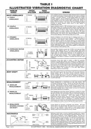
Transcription
European Archives of Oto-Rhino-Laryngology (2021) 06324-wHEAD AND NECKAccuracy of the preoperative diagnostic workup in patients with headand neck cancers undergoing neck dissection in terms of nodalmetastasesAngéla Horváth1 · Péter Prekopp1 · Gábor Polony1 · Eszter Székely2 · László Tamás1 · Kornél Dános1Received: 16 July 2020 / Accepted: 24 August 2020 / Published online: 29 August 2020 The Author(s) 2020AbstractPurpose The presence of cervical lymph node metastases is one of the most influential prognostic factors in head and necksquamous cell carcinomas. The management of clinically N0 neck in patients with head and neck cancer remains controversial: elective neck dissection has relatively high morbidity, adversely affecting the quality of life, however, abandoningelective neck dissection is known to compromise overall survival in numerous primaries. The purpose of this study was toevaluate the accuracy of the conventional imaging modalities (CT, MRI, US) and fine-needle aspiration cytology (FNAC)in the detection of lymph node metastases in the neck.Methods Sixty two patients were included in the study, who underwent primary tumor resection and neck dissection. Preoperative nodal status was compared with postoperative histopathology nodal status. In our retrospective study, we reviewed thepatient documentation. Statistical analysis of the data—with descriptive statistics and correlation analysis—was performedwith Chi-square test.Results The sensitivity of conventional imaging modalities and FNAC were 82.8% and 81.8%, respectively, while specificity were 73.9% and 100%, respectively. Positive predictive value calculated for imaging modalities and FNAC were 82.8%,100%, respectively, while negative predictive values were 73.9% and 66.6%, respectively.Conclusion Neither the sensitivity of imaging modalities (CT, MRI, US) nor FNAC reached 100%, none of these methodscan definitively exclude the presence of regional tumor metastasis. According to these data, no permissive alteration shouldbe allowed from the current guidelines (e.g. NCCN) based on imaging/FNAC examinations regarding the need for electiveneck dissection.Keywords Imaging · Head and neck · Squamous cell carcinoma · Ultrasound · FNAC · Neck dissectionPurposeThe presence of cervical lymph node metastases is one of themost influential prognostic factors in head and neck squamous cell carcinomas. Adequate treatment based on precisepreoperative diagnostic work-up is crucial for achievingthe desired oncologic outcome [1]. The indication for neck* Kornél ment of Otorhinolaryngology, Head and NeckSurgery, Semmelweis University, Szigony Str. 36,Budapest 1083, Hungary22nd Department of Pathology, Semmelweis University,Budapest, Hungarydissection can be either therapeutic (for clinically N necks)or elective (for clinically N0 necks). Most currently available guidelines recommend comprehensive neck dissection(levels I–V.) if preoperative examinations (physical examination, imaging, FNAC) reveal pathologic lymph nodes.Based on classic textbooks, elective treatment of the neckis recommended in clinically negative (cN0) necks whenthe probability of occult micrometastases exceeds 15–20%,which is embraced by the therapeutic guidelines, easing theeveryday clinical decision making for certain tumor sitesand stages [2].Unilateral lymph node dissection is usually recommended, but bilateral dissection is warranted in tumorsreaching the midline and due to the bilateral lymphaticdrainage in tumors in certain locations (e.g. base of thetongue, soft palate, supraglottic larynx).13Vol.:(0123456789)
2042Management of the N0 neck is still a controversial area inthe surgical treatment of head and neck tumors. It is knownthat the number of elective neck dissections in which thepostoperative histopathological examinations reveal nometastatic lymph nodes is relatively high, however, themorbidity—such as hypesthesia, lymphedema, decreasedhead/neck, or arm mobility—of these procedures are alsosubstantial in a varying degree, resulting in a decreasedquality of life [3]. Moreover, there are cases when regionalrecurrence occurs over the years, adversely affecting overallsurvival. For this reason, careful preoperative investigation,diagnostic algorithms, and therapeutic recommendations areextremely important.In our study, we aimed to evaluate the reliability of thepreoperative investigations of our patients who underwentneck dissections by calculating the specificity, sensitivity,negative-, and positive predictive values regarding the presence/absence of metastatic lymph nodes.MethodsAll patients enrolled in our study were diagnosed (histologically) and treated between September 2012 and August2017 at the Department of Otolaryngology and Head-NeckSurgery, Semmelweis University, Hungary. The patientsunderwent upfront surgical treatment (resection of the primary and neck dissection) for cancers of the oral cavity,oropharynx, hypopharynx, or larynx.Patients who were not diagnosed (by biopsy) at our tertiary referral center, had primaries in other head and necksites (e.g. nasopharynx, nasal cavity, paranasal sinuses, salivary gland, or skin, furthermore those unknown primaries),or had previous head and neck oncological treatment wereexcluded from our study, thus, to maintain sample homogeneity, we had to exclude a large number of patients whounderwent salvage surgery or were referred to our department from other hospitals.The patients underwent head and neck imaging (CT orMRI) to determine the stage of their disease. All imagingstudies were performed—according to the Hungarian regulations—at the regional imaging center indicated by thepatient’s location (home addresses). Then, the patients werereferred to the US-guided FNAC clinic of the 2nd Department of Pathology, Semmelweis University to obtain cytologic confirmation of any pathologic lymph node detectedby ultrasound-expert cytologists.In our retrospective study, we evaluated the patients’medical records (discharge summaries, multidisciplinarytumor board (MTB) reports, histological reports, and imaging studies). The results of the preoperative examinationswere correlated to the histopathological results of the neckdissection specimens. Both preoperative examination results13European Archives of Oto-Rhino-Laryngology (2021) 278:2041–2046(cTNM) and pathological TNM (pTNM) were evaluatedaccording to the UICC 2010 version 7 TNM classification[4]. When reviewing the radiological findings, lymph nodeswere considered abnormal if they had previously been considered abnormal by the radiologist if they were larger than10 mm in their shortest diameter, contained central necrosis,or had a round shape [5].Bilateral neck dissections were performed in 12 caseswhere the preoperative FNAC results were the same for thetwo sides of the neck. However, in two cases, discordant(one side positive, the other side negative) FNAC resultswere obtained, while bilateral metastases were reported inthe pathological report of the neck dissection specimen.Both cases were considered false negatives in our study.Both FNAC and imaging were done as part of the clinical routine, so they were performed by various physicians(radiologists and cytologists).Statistical procedureThe statistical analysis of the data—with descriptive statistics and correlation analysis—was performed with the Chisquare test (Fisher’s exact test) using SPSS Statistics for Macv.22.0 (IBM Corp., Armonk, NY).ResultsWe identified 62 patients who met the criteria describedabove. In terms of their gender distribution, we examined54 men and 8 women. The mean age of the patients at thetime of diagnosis was 61 years (43–77 years). Regarding thelocalization of the primaries, most patients had hypopharyngeal tumors (32.3%), followed (in descending order of frequency) by supraglottic (22.6%), transglottic (21%), glottic (9.7%) laryngeal cancer, oropharyngeal (9.7%), oral(3.2%), and subglottic laryngeal (1.6%) tumors, respectively(Table 1).Regarding preoperative stages, T1 (T1a and T1b forglottic cancers) stage tumors were the least common, onlyTable 1 Distribution of tumorsby 322.69.7211.6
European Archives of Oto-Rhino-Laryngology (2021) 278:2041–2046Table 2 Types of surgeries performedTherapy%Partial laryngectomy with ipsilateral neck dissectionTotal laryngectomy with ipsilateral neck dissectionPharyngotomy with ipsilateral neck dissectionTransoral resection with ipsilateral neck dissectionPartial laryngectomy bilateral neck dissectionsTotal laryngectomy bilateral neck dissections27.438.78.16.54.814.52043Table 4 Relationship between MRI report and pathological lymphnode status% (No)Pathology lymph node status(pN)Lymph node status described onthe MRINegativePositiveNegativePositive80 (4)020 (1)100 (1)Table 3 Relationship between CT report and pathological lymphnode statusTable 5 Relationship between US report and pathological lymphnode status% (No)Pathology lymph node status (pN)% (No)Lymph node status described onthe CTNegativePositivePathology lymph node status(pN)NegativePositiveNegativePositive72.2 (13)15.6 (5)27.8 (5)84.4 (27)Lymph node status described onthe USNegativePositive0 (0)50 (1)0 (0)50 (1)4.8%, while T2, T3 stage tumors accounted for 32.3% 32.3%, slightly less than T4a and T4b stages: 30.6%.In terms of preoperative N stage of the tumors, 53.2%were N0, 16.1% were N1, 16.1% were N2a, 8.1% wereN2b, 4.8% were N2c, and 1.6% of them were stage N3,so 53.2% of the patients received elective neck dissection and 46.7% received therapeutic, comprehensive neckdissection.The most common therapies recommended by MTB weretotal laryngectomy with ipsilateral neck dissection (ND)(38.7%), partial laryngectomy with ipsilateral ND (27.4%),and total laryngectomy with bilateral NDs (14.5%). The procedures performed are listed in Table 2.Concerning the distribution of imaging techniques usedfor head and neck cancer staging, the majority of patientsunderwent CT scan (84%), 10% had MRI scan, and 3.2%only had ultrasound scan.As anticipated, we found a significant (p 0.001) correlation between the nodal stage described by imaging (CT,MRI, US) and the histopathology report (pN1, pN2a, pN2b,pN2c, pN3 summarized as pN and N0 as pN ). However,the discordance was noteworthy: 73.9% of the radiologicN0 cases were truly negative, while 26.1% had false negative preoperative imaging results in terms of the nodal stage(Tables 3, 4, 5, 6).Although there was a significant association betweenFNAC results and histopathologic lymph node status(p 0.001), the rate of false-negative cases was surprisinglyhigh: only 66.7% of neck statuses considered negative byFNAC were proven to be negative by the histopathologicexaminations of the surgical specimens. As expected, 100%Table 6 Relationship between imaging study report and pathologicallymph node status% (No)Pathology lymph nodestatus (pN)Lymph node status described on theimaging techniques combinedNegativePositiveNegativePositive73.9 (17)17.1 (6)26.1 (6)82.9 (29)Table 7 Relationship betweenFNAC results and pathologicallymph node status% (No)Pathology lymphnode status (pN)FNACNegativePositiveNegativePositive66.7 (12)0 (0)33.3 (6)100 (27)of the lymph nodes diagnosed positive by the FNAC werealso positive histopathologically (Table 7).Based on the above, the sensitivity of imaging modalitieswas calculated to be 82.8%, while specificity was 73.9%,the positive predictive value was 82.8%, and the negativepredictive value was 73.9%. On the other hand, regardingthe FNAC, the sensitivity, specificity, negative and positive(Table 8).The clinical TNM stage (along with clinical lymph nodestatus—cN) is finally established by the multidisciplinarytumor board based on physical examination, imaging, andFNAC results. In our study, 96.6% of clinically N cases13
2044European Archives of Oto-Rhino-Laryngology (2021) 278:2041–2046Table 8 Percentage values for CT separately, imaging techniquescombined, and FNAC examinations%CT*Imaging techniques combinedFNACSensitivitySpecificityPositive predictive valueNegative predictive 6*For MRI and US, specificity and sensitivity could not be calculatedseparately because of low number of participantswere proven to be pathologically positive. However, only69.7% of cases preoperatively evaluated as N0 had pN0 stagebased on the histopathological examination of the dissectionspecimen, accordingly, 30.3%. of cases with cN0 stages werefalse negatives.DiscussionPrecise staging workup is crucial before decision makingregarding the treatment of head and neck cancer patients. Inour work, we aimed to determine the diagnostic accuracy ofpreoperative imaging modalities (CT, MRI, US) and FNACin terms of clinical and pathological nodal stages. Previous reports found different diagnostic reliability of certaindiagnostic modalities.Knappe and colleagues compared the US-guided fineneedle aspiration cytology results with the histopathologysamples of the patients undergone neck dissections. Theyfound FNAC sensitivity to be 89.2% and specificity to be98.1% [6].According to Takes et al., the US-guided FNAC had 77%sensitivity and 100% specificity in the detection of cervicallymph node metastases. This research highlights that themethod has low inter-observer variability—and therefore itswidespread use is encouraged [7].Dammann et al. evaluated the efficiency of CT, MRI, andFDG-PET in the preoperative staging of SCC of the headand neck region. They found CT sensitivity to be 80%, specificity to be 93%, while MRI sensitivity was 93%, specificitywas 95%. In the case of ambivalent cases, the study recommended PET for an additional diagnostic procedure [8].Adams et al. compared the accuracy of conventionalimaging studies (US, CT, MRI) with FDG-PET. CT showed82% sensitivity and 85% specificity, while MRI had 80%sensitivity and 79% specificity. The ultrasound showed alower sensitivity with 72%. FDG-PET and conventionalimaging showed a statistically significant correlation in thedetection of cervical lymph node metastases (PET vs. CT,p 0.017; PET vs. MRI, p 0.012; PET vs. US, p 0.0001)[9].13W. van den Brekel et al. performed a study evaluatingthe value of US and US-guided FNAC for the assessmentof N0 neck status. US detected the occult lymph nodemetastasis with 60% sensitivity and 77% specificity, whilethe US-guided FNAC showed 76% sensitivity and 100%specificity. Due to this high accuracy, this work consideredUS-guided FNAC as an important technique in the N0neck examination [10].De Bondt and colleagues performed a meta-analysiscomparing US, US-guided FNAC, CT, and MRI in thedetection of lymph node metastases in head and neck cancer patients. There was a large variability not only in theaccuracy of the techniques but also in the cut-off levels inthe size and morphology of the lymph nodes considered aspositive. US-guided FNAC showed the highest diagnosticodds ratios (DOR 260) compared to US (DOR 40), CT(DOR 14), MRI (DOR 7). US showed the highest sensitivity with 87%, while the highest specificity was linkedto US-guided FNAC (98%). The meta-analysis concludedthat the US-guided FNAC showed the best diagnostic performance, while the US, CT, and MRI were less effectivein this regard [11].In Sumi et al.’s research, CT and MRI were compared byexamining changes in lymph node parenchyma (cancer nest,necrosis, and keratinization). Small-sized (shorter axial diameter less than 10 mm) and large-sized (shorter axial diameter10 mm or larger) lymph nodes were evaluated separately. Inthe detection of small metastatic lymph nodes, MRI performedsignificantly better, sensitivity, specificity, positive predictivevalue, and negative predictive value were 83%, 89%, 89%,and 84%, respectively, while those for CT-scan were 68%,79%, 79%, and 72%, respectively. There was no significantdifference when examining large lymph nodes between thetwo imaging techniques (MRI: sensitivity 100%, specificity98%, positive predictive value 99%, negative predictive value100%, CT: sensitivity 98%, specificity 89% positive predictivevalue 95%, negative predictive value 97%) [12].In Akoglu et al.’s paper, the diagnostic efficacy ofconventional imaging (US, CT, MRI) in the detection ofcervical lymph node metastasis was compared. The following results were obtained in their research: CT sensitivity 77.7%, specificity 85.7%, positive predictive value91.3%, negative predictive value: 66.6%, MRI sensitivity59.2%, specificity 92.8%, positive predictive value 94.1%,negative predictive value: 54.1%, US sensitivity 81.4%,specificity 64.2%, positive predictive value 81.4%, negative predictive value: 64.2%. US and CT performed betterthan MRI, however, there was no significant differencebetween them. They also concluded that because of thelow negative predictive value, neither method could beused to accurately detect cervical metastases [13].In their retrospective research, Yoon et al. investigatedhow effectively CT, MRI, US and FDG-PET-CT, and a
European Archives of Oto-Rhino-Laryngology (2021) 278:2041–2046Table 9 Sensitivity andspecificity of FNAC, CT, MRIexpressed in percentageAuthorsKnappe et al.Takes et al.Dammann et al.Adams et al.Brekel et al.De Bondt et al.Sumi et al.Akoglu et al.Yoon et al.Our research2045Type of the studyFNACProspective, single institutionProspective, multi-instituteProspective, single institutionProspective, single institutionProspective, single institutionMeta-analysisRetrospective,single institutionProspective, single institutionRetrospective, multi-instituteRetrospective, single 85.799.473.9818359.277638992.899.4combination of these modalities detect cervical lymph nodemetastasis. As a result, they had a sensitivity of 77% and aspecificity of 99.4% for CT and MRI, sensitivity of 78.4%,and specificity of 98.5% for US. In conclusion, the results ofthe study showed that sensitivities and specificities of CT,MR, US, and PET/CT appeared to be similar in the detection of cervical lymph node metastases. The use of the fourtechniques combination yielded improved sensitivity compared with the single use of these techniques, but without astatistically significant difference [14].Interpreting our results, we can see that neither the sensitivity of the imaging methods (CT, MRI, US) nor the sensitivity of fine-needle aspiration reached 100%. Also, the rateof false-negative results was relatively high for both methods: 26.1% for imaging techniques and 33.3% for FNAC.Thus neither method can definitively establish the presenceof cervical metastasis. This is especially important in T1,T2 stage tumors because the N0 cervical stage is the mostcommon in these cancers.Our results do not differ substantially from those previously published, underlining that no current diagnosticmethod is reliable enough to exclude the presence of metastatic cervical lymph nodes (Table 9).Compliance with ethical standardsConclusionReferencesWe cannot decide not to perform an elective neck dissectionbased on the ’negative’ result of the preoperative stagingworkup, current therapeutic guidelines (e.g. NCCN) shouldbe followed in therapeutic decision-making.Funding K.D. was supported by the Hungarian Talent Program, Individual Scholarship Award 2019 (NTP-NFTÖ-19-B-0045). Open accessfunding provided by Semmelweis University.MRIConflict of interest The authors declare that they have no conflict ofinterest.Ethical approval All procedures performed in studies involving humanparticipants were in accordance with the ethical standards of the institutional and/or national research committee and with the 1964 HelsinkiDeclaration and its later amendments or comparable ethical standards.This research was approved by the Semmelweis University’s Regional,Institutional Scientific and Research Ethics Committee (SE TUKEB105/2014).Informed consent Informed consent was obtained from all individualparticipants included in the study.Open Access This article is licensed under a Creative Commons Attribution 4.0 International License, which permits use, sharing, adaptation, distribution and reproduction in any medium or format, as longas you give appropriate credit to the original author(s) and the source,provide a link to the Creative Commons licence, and indicate if changeswere made. The images or other third party material in this article areincluded in the article’s Creative Commons licence, unless indicatedotherwise in a credit line to the material. If material is not included inthe article’s Creative Commons licence and your intended use is notpermitted by statutory regulation or exceeds the permitted use, you willneed to obtain permission directly from the copyright holder. To view acopy of this licence, visit http://creat iveco mmons .org/licen ses/by/4.0/.1. De Virgilio A et al (2012) The oncologic radicality of supracricoidpartial laryngectomy with cricohyoidopexy in the treatment ofadvanced N0–N1 laryngeal squamous cell carcinoma. Laryngoscope 122(4):826–8332. Shah JP (2019) Jatin shah’s head and neck surgery and oncology,5th edn. Elsevier Inc, St. Louis3. Teymoortash A et al (2010) Postoperative morbidity after differenttypes of selective neck dissection. Laryngoscope 120(5):924–9294. Sobin LH (2010) TNM classification of malignant tumours, vol7. Wiley, Hoboken, p 309 (Chichester, West Sussex, UK)5. Anzai Y, Brunberg JA, Lufkin RB (1997) Imaging of nodal metastases in the head and neck. J Magn Reson Imaging 7(5):774–78313
20466. Knappe M, Louw M, Gregor RT (2000) Ultrasonography-guidedfine-needle aspiration for the assessment of cervical metastases.Arch Otolaryngol Head Neck Surg 126(9):1091–10967. Takes RP et al (1996) Regional metastasis in head and neck squamous cell carcinoma: revised value of US with US-guided FNAB.Radiology 198(3):819–8238. Dammann F et al (2005) Rational diagnosis of squamous cellcarcinoma of the head and neck region: comparative evaluation of CT, MRI, and 18FDG PET. AJR Am J Roentgenol184(4):1326–13319. Adams S et al (1998) Prospective comparison of 18F-FDGPET with conventional imaging modalities (CT, MRI, US) inlymph node staging of head and neck cancer. Eur J Nucl Med25(9):1255–126010. van den Brekel MW et al (1991) Occult metastatic neck disease:detection with US and US-guided fine-needle aspiration cytology.Radiology 180(2):457–46111. de Bondt RB et al (2007) Detection of lymph node metastases inhead and neck cancer: a meta-analysis comparing US, USgFNAC,CT and MR imaging. Eur J Radiol 64(2):266–27213European Archives of Oto-Rhino-Laryngology (2021) 278:2041–204612. Sumi M et al (2007) Diagnostic performance of MRI relative toCT for metastatic nodes of head and neck squamous cell carcinomas. J Magn Reson Imaging 26(6):1626–163313. Akoglu E et al (2005) Assessment of cervical lymph node metastasis with different imaging methods in patients with head and necksquamous cell carcinoma. J Otolaryngol 34(6):384–39414. Yoon DY et al (2009) CT, MR, US,18F-FDG PET/CT, and theircombined use for the assessment of cervical lymph node metastases in squamous cell carcinoma of the head and neck. Eur Radiol19(3):634–642Publisher’s Note Springer Nature remains neutral with regard tojurisdictional claims in published maps and institutional affiliations.
Keywords Imaging · Head and neck · Squamous cell carcinoma · Ultrasound · FNAC · Neck dissection Purpose The presence of cervical lymph node metastases is one of the most influential prognostic factors in head and neck squa-mous cell carcinomas. Adequate treatment based on precise preoperative

