Transcription
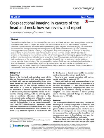
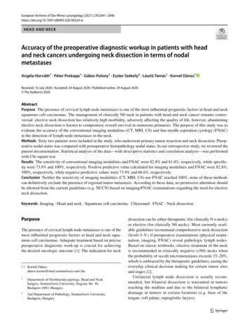
6Clinical Radiation TherapyMReadings: MR in RTwww.siemens.com/magnetom-world-rtMRI in Head and Neck Radiotherapy PlanningHouda Bahig, M.D.1; Karim Boudam, Ph.D.1; David Landry, M.D.2; Edith Filion, M.D.1; Olivier Ballivy, M.D.1;David Roberge, M.D.1; Jean-Charles Côté, Ph.D.1; Phuc-Félix Nguyen-Tan, M.D.112Department of Radiation Oncology, Centre Hospitalier de l’Université de Montréal, Montreal, Quebec, CanadaDepartment of Radiology, Centre Hospitalier de l’Université de Montréal, Montreal, Quebec, CanadaThe introduction of intensity modulated radiotherapy (IMRT)as standard of care as well as the sustained improvementsin image-guidance have significantly increased the precisionand complexity of head and neck cancer (HNC) radiotherapy(RT) planning. As have many other institutions, the CentreHospitalier de l’Université de Montréal (CHUM), a highvolume center for head and neck oncology, has adopted amulti-modality imaging approach for RT planning in locallyadvanced HNC. This approach includes routine acquisition offluorodeoxyglucose-positron emission tomography (FDG-PET)and magnetic resonance imaging (MRI) using RT-dedicatedtechnologies, combined with contrast-enhanced planningcomputed tomography (CT). While the use of IMRT allowsfor improved conformity of dose distribution, it requiresaccurate tumor volume definition in order to preventtarget-miss or unnecessary dose to organs at risk. In thisreport, we describe CHUM’s planning MRI workflow forlocally advanced HNC and we discuss the current role ofMRI in HNC planning.MRI in head and neck cancer – planning workflowat CHUMThe current approach at CHUM involves the systematicacquisition of a planning CT for dose calculation, as wellas MRI sequences in treatment position for improvementof soft-tissue delineation and optimal registration withplanning CT for HNC cases. This is of particular importancein HNC where differences in imaging planes and neck flexionFigure 1: T2-weighted MRI allows to detect a base of tongue lesion that is occult on CT scan.1A1B1CThis patient presented with a right T1N2b squamous cell cancer of the base of tongue. (1A) Planning contrast-enhanced CT scan showsa large level IIA necrotic lymph node but fails to detect primary tumor; (1B, C) axial T2-weighted MRI sequence shows a suspicious hyperintensity at the right base of tongue measuring 1.6 x 1.3 cm and corresponding to the known primary tumor detected on fiberopticnasopharyngoscopy.
MReadings: MR in RTwww.siemens.com/magnetom-world-rtbetween planning and diagnostic imaging can be major.MRI planning examinations are obtained on a RT-dedicated70 cm open bore 1.5 Tesla system (MAGNETOM Aera,Siemens Healthcare, Erlangen, Germany). The imagesare acquired in treatment position with a head and neckthermoplastic mask fixed to a custom hard foam flattable insert. Due to the incompatibility between the headand neck mask and the standard head coil, surfaceradiofrequency coils are used [1]; this typically involves aspine array coil posteriorly and a large 18-channel flexiblearray coil anteriorly (Siemens Healthcare, Erlangen,Germany).Our institutional planning MRI protocols have been adaptedto RT planning through optimization of resolution andgeometric distortions, resulting in scanning parameters thatdiffer from those used in diagnostic radiology. All sequenceswere corrected for geometric distortion using the built in 3Dcorrection algorithm. Parameters of the sequences currentlyused in our standard workflow are as follows:(1) Transverse T2-weighted Turbo Spin Echo (TSE) sequence:Repetition time (TR) / echo time (TE) 5610/80 ms, fieldof-view (FOV) 19 cm, voxel resolution 0.6 mm x 0.6 mm x3.0 mm, matrix 224 x 320 and bandwidth 191 Hz/pixel.In patients presenting dental restorations, a modifiedmetallic artifact protocol is used, with the followingparameters: TR/TE 5000/91 ms, FOV 20 cm, voxelresolution 0.6 x 0.6 x 2.0 mm, matrix 320 x 320 andbandwidth 488 Hz/pixel.(2) Transverse T1-weighted TSE sequence: TR/TE 689/23 ms,FOV 19 cm, voxel resolution 0.6 x 0.6 x 3.0 mm, matrix224 x 320, bandwidth 200 Hz/pixel. Parameters of themodified metallic artifact protocol: TR/TE 626/9 ms,FOV 20 cm, voxel resolution 0.6 x 0.6 x 2.0 mm,matrix 320 x 320, and bandwidth 504 Hz/pixel.(3) Transverse post-gadolinium T1-weighted fat saturatedTSE sequence: TR/TE 739/23 ms, FOV 19 cm,voxel resolution 0.6 x 0.6 x 3.0 mm, matrix 224 x 320,bandwidth 200 Hz/pixel. Parameters of the modifiedmetallic artifact protocol: TR/TE 654/9 ms, FOV 20 cm,voxel resolution 0.6 x 0.6 x 2.0 mm, matrix 320 x 320and bandwidth 504 Hz/pixel.In addition to anatomic sequences, a focused diffusionweighted sequence targeting gross tumor volume (GTV)is also routinely obtained before gadolinium injection,using a transverse short tau inversion recovery-echo planarimaging (STIR EPI) sequence with the following parameters:TR/TE 6900/81 ms, FOV 26 cm, voxel resolution 2.0 x 2.0 x5.0 mm, matrix 119 x 128, bandwidth 1302 Hz/pixel. Threeb-values are applied: 0, 500, 1000 s/mm2, with diffusiongradient encoding in 3 orthogonal directions and combinedinto a trace image.After their acquisition, MRI sequences are co-registeredwith the planning CT. Primary and nodal GTV delineation isperformed using multimodality information from contrastand non-contrast CT, MRI as well as FDG-PET. OurRadiation Therapy Clinicalinstitutional protocol involves systematic formalinterpretation of MRI imaging of all patients by an experthead and neck radiologist.Advantages of MRI in HNC planningMRI is now routinely integrated in the HNC RT planningworkflow [2, 3]. While planning CT provides the geometricintegrity and relative electron density crucial for dosecalculation, MRI co-registration to the planning CT hasbecome indispensable for precise contouring in HNC owingto the improved soft tissue contrast. The use of an RTdedicated MRI has the advantage of increasing accessibilityand allowing optimal scheduling within radiation oncology,without encroaching on diagnostic time slots. In addition,our 70 cm open bore RT MRI allows for acquisition of imagesin treatment position with immobilisation devices in place.For optimal RT planning imaging, major particularitiesof an RT dedicated MRI system include use of: (a) adaptedplanning MRI acquisition protocols, (b) compatibleimmobilisation devices, (c) flat table tops, and (d) surfacecoils rather than standard MRI head coils [4–6]. In addition,when looking forward to MR-only planning, in-room mobilelasers may be required. Use of planning MRI in HNC wasshown to increase the precision of CT-to-MR registrationcompared to use of diagnostic MRI [7, 8]. In a studyincluding 22 patients with oropharyngeal cancer, Hanveyet al. showed that MRI in treatment position was associatedwith a reduction of mean geometric error from 7 mm to2 mm which translated in significant improvement ofdose distribution [8]; data on the clinical impact of thisimprovement is still needed.The excellent soft-tissue contrast of MRI is of particularimportance in HNC where discrimination between tumorand surrounding healthy tissues is as challenging as it iscrucial to avoid unnecessary dose to organs at risk. MRI hasbeen associated with increased accuracy of GTV definition inoral cavity, oropharynx, and nasopharynx [9, 10]. In addition,MRI multi-planar imaging helps cranio-caudal tumordelimitation [11–14]. Importantly, use of morphologicalMRI in RT planning has been associated with reducedinter-observer variability for both GTV and organs at riskcontouring [7, 12, 15]. In a prospective study of 10 patientswith oropharynx cancer using multimodality assessmentbased on MRI, FDG-PET, and CT – MRI had the lowest interobserver variability [16]; this is critical as delineationvariability was shown to have a large impact on dose toboth tumor and organs at risk [17]. International head andneck consensus guidelines, published in 2015, stronglyrecommend the use of MRI for RT planning for oral cavity,oropharynx, and nasopharynx tumors, as well as fordelineation of several organs at risk (brainstem, spinal cord,pituitary gland, lacrimal glands, optic structures, parotidglands, and pharyngeal constrictor muscles) [18]. PreciseMRI-based delineation of organs at risk is particularlyuseful when the GTV is in the vicinity of critical structures.Figure 1 shows an example of a patient with stage T1N2bsquamous cell carcinoma of the base of tongue who7
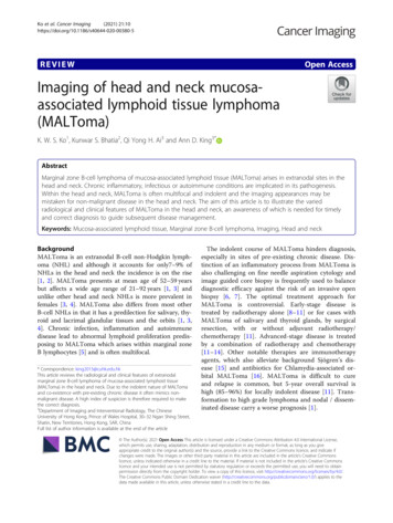
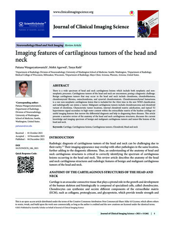
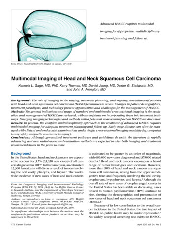
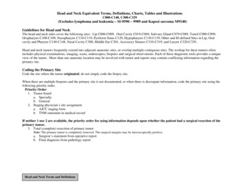
8Clinical Radiation TherapyMReadings: MR in RTwww.siemens.com/magnetom-world-rtpresented a radio-graphically occult primary tumor oncontrast-injected CT. This tumor was however detectedon a T2-weighted MRI as a suspicious heterogeneous signal.Figure 2 shows an example of a large T4aN2c squamous cellcancer of the base of tongue with anterior extension to theextrinsic muscles of tongue. As can be observed, the limitsof the tumor are better defined on MRI.The advantage of MRI for delineation of nodal diseaseis more controversial [19, 20]. Anatomical MRI mayhowever offer an advantage in the particular contextof retropharyngeal lymph nodes. In a study comparingthe diagnostic accuracy of CT versus MRI for detection ofmetastatic retropharyngeal lymph nodes in 38 patientswith nasopharynx or oropharynx cancers, the twomodalities were found to have similar specificity butFigure 2: MRI improving delineation of a base of tongue tumor.2A2B2CThis patient presented with a left T4aN2c squamous cell cancer of the base of tongue. (2A) Planning contrast-enhanced CT scan showsa large base of tongue mass; (2B, C) axial contrast-injected T1-weighted MRI sequence shows a large 5.4 cm base of tongue lesion withanterior extension to the extrinsic muscles of the tongue; anterior, lateral and posterior limits of the tumor are better appreciated on MRI.Figure 3: MRI improving detection and delineation of retropharyngeal lymph nodes.3A3B3CThis patient presented with a T2N2c squamous cell cancer of the oropharynx. While bilateral retropharyngeal lymph nodes are suspectedon contrast-injected CT scan (3A), gadolinium-enhanced T1-weighted MRI allows better visualisation and delineation of bilateralretropharyngeal lymph nodes (3B, C).
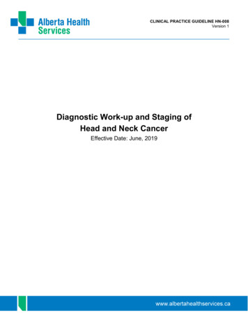
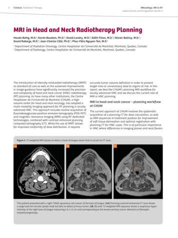
MReadings: MR in RTwww.siemens.com/magnetom-world-rtMRI had a superior sensitivity [21]. Figure 3 shows theexample of a patient presenting with a T2N2c squamouscell cancer of the oropharynx with bilateral retropharyngeallymphadenopathies, better observed on gadoliniumenhanced T1-weighted MRI sequence.The advantage of MRI in HNC RT planning is perhapsmost eloquent in the context of base of skull tumors,where the use of MRI has been associated with not onlydecreased inter-observer variation [22], but also increasedidentification of intracranial and perineural spreads whichare poorly visualized on CT scan [22–24]. In a study byChung et al. [9] involving 258 patients with nasopharyngealcarcinoma, MRI had significantly higher detection rateof intracranial and pterygo-palatine fossa infiltrationscompared to CT, which translated into both improvedtumor delineation and staging. In addition, althoughbone cortex erosion is often better appreciated on CT, MRImay be superior for detection of skull base invasion [25].Figure 4 shows post-operative planning CT and gadoliniumenhanced T1-weighted MRI from a patient with a partiallyresected nasopharyngeal adenoid cystic cancer. The imagesillustrate improved tumor delineation, as well as base ofskull and perineural extensions.Lastly, use of MRI is particularly beneficial in patientspresenting dental artifacts. Dental artifacts are a commonproblem in HNC RT planning, given that poor dentitionRadiation Therapy Clinicalshares risk factors with HNC. High attenuation metalobjects1 such as dental restorations, surgical plates orpins can cause significant scatter artifacts and, as aconsequence, can severely impair CT-based oral cavity ororopharynx primary tumor delineation [26]. Variations inmagnetic field strength at the interface between dentalmaterial and soft tissues can also cause artifacts, but imagequality is affected to a lesser extent [27]. Figure 5 showsplanning CT and gadolinium-enhanced T1-weighted MRI(modified metal artifact protocol) from a patient with aT4N0 squamous cell cancer of the oropharynx. While theprimary lesion is poorly visualized on planning CT, MRIshows a well-defined right oropharynx lesion measuring4.4 cm with extension to the median pterygoid muscle,buccal space, soft palate, and uvula.In conclusion, the use of MRI has become an essential partof HNC RT planning owing to the increased accuracy ofco-registration with planning CT and improved tumorand organs at risk delineation, particularly for oral cavity,oropharynx, and skull base sites. However, planning MRI1The MRI restrictions (if any) of the metal implant must be considered prior to patientundergoing MRI exam. MR imaging of patients with metallic implants brings specificrisks. However, certain implants are approved by the governing regulatory bodiesto be MR conditionally safe. For such implants, the previously mentioned warningmay not be applicable. Please contact the implant manufacturer for the specificconditional information. The conditions for MR safety are the responsibility of theimplant manufacturer, not of Siemens Healthcare.Figure 4: Improved assessment of soft tissue, base of skull, and perineural invasion in a case of nasopharynx adenoid cystic cancer.4A4B4C4D4E4F4G4HThis patient presented with a left T4N0 adenoid cystic cancer of the nasopharynx with cranial nerve involvement and positive biopsy at theclivus, status post partial resection. Planning CT (4A, B) shows a post-operative left parapharyngeal residual lesion; gadolinium-enhancedT1-weighted MRI allows to better appreciate extensions to the retropharynx, medial and lateral pterygoid plates, and clivus (4E, F), as wellas perineural dissemination along extracranial V3 path (4G, H).9
10Clinical Radiation TherapyMReadings: MR in RTwww.siemens.com/magnetom-world-rtFigure 5: MRI improves oropharyngeal tumor delineation in a patient with important artifacts secondary to dental amalgams.5A5B5CThis patient presented with a right T4N0 squamous cell cancer of the oropharynx that can hardly be seen on planning CT due to importantdental artifacts (5A); on T1-weighted MRI, a well delineated 4.4 cm oropharynx lesion, invading the right median pterygoid muscle, joiningthe right buccal space and extending to the soft palate and uvula is demonstrated (5C, D).remains associated with several challenges including themanagement of geometric distortions, the need for MRIcompatible immobilisation devices that maintain imagequality, the prolonged time of acquisition, and the increaseduse of resources. In addition, there remains uncertaintyas to which imaging modality is closest to ground-truth.Considering the low concordance between CT-, FDG-PET-,and MRI-based delineations [22], MRI currently remainsa complementary imaging modality to be used incombination with FDG-PET and physical examination forsafe target volume delineation. Synthetic CT solutions,deriving relative electronic density data from MRI imaging,are currently being evaluated at the CHUM and, in theupcoming years, will likely lead to a more widespreadadoption of MR-based workflow in HNC [28–31]. In addition,the potential value of functional MRI in HNC radiotherapyfor predicting tumor response and spatiotemporalmapping of radioresistant tumor areas is currently underinvestigation [25, 32–35]. With the emergence of morerobust data on functional imaging biomarkers, diffusionweighted and dynamic contrast-enhanced MRI maybecome crucial tools to the promising avenues of dosepainting and adaptive radiotherapy.References1 Ahmed M, Schmidt M, Sohaib A, Kong C, Burke K, Richardson C,et al. The value of magnetic resonance imaging in target volumedelineation of base of tongue tumours--a study using flexiblesurface coils. Radiotherapy and oncology : journal of the EuropeanSociety for Therapeutic Radiology and Oncology. 2010;94(2):161-7.2 Khoo VS, Joon DL. New developments in MRI for target volumedelineation in radiotherapy. The British journal of radiology.2006;79 Spec No 1:S2-15.3 Dirix P, Haustermans K, Vandecaveye V. The value of magneticresonance imaging for radiotherapy planning. Seminars in radiationoncology. 2014;24(3):151-9.4 Paulson ES, Erickson B, Schultz C, Allen Li X. Comprehensive MRIsimulation methodology using a dedicated MRI scanner in radiationoncology for external beam radiation treatment planning.Medical physics. 2015;42(1):28-39.5 Schmidt MA, Payne GS. Radiotherapy planning using MRI.Physics in medicine and biology. 2015;60(22):R323-61.6 Metcalfe P, Liney GP, Holloway L, Walker A, Barton M, Delaney GP,et al. The potential for an enhanced role for MRI in radiationtherapy treatment planning. Technology in cancer research &treatment. 2013;12(5):429-46.7 Prestwich RJ, Sykes J, Carey B, Sen M, Dyker KE, Scarsbrook AF.Improving target definition for head and neck radiotherapy: a placefor magnetic resonance imaging and 18-fluoride fluorodeoxyglucosepositron emission tomography? Clinical oncology (Royal College ofRadiologists (Great Britain)). 2012;24(8):577-89.8 Hanvey S, McJury M, Tho LM, Glegg M, Thomson M, Grose D, et al.The influence of MRI scan position on patients with oropharyngealcancer undergoing radical radiotherapy. Radiation oncology(London, England). 2013;8:129.9 Chung NN, Ting LL, Hsu WC, Lui LT, Wang PM. Impact of magneticresonance imaging versus CT on nasopharyngeal carcinoma:primary tumor target delineation for radiotherapy. Head & neck.2004;26(3):241-6.10 Weber AL, Romo L, Hashmi S. Malignant tumors of the oral cavityand oropharynx: clinical, pathologic, and radiologic evaluation.Neuroimaging clinics of North America. 2003;13(3):443-64.11 Emami B, Sethi A, Petruzzelli GJ. Influence of MRI on target volumedelineation and IMRT planning in nasopharyngeal carcinoma.
MReadings: MR in 920212223242526International journal of radiation oncology, biology, physics.2003;57(2):481-8.O'Daniel JC, Rosenthal DI, Garden AS, Barker JL, Ahamad A,Ang KK, et al. The effect of dental artifacts, contrast media, andexperience on interobserver contouring variations in head and neckanatomy. American journal of clinical oncology. 2007;30(2):191-8.Tien RD, Robbins KT. Correlation of clinical, surgical, pathologic,and MR fat suppression results for head and neck cancer.Head & neck. 1992;14(4):278-84.Phillips CD, Gay SB, Newton RL, Levine PA. Gadolinium-enhancedMRI of tumors of the head and neck.Head & neck. 1990;12(4):308-15.Rasch C, Keus R, Pameijer FA, Koops W, de Ru V, Muller S, et al.The potential impact of CT-MRI matching on tumor volumedelineation in advanced head and neck cancer. Internationaljournal of radiation oncology, biology, physics. 1997;39(4):841-8.Bird D, Scarsbrook AF, Sykes J, Ramasamy S, Subesinghe M,Carey B, et al. Multimodality imaging with CT, MR and FDG-PET forradiotherapy target volume delineation in oropharyngealsquamous cell carcinoma. BMC cancer. 2015;15:844.Rasch C, Steenbakkers R, van Herk M. Target definition in prostate,head, and neck. Seminars in radiation oncology. 2005;15(3):136-45.Brouwer CL, Steenbakkers RJ, Bourhis J, Budach W, Grau C,Gregoire V, et al. CT-based delineation of organs at risk in the headand neck region: DAHANCA, EORTC, GORTEC, HKNPCSG, NCIC CTG,NCRI, NRG Oncology and TROG consensus guidelines. Radiotherapyand oncology: journal of the European Society for TherapeuticRadiology and Oncology. 2015;117(1):83-90.Liao LJ, Lo WC, Hsu WL, Wang CT, Lai MS. Detection of cervicallymph node metastasis in head and neck cancer patients withclinically N0 neck-a meta-analysis comparing different imagingmodalities. BMC cancer. 2012;12:236.Sun J, Li B, Li CJ, Li Y, Su F, Gao QH, et al. Computed tomographyversus magnetic resonance imaging for diagnosing cervical lymphnode metastasis of head and neck cancer: a systematic review andmeta-analysis. OncoTargets and therapy. 2015;8:1291-313.Kato H, Kanematsu M, Watanabe H, Mizuta K, Aoki M.Metastatic retropharyngeal lymph nodes: comparison of CT andMR imaging for diagnostic accuracy.European journal of radiology. 2014;83(7):1157-62.Thiagarajan A, Caria N, Schoder H, Iyer NG, Wolden S, Wong RJ,et al. Target volume delineation in oropharyngeal cancer: impact ofPET, MRI, and physical examination. International journal ofradiation oncology, biology, physics. 2012;83(1):220-7.Gandhi D, Gujar S, Mukherji SK. Magnetic resonance imaging ofperineural spread of head and neck malignancies. Topics inmagnetic resonance imaging: TMRI. 2004;15(2):79-85.Geets X, Daisne JF, Arcangeli S, Coche E, De Poel M, Duprez T, et al.Inter-observer variability in the delineation of pharyngo-laryngealtumor, parotid glands and cervical spinal cord: comparisonbetween CT-scan and MRI. Radiotherapy and oncology: journalof the European Society for Therapeutic Radiology and Oncology.2005;77(1):25-31.Zhang SX, Han PH, Zhang GQ, Wang RH, Ge YB, Ren ZG, et al.Comparison of SPECT/CT, MRI and CT in diagnosis of skull basebone invasion in nasopharyngeal carcinoma. Bio-medical materialsand engineering. 2014;24(1):1117-24.Cooper JS, Mukherji SK, Toledano AY, Beldon C, Schmalfuss IM,Amdur R, et al. An evaluation of the variability of tumor-shapedefinition derived by experienced observers from CT images ofsupraglottic carcinomas (ACRIN protocol 6658). Internationaljournal of radiation oncology, biology, physics. 2007;67(4):972-5.Radiation Therapy Clinical27 Klinke T, Daboul A, Maron J, Gredes T, Puls R, Jaghsi A, et al.Artifacts in magnetic resonance imaging and computedtomography caused by dental materials. PloS one.2012;7(2):e31766.28 Korhonen J, Kapanen M, Keyrilainen J, Seppala T, Tuomikoski L,Tenhunen M. Influence of MRI-based bone outline definitionerrors on external radiotherapy dose calculation accuracy inheterogeneous pseudo-CT images of prostate cancer patients.Acta oncologica (Stockholm, Sweden). 2014;53(8):1100-6.29 Korhonen J, Kapanen M, Keyrilainen J, Seppala T, Tenhunen M.A dual model HU conversion from MRI intensity values within andoutside of bone segment for MRI-based radiotherapy treatmentplanning of prostate cancer. Medical physics. 2014;41(1):011704.30 Gudur MS, Hara W, Le QT, Wang L, Xing L, Li R.A unifying probabilistic Bayesian approach to derive electrondensity from MRI for radiation therapy treatment planning.Physics in medicine and biology. 2014;59(21):6595-606.31 Hsu SH, Cao Y, Huang K, Feng M, Balter JM. Investigation of amethod for generating synthetic CT models from MRI scans ofthe head and neck for radiation therapy. Physics in medicine andbiology. 2013;58(23):8419-35.32 Quon H, Brizel DM. Predictive and prognostic role of functionalimaging of head and neck squamous cell carcinomas. Seminars inradiation oncology. 2012;22(3):220-32.33 Vandecaveye V, De Keyzer F, Nuyts S, Deraedt K, Dirix P,Hamaekers P, et al. Detection of head and neck squamous cellcarcinoma with diffusion weighted MRI after (chemo)radiotherapy:correlation between radiologic and histopathologic findings.International journal of radiation oncology, biology, physics.2007;67(4):960-71.34 Wang H, Balter J, Cao Y. Patient-induced susceptibility effect ongeometric distortion of clinical brain MRI for radiation treatmentplanning on a 3T scanner. Physics in medicine and biology.2013;58(3):465-77.35 Powell C, Schmidt M, Borri M, Koh DM, Partridge M, Riddell A, et al.Changes in functional imaging parameters following inductionchemotherapy have important implications for individualisedpatient-based treatment regimens for advanced head and neckcancer. Radiotherapy and oncology: journal of the EuropeanSociety for Therapeutic Radiology and Oncology. 2013;106(1):112-7.ContactHouda Bahig, M.D.Radiation OncologistCentre Hospitalier de l’Université de Montréal1560 Sherbrooke Street EastH2L 4M1Montreal, QCCanadahouda.bahig.chum@ssss.gouv.qc.ca11
between planning and diagnostic imaging can be major. MRI planning examinations are obtained on a RT-dedicated 70 cm open bore 1.5 Tesla system (MAGNETOM Aera, Siemens Healthcare, Erlangen, Germany). The images are acquired in treatment position with a head and neck thermopl