Transcription
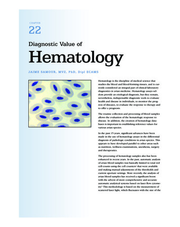
22 Hematology.qxd8/23/20051:05 PMPage 587CHAPTER22Diagnostic Value ofHematologyJAIME SAMOUR, MVZ, P hD, D ipl ECAMSHematology is the discipline of medical science thatstudies the blood and blood-forming tissues, and is currently considered an integral part of clinical laboratorydiagnostics in avian medicine. Hematology assays seldom provide an etiological diagnosis, but they remain,nevertheless, indispensable diagnostic tools to evaluatehealth and disease in individuals, to monitor the progress of diseases, to evaluate the response to therapy andto offer a prognosis.The routine collection and processing of blood samplesallows the evaluation of the hematologic response todisease. In addition, the creation of hematology databases is important in establishing reference values forvarious avian species.In the past 15 years, significant advances have beenmade in the use of hematology assays in the differentialdiagnosis of pathologic conditions in avian species. Thisappears to have developed parallel to other areas suchas nutrition, wellness examinations, anesthesia, surgeryand therapeutics.The processing of hematology samples also has beenenhanced in recent years. In the past, automatic analysisof avian blood samples was basically limited to total redcell counts using the cell countersa that were availableand making manual adjustments of the thresholds andcurrent aperture settings. More recently, the analysis ofavian blood samples has received a significant boostwith the advent of more comprehensive and accurateautomatic analytical systems based on laser flow cytometry.b This methodology is based on the measurement ofscattered laser light, which fluctuates with the size of the
22 Hematology.qxd5888/23/20051:05 PMPage 588C l i n i c a l Av i a n M e d i c i n e - Vo l u m e I ITable 22.1 Materials Needed for Blood SampleCollection Syringe, 1-ml or 3-ml,disposable Needle, 27-gauge to 23gauge, bent, disposable Cotton Alcohol EDTA or heparin pediatric0.5-ml or 1-ml bloodtubes, or sodium citratetubescell, the complexity of the cell (eg, overall shape,nucleus-to-cytoplasm ratio, granulation), and the sizeand shape of the nucleus after blood cells are exposedto a laser beam. The laser flow cytometry unit producesa graphic display containing a total optical white cellcount, white cell differential count expressed in percentage, absolute values and total red cell count, hemoglobin measurement by the cyanmethemoglobin method,thrombocyte count, and white cell count by cell-lysingimpedance measurement of cell nuclei.14 However, theuse of laser flow cytometric technology in avian speciesis not free from deficiencies.There are certain pathological conditions in which thepresence of enlarged thrombocytes (commonly referredto as megathrombocytes) in the blood film appear to bea characteristic hemoresponse. For instance, in thehoubara bustard (Chlamydotis undulata macqueenii),the mean thrombocyte measurements in birds undergoing chronic inflammation (severe shoulder injury as aresult of repeated crashing against the enclosure wall)were 9.22 0.21 µm length and 8.10 0.19 µm widthcompared with 5.47 0.12 µm length and 4.96 0.10µm width in clinically normal birds.9 The mean diameterof lymphocytes in clinically normal houbara bustards is7.7 µm;51 however, there are other species, such as thekori bustard (Ardeotis kori), in which the presence oflarge and small thrombocytes in the same blood filmappears to be normal.51 It is, therefore, probable that asample containing megathrombocytes would yield ahigh lymphocyte count under an automatic analyticalsystem, as it would be impossible for even a sophisticated unit to differentiate between lymphocytes andmegathrombocytes. When dealing with such species, thesoftware would require some adjustments in order toproperly differentiate these cells. This would obviouslyimply the need to carry out extensive calibration basedon repeated manual assessments on a significant number of samples.Furthermore, in certain species it is relatively commonto find large and small lymphocytes in the same bloodfilm.4,12,26,31,35 This phenomenon has been observed inmany psittacine species. Clinically normal kori bustards,for instance, demonstrated a mean diameter for smalllymphocytes of 7.2 0.12 µm, whereas the mean diameter of large lymphocytes was 10.7 0.16 µm.31 There-fore, total white blood cell counts and differential whiteblood cell counts cannot be accepted as reliable in everyclinical case and in every species if the values were estimated by laser flow cytometry. It is, therefore, highly recommended to re-evaluate these samples using manualmethods. Clearly, the clinician must be fully familiar withthe materials and methods of hematology analysis inorder to assess and understand the results.Blood Sample CollectionIt is essential that blood samples be obtained from avianspecies by or under the supervision of a veterinarianwho is experienced with avian venipuncture. The assistant, if one is used, also should be comfortable withrestraint and handling techniques. The techniques usedvary according to personal preferences and the speciesbeing handled. Materials needed for blood sample collection, ie, syringes, slides, tubes, should be labeled inadvance and readily accessible (Table 22.1).METHODThe total blood volume in clinically normal birds is inthe range of 6 to 11 ml per 100 g of body weight.54Thus, a bird weighing 250 g would have approximately15 to 27.5 ml of blood, of which, in a clinically normalindividual, up to 10% (1.5-2.7 ml) can be safely withdrawn without having any detrimental effect on thepatient. However, 0.2 to 0.3 ml of blood is generally sufficient to carry out a comprehensive hematology examination in a bird.In birds, blood samples are commonly collected usingthe right jugular vein (v. jugularis dextra), as this is generally larger than the left jugular vein in most avianspecies (Fig 22.1a). Other preferred sites include thebasilic vein (Fig 22.1b) (v. cutanea ulnaris superficialis)and the caudal tibial vein (v. metatarsalis plantarissuperficialis) (Fig 22.1c).The methodology used for the collection of bloodsamples varies according to the species and the siteselected (Fig 22.1d-h). For example, in long-leggedbirds such as large bustards, cranes and storks, thejugular or caudal tibial veins are very often used. In theauthor’s opinion, blood samples should be collectedfrom the heart or the occipital sinus only if these birdsare under anesthesia and are to be euthanized. It is apoor practice to collect blood samples from clippednails, as cell distribution and cell content is invariablyaffected.The author prefers obtaining blood samples from mostbird species from 200 to 4000 g using a basilic vein
22 Hematology.qxd8/23/20051:05 PMPage 589Chapter 22 D I A G N O S T I C V A L U E O F H E M A T O L O G YFig 22.1a Right jugular being blocked in a love bird and asyringe with a 27 ga needle that has been bent to allowvenipuncture.Fig 22.1c Caudal tibial vein in a love bird.while the bird is in dorsal recumbency, although mostpractitioners in the US dealing with psittacine speciesprefer jugular venipuncture. In most avian species, theoptimal area for collecting a blood sample from a basilicvein is along the medial section of the vein. The preferred side is from the right wing if the practitioner isright-handed, while the left wing is the preferred side ifthe practitioner is left-handed. Venipuncture immediately above the elbow joint is not recommended, ashemostasis is difficult to achieve at this site in mostcases. The application of digital pressure with the thumbat the proximal humerus would help in raising the vein,making it clearly visible running parallel to the externalaspect of the humerus. After separating the feathers andpreparing the site with an alcohol swab, the bent needleis gently inserted into the vein at an approximately 45 angle. The sample can now be collected, taking precaution not to exert high negative pressure while withdrawing with the syringe because this will invariably result inthe collapse of the vein.While withdrawing the sample, it is recommended tocontinue maintaining pressure on the proximal589Fig 22.1b The blocked basilic vein showing the torturousnature of the vein making cannulation with a hypodermic needle difficult at best.humerus to ensure a raised and well-defined vein. Themethod of approaching the basilic vein dorsally byentering under the adjacent tendon prevents hematomaformation because the underlying tissue exerts pressureon the venipuncture site when the needle is withdrawn.It is essential to avoid sudden movements that canalarm the bird and trigger a struggle, as this can easilylacerate the vein and result in a hematoma or, worse, asevere hemorrhage. After collection, a small ball of drycotton should be placed over the venipuncture site andthe wing closed to maintain pressure over the site for afew seconds. It is strongly advisable to check thevenipuncture site before releasing the bird back to itsenclosure to ensure no post-collection hemorrhage hasoccurred. Any sample that contains clots should berejected, as the processing of such samples wouldinvariably lead to imprecise and therefore misleadingresults.STORAGE OF BLOOD SAMPLESAfter collection, the needle should be removed and theblood gently deposited into a 0.5 to 1.0 ml commerciallyavailable pediatric blood storage tube containing theanticoagulant agent ethylenediamine tetra-acetic acid(EDTA) (1.5 mg/ml of blood) or lithium heparin (1.8mg/ml of blood). Squirting the sample through theneedle is a poor practice, as it may cause severe disruption to the fragile blood cells. For general hematologyanalysis, EDTA is the anticoagulant of choice, as it is notpossible to estimate fibrinogen or to count white bloodcells accurately in heparinized samples.In some avian species, however, storing blood samplesin tubes containing EDTA causes progressive red cellhemolysis and is not recommended; in these cases, it ispreferable to use heparinized tubes. This is the casewith some species of Corvidae such as the jackdaw
22 Hematology.qxd5908/23/20051:06 PMPage 590C l i n i c a l Av i a n M e d i c i n e - Vo l u m e I IFigs 22.1d-h Small Birds - Basilic VeinFig 22.1d The ventral surface of the wing of a cadaver withthe covert feathers removed to show the location for superficialulnaris vein lancing (arrow). Lancing the basilic vein in birdsunder 100 gms avoids subcutaneous hematomas and the possibility of death due to exsanguination. The site has a series ofnatural depressions over the vein that serve to allow the blood topool. The depressions are formed by the insertion of the secondary feathers intermittently elevating and depressing the wing dermis just caudal to the border of the flexor carpi ulnaris muscle.Fig 22.1e Lancet used in a finger stick technique for bloodsampling for glucose analysis in humans. The lancet still has themetal tip in place in the plastic cap.Fig 22.1g The lancet readied at the site to lance thesuperficial ulnaris vein.Fig 22.1f Lancet with the cap removed and the 1 mm tipexposed.Fig 22.1h The superficial ulnaris vein has been lanced andblood is being drawn into a micro capillary tube. A cotton ball isapplied to the site until hemostasis is achieved.Fig 22.2a The improved Neubauer counting chamber andthe method for counting red blood cells. The total red blood cellcount is performed by counting the number of cells contained inthe 25 groups of 16 small squares (shaded) at the 4 cornersquares and center square in the central area of the chamber.Closely ruled triple lines (illustrated in the drawing as thick lines)separate these squares.
22 Hematology.qxd8/23/20051:06 PMPage 591Chapter 22 D I A G N O S T I C V A L U E O F H E M A T O L O G Y591tubes is recommended when sending samples to a commercial laboratory for processing using laser flowcytometry.14,19Commercially available collection tubes usually haveprinted labels. A pencil or ballpoint pen is used to enterthe date and identification of the bird, preferably priorto filling the tube with the collected blood sample.Always remember the rule of thumb in clinical pathology: label tubes, not lids.TRANSPORTATION OF BLOODSAMPLESFig 22.2b The counting system. Count cells that touch thecenter triple line (seen here as a thick line) of the rules to theleft and bottom; do not count cells that touch the center tripleline of the rules to the right and top.In avian practice, hematology samples are commonlysent to commercial laboratories for processing (Table22.2). Therefore, it is essential to be familiar with and tosubmit samples in full compliance with current localmail and courier regulations.Table 22.2 Special Considerations When SubmittingBlood Samples to a Laboratory Seal the lid of the tube using waterproof tape in order toprevent any leakage. Wrap the tube using an absorbent packing material, eg,cotton, to soak up any potential leakage and to protect itfrom breakage. Fasten it securely with tape, preferablycommercially available printed tape with the legend“Pathological specimen. Fragile handle with care” orsimilar tape. The tube should then be placed within two leak-proofplastic bags. Fasten the double wrapping securely withprinted tape. Place submission form in a separate plastic bag. The package should then be placed within a postalapproved commercially available transport containermade of aluminum, polystyrene, plastic or cardboard. Attach recipient and sender labels directly to the container using tape, or place container within a paddedenvelope and address it accordingly.Table 22.3 Priorities When ProcessingHematology Samples Blood film (differential,white and red cell morphology, hemoparasites) Packed cell volume (PCV) White cell count (WBC)Hemoglobin (Hb)Red cell count (RBC)Fibrinogen(Corvus monedula) and raven (Corvus corax); Gruidaesuch as the black-necked crowned crane (Balearicapavonina) and gray-necked crowned crane (Balearicaregulorum); Cracidae such as the black curassow (Craxalector); Phasianidae such as the brush turkey (Alecturalathami); Bucerotidae such as the crowned hornbill(Tockus alboterminatus); and the ostrich (Struthiocamelus).3,25 Storing blood samples in sodium citrateHematologyLaboratory AnalysisAlthough the hematology laboratory analysis describedin this chapter were developed primarily for testinghuman blood and are in full compliance with the recommendations of the International Committee forStandardization in Hematology,34 these have beenadapted and used successfully in avian hematology.Ideally, laboratory analysis should be carried out within3 to 4 hours after collection. Many laboratories in theUSA request that a smear be made immediately and sentalong with the EDTA tube. If this is not possible, samples should be refrigerated at 8 to 12 C or within a suitable container for processing within 24 to 48 hours.Refrigerated samples are not ideal for hematology testing, as the cells invariably suffer some changes. Only anexperienced hematologist would be able to differentiatethese changes from true hemoresponses to particularmedical disorders. Samples should not be exposed toextreme environmental conditions or excessive shaking,as this will affect the quality of the sample. Any form ofmouth pipetting with a Thoma pipette or any otherpipette with or without tubing is not acceptable withinclinical laboratory practices.The amount of blood available for testing from smallbirds (eg, 80 g) is very often limited, making it impossible to carry out a full range of analyses. The clinicianshould bear this in mind and request the analysis inorder of priority (Table 22.3).
22 Hematology.qxd5928/23/20051:06 PMPage 592C l i n i c a l Av i a n M e d i c i n e - Vo l u m e I ITHE TOTAL RED BLOOD CELLC O U N T ( R B C X 1 0 12 / L )The total red blood cell count is in itself an importanthematology assay, but it also is essential for the estimation of mean corpuscular volume (MCV) and mean corpuscular hemoglobin (MCH). Many laboratories preferto estimate RBC using an automatic system, as this ismore precise than manual methods. The materials, solutions and method described in Table 22.4, together withFig 22.2a,b, apply to a manual technique.HEMOGLOBIN ESTIMATION (H b g/dl )In avian species, estimation of hemoglobin is hamperedby the presence of nuclei in the erythrocytes. Hemoglobin estimation relies on the colorimetric measurementof hemoglobin released after the lysing of the erythrocytes. Hemoglobin can be estimated using automaticmethods or manual methods (Table 22.5). Commerciallaboratories that estimate hemoglobin using an automatichematology analyzer have to take into consideration thephotometric interference of the free nuclei after lysing ofthe erythrocyte. In the manual method, it is essential toremove the nuclei from the preparation because its presence could yield unreliable results. The nuclei can bedeposited by low-speed centrifugation, but because somehemoglobin remains attached to the nuclei, colorimetricreadings are commonly low. This can be overcome byestimating hemoglobin as cyanmethemoglobin usingalkaline Drabkin’s cyanide-ferricyanide solution or as oxyhemoglobin using ammonia solution. In both cases, theestimation is carried out using a spectrophotometer atthe absorbance reading of 540 nm. A calibration graphshould be made using commercially available hemoglobin standards to express hemoglobin as oxyhemoglobin.Conversely, hemoglobin can be estimated directly as oxyhemoglobin using a commercially available hemoglobinometer. This is the preferred method used and recommended by the author.PACKED CELL VOLUME ESTIMATIONP C V % ( H E M A T O C R I T H ct L/L)Packed cell volume (PCV) is an important hematologicassay because it provides an easy and objective way ofestimating the number of erythrocytes in the sample. Italso is essential for the calculation of the mean corpuscular volume (MCV) and mean corpuscular hemoglobinconcentration (MCHC). In avian species, PCV is best estimated using the microhematocrit method described(Fig 22.3). The use of plain microcapillary tubes ispreferable, since the same tube can be subsequentlyused to estimate fibrinogen.- 100- 90- 80PlasmaB- 70Fibrinogen- 60WBC, thrombocytes- 50- 40CRBC- 30- 20- 10A-0Fig 22.3 Hematocrit reader and method for estimatingpacked cell volume. After centrifugation, position the capillarytube on the rack. Align tube (at the bottom, with the demarcation line between the sealing compound and the red blood cells)with line A. Slide the rack to the right or to the left, align themarginal meniscus at the top of the plasma column with line B.Position line C at the interface of the buffy layer and red cells,and read value on the scale.Mean Corpuscular Values (Red Cell Absolute Values)Mean corpuscular volume (MCV)Mean corpuscular volume (MCV) is the expression of theaverage volume of individual erythrocytes calculatedwith the following formula:MCV (PCV x 10)/RBC MCV femto liters (fl)Mean corpuscular hemoglobin (MCH)Mean corpuscular hemoglobin (MCH) is the expressionof the average hemoglobin content of a single erythrocyte calculated with the following formula:MCH (Hb x 10)/RBC MCH picogram (pg)Mean corpuscular hemoglobin concentration(MCHC)Mean corpuscular hemoglobin concentration (MCHC) isthe expression of the volume within the erythrocyteoccupied by the hemoglobin and is calculated with thefollowing formula:MCHC (Hb x 100)/PCV MCHC (g/L)TOTAL WHITE BLOOD CELL COUNT( W B C X 1 0 9/ L )The total white blood cell count (WBC) is one of themost important hematology assays in the assessment ofhealth and disease in an individual. The WBC also is useful because it is used together with the differential whitecell count to calculate the absolute number of eachwhite blood cell within a blood sample. The materials,solutions and method described below, Table 22.6 andFig 22.4, apply to the technique.THE BLOOD FILMThe examination of stained blood films is the single mostimportant assay in hematology analysis. An adequately
22 Hematology.qxd8/23/20051:06 PMPage 593Chapter 22 D I A G N O S T I C V A L U E O F H E M A T O L O G Y593Table 22.4 Manual Total Red Blood Cell (RBC) Count Hematology TestcMaterials and EquipmentMethod Label sample tubes using a permanent marker. Use an automatic dispenser to transfer 4 ml of eitherformol citrate solution or Natt and Herrick’s solution intosample tube. Wait for 5 minutes to allow working solution to reachroom temperature. Aspirate 20 µl of whole blood from storage tube usingmicropipette, wipe side of pipette tip carefully using tissue and dispense on the side of sample tube to make adilution of 1:200. Avoid touching the distal opening of the pipette tip withthe tissue, as this will cause capillary shift of blood intothe tissue. Avoid immersing the pipette tip into the diluting fluid.This is a poor laboratory practice. Place sample tube in roller mixer and wait for 3 minutes. Clean Neubauer hemocytometer and coverslip using adry, lint-free cloth or laboratory lens tissue. Place coverslip onto hemocytometer and slide gently overit, making sure Newton’s rings (colored interference pattern) appear on both sides of the contact surfaces. Withdraw a small aliquot of the diluted sample using aplain capillary tube. Fill up one side of the hemocytometer by touching gentlythe intersection between coverslip and hemocytometerwith the loaded capillary tube. Avoid air bubbles andunderfilling or overfilling. Place filter paper at the bottom of the Petri dish. Positiontwo toothpicks on either side of the dish. Wet filter paperlightly with distilled water. Rest hemocytometer on toothpicks. Cover Petri dish. Leave for 5 minutes for the cellsto settle down. The hemocytometer is now ready for use. Count cells contained in the four corner and centralsquares in the mid section of the hemocytometer.Following the “L” rule: count cells that touch the centertriple line of the ruling on the left and the bottom sides;do not count cells that touch the center triple line of theruling on the right and the top sides (see Figs 22.2a,b). Calculate red blood cell count using:N/100 RBC x 1012/LAutomatic dispenser, 0-50 mlDisposable sample tube with lid, 5 mlMicropipette, 20 µl and corresponding tipRoller mixerPlain capillary tubesImproved Neubauer hemocytometer and coverslipLaboratory lens tissuePetri dish 8.5 cm diameterFilter paper 8.5 cm diameterToothpickDistilled waterMicroscope, preferably with phase contrast capabilityTest SystemscThe Unopette 365851 system is probably the most popularmethod used for manual red blood cell count in avianspecies. It uses 10 µl of whole blood in 1.9 ml of 0.85%saline, resulting in a 1:200 dilution. The two other commonly used systems are based on using either formolcitrate solution (Dacie’s fluid) or Natt and Herrick’s solution, depending on whether the examination is carried outwith or without phase contrast microscopy. Dacie’s formolcitrate solution is the least known diluting fluid, but oneused and recommended by the author.Working Solutions1. BD Unopette 365851c red blood count manualhematology test2. Natt and Herrick’s solution(for use without phase contrast microscopy)NaClNa2SO43.88 g2.5 gNa2HPO4 12 H2O2.91 gKH2PO40.25 gFormaldehyde 40%7.5 mlMethyl violet 2BDistilled water0.1 gto 1000 mlNote: Allow solution to stand overnight. Filter before use.3. Formol citrate solution or Dacie’s fluid(for use with phase contrast microscopy)Formaldehyde 10%10 mlTrisodium citrate31.3 gDistilled water100 mlNote: Refrigerate at 8 to 12 C.Note: N number of cells counted in 160 small squares.
22 Hematology.qxd5948/23/20051:08 PMPage 594C l i n i c a l Av i a n M e d i c i n e - Vo l u m e I ITable 22.5 Hemoglobin EstimationMaterials and Equipment Automatic dispenser, 0-50 ml Disposable sample tube withlid, 5 ml Micropipette, 20 µl and tip Roller mixer ToothpicksCuvette, 10 mm2Laboratory lens tissueHemoglobinometerWorking SolutionAmmonia solutionAmmonia solution(0.88 specific gravity)Distilled water4 mlto 1000 mlNote: Refrigerate at 8 to 12 C.Method Label sample tubes using a permanent marker. Use an automatic dispenser to transfer 4 ml of ammoniasolution into sample tube. Wait for 5 minutes to allow working solution to reachroom temperature. Aspirate 20 µl of whole blood from storage tube usingmicropipette, wipe side of pipette tip carefully using tissue and dispense on the side of sample tube. Avoid touching the distal opening of the pipette tip withthe tissue, as this will cause capillary shift of blood intothe tissue. Avoid immersing the pipette tip into the diluting fluid.This is a poor laboratory practice. Place sample tube in roller mixer and wait for 3 minutes. Decant approximately 3.5 ml of the diluted bloodinto cuvette. Remove cell nuclei jelly using toothpicks. Do not touch the clear reading walls of the cuvettewith bare fingers. Clean clear reading walls of cuvette using laboratorylens tissue. Zero hemoglobinometer using ammonia solutionas blank. Reading expressed as Hb g/dl.prepared blood film provides the differential white bloodcell count and absolute white blood cell count, thethrombocyte count and the hemoparasite examination.Preparation of the Blood FilmBlood films can be made from a drop of fresh, non-anticoagulated blood directly from the tip of the syringe.Conversely, films can be made from blood stored inEDTA within 2 to 3 hours after collection. There are twogenerally accepted methods for the preparation of bloodfilms in hematology: the slide-to-slide technique (Fig22.5, Table 22.7) and the coverslip-to-slide technique(Fig 22.6, Table 22.8). A two cover-slip technique is notdescribed here. The most popular method among avianFig 22.4 Improved Neubauer counting chamber and themethod for counting white blood cells. The total white blood cellcount is performed by counting the number of cells contained in4 groups (shaded areas) of 16 large squares at the four cornersquares of the chamber. Closely ruled triple lines (illustrated inthe drawing as thick lines) separate these squares.Fig 22.5 The slide-to-slide technique. Move spreader slidebackward to gently touch the drop of blood, allowing it to runacross the edge of the slide. Move forward to make smear. Moveslowly if blood runs slowly, move quickly if blood runs quickly.clinicians is the coverslip-to-slide technique, as smudging of blood red cells is generally minimized.Fixation and Staining of the Blood FilmIt is commonly accepted that blood films can be prepared and be fixed and stained at a later date. This isincorrect; blood films should at least be fixed immediately after preparation, particularly if made in a hot andhumid environment or under cold and freezing conditions. Blood films should not be exposed to direct sunlight, moisture of any kind or vapor from chemicals(formaldehyde in particular), as this would invariablyaffect cell morphology.
22 Hematology.qxd8/23/20051:08 PMPage 595Chapter 22 D I A G N O S T I C V A L U E O F H E M A T O L O G Y595Table 22.6 Total White Blood Cell (WBC) Count Hematology TestMaterials and EquipmentMethod Label sample tubes using a permanent marker. Use an automatic dispenser to transfer 1.9 ml of 1%ammonium oxalate solution into sample tube. Wait for 5 minutes to allow working solution to reachroom temperature. Aspirate 100 µl of whole blood from storage tube usingmicropipette, wipe side of pipette tip carefully using tissue and dispense on the side of sample tube. Avoid touching the distal opening of the pipette tip withthe tissue, as this will cause capillary shift of blood intothe tissue. Avoid immersing the pipette tip into the diluting fluid.This is a poor laboratory practice. Place sample tube in roller mixer and wait for 3 minutes. Clean Neubauer hemocytometer and coverslip using alaboratory lens tissue or dry, lint-free cloth. Place coverslip onto hemocytometer and slide gently overit, making sure Newton’s rings (colored interference pattern) appear on both sides of the contact surfaces. Withdraw a small aliquot of the diluted sample using aplain capillary tube. Fill up one side of the hemocytometer by gently touchingthe intersection between coverslip and hemocytometerwith the loaded capillary tube. Avoid air bubbles andunderfilling or overfilling. Place filter paper at the bottom of the Petri dish. Positiontwo toothpicks on either side of the dish. Lightly wet filterpaper with distilled water. Rest hemocytometer on toothpicks. Cover Petri dish. Leave for 5 minutes for the cellsto settle down. The hemocytometer is now ready for use. Count cells contained in the four outer large squares ofthe hemocytometer. Calculate total white blood cell count using:N/20 WBC x 109/LAutomatic dispenser, 0-50 mlDisposable sample tube with lid, 5 mlMicropipette, 100 µl and tipRoller mixerPlain capillary tubesImproved Neubauer hemocytometer and coverslipLaboratory lens tissuePetri dish, 8.5 cm diameterFilter paper, 8.5 cm diameterToothpicksDistilled waterMicroscope, preferably with phase contrast capabilityTest SystemsThe Unopette 365877d system was originally developed forthe estimation of eosinophils in human hematology, but ithas proved useful for determining the total white cell countin avian species. This system uses 25 µl of whole blood into0.775 ml of 1% Phloxine B diluent resulting in a 1:32 dilution, and is the system used by most practitioners in theUSA.3,19 The method described below is based on the use ofammonium oxalate solution, which is the method used andrecommended by the author.Working Solutions1. BD Unopette 365877d eosinophil countmanual hematology testd2. Ammonium oxalate solution 1%Ammonium oxalateDistilled water10 gto 1000 mlNote: Refrigerate at 8 to 12 C.Note: N number of cells counted in 64 small squares.Table 22.7 Method for Slide-to-Slide
Aug 22, 2013 · Diagnostic Value of Hematology is the discipline of medical science that studies the blood and blood-forming tissues, and is cur-rently considered an integral part of clinical laboratory diagnostics in avian medicine. Hematology assays sel-dom provide an etiological diagnosis, but they remain, nevertheles
