Transcription
LAB 4 – Microscopy & CellsObjectives1. Explain each part of the compound microscope and its proper use.2. Examine a variety of cells with the compound microscope and estimate cell size.3. Examine larger specimens with the stereoscopic dissecting microscope.IntroductionIn this laboratory you will be learning how to use one of the most important tools in biology – thecompound light microscope – to view a variety of specimens. You will also use a slightly differenttype of light microscope called a stereoscopic dissecting microscope. The first lens used tomagnify things was developed in the first century A.D. These were pieces of glass shaped in aconvex form – thicker in the middle and tapering off to the sides – and were the first magnifyingglasses that could increase the image of an object about 10 to 20X. The creation of glass lensesimproved dramatically at the end of the 16th century, vastly improving the magnifying power. By1609, Galileo Galilei refined the methods of lens making in an effort to view objects in the sky.About half a century later, the Dutchman Anton van Leeuwenhoek further improved the art oflens making, allowing him to view objects in pond water that had never been viewed by humans– microorganisms – life at a tiny level. At the same time, an English physicist named Robert Hookeimproved the technology of van Leeuwenhoek and confirmed the existence of tiny organisms inpond water. He also famously examined a piece of cork and observed tiny boxes arranged in sucha way that they looked like the “cells” (rooms) in a monastery if you removed the roof and lookedin from above. Today the best compound light microscopes are able to magnify objects up to2,500X without losing their resolution – the sharpness of the image itself.Part 1: THE COMPOUND LIGHT MICROSCOPEThe Parts of the Compound Light MicroscopeExercise 1A – Getting familiar with the microscopeYou will first get acquainted with the major parts of the compound light microscope before learning theproper way to use it. Get a microscope from the cabinet below your lab bench, being sure to handle it bythe arm and base (refer to image on page 2), and place it on the bench in front of you. Remove the coverand place it below, out of the way, and then plug in the microscope. The ocular lens (eyepiece) and stageshould be facing you. Read the description of each part of your microscope on the next two pages beingsure to follow all instructions, and then complete the matching exercise on your worksheet.51
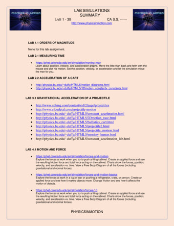
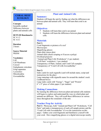
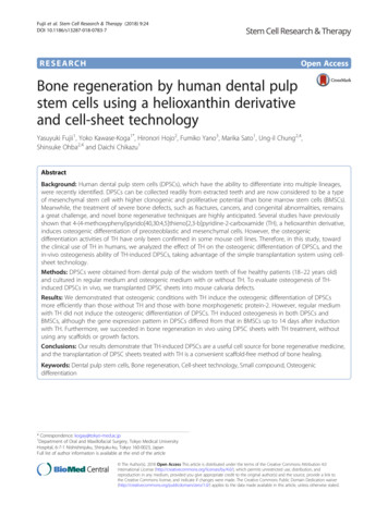
ocular lensesarmrevolving nosepieceobjective lensmechanicalstage knobsstage rse focusfine focusOCULAR LENS (eyepiece) – Your microscope will have either one (monocular) or two (binocular) ocularlenses. These are the lenses you will look through when examining a specimen with the microscope. Takea look at the side of your ocular lens and you will notice a label of “10X”. This indicates that each ocularlens magnifies the image by a factor of 10 or 10X.OBJECTIVE LENSES – Notice the set of objective lenses on the revolving nosepiece. These lenses allowyou to change the degree of magnification. Some of our microscopes have four objective lenses while othershave only three. The degree of magnification for each objective lens is indicated on its side. Let’s take alook at each progressing from the shortest to longest objective lenses, being sure to rotate the revolvingnosepiece to click each objective lens into position above the stage before examining it:4X – This objective magnifies the image by a factor of 4. It is referred to as the “scanningobjective” since it is used to scan the slide to locate the specimen before viewing it athigher magnification. Your microscope may not have this objective lens, in which caseyou can begin with the 10X objective.10X – This objective magnifies the image by a factor of 10 and is referred to as the “low power”objective.40X – This objective magnifies the image by a factor of 40 and is referred to as the “high power”objective.100X – This objective magnifies the image by a factor of 100. It is referred to as the “oilimmersion objective” since it requires a drop of immersion oil on the slide to providegood resolution. You will not be using this objective lens.For now, make sure that the low power objective is clicked into position above the stage, and keep in mindthat you will only be using the low power and high power objectives. Also keep in mind that the totalmagnification of any image you see through the ocular lens is the product of the objective and ocular lensmagnifications (for example, when using the lower power lens the total magnification is: 10X ocular lens x10X low power objective lens 100X).52
STAGE and STAGE CLIP – The stage is the flat surface upon which you will place each slide you will examine.Notice that there is a moveable stage clip that can be used to secure the slide on the stage. Open and closethe stage clip to see how it will snugly hold your slide in position.MECHANICAL STAGE KNOBS – To move the slide on the stage when it is secured in the stage clip, you willuse the mechanical stage knobs on the underside of the stage to move the slide backward/forward andright/left. Adjust each knob to see how one knob controls backward/forward movement and the otherknob controls right/left movement.COARSE FOCUS and FINE FOCUS KNOBS – In order for a specimen on a slide to be in focus, the distancebetween the specimen and the objective lens must be just right. The coarse focus knob, the larger of thetwo, will move the stage or objective lens (depending on the microscope) up and down quickly and quitevisibly, altering the distance between them. It is very important that the coarse focus knob is only used withthe low power or scanning objective lenses, otherwise the microscope or objective lenses could be damaged.Adjust the coarse focus knob to observe how quickly the focal distance changes. In contrast, the fine focusknob will move the stage or objective lens such a small amount that it is hardly noticeable to the naked eye.This is the knob you will use to get the perfect focal distance so the image will be crystal clear.CONDENSER LENS – Just underneath the stage is the condenser lens. This lens serves to capture and focuslight from the lamp below onto the slide mounted on the stage. On many microscopes the condenser lenscan be adjusted up or down with a knob beneath the stage. Examine the condenser on your microscope tosee if it is adjustable. If so, be sure to adjust it as high (close to the stage) as possible since, for our purposes,this is where it should be set.DIAPHRAGM – The diaphragm is located within the condenser and is one of the most important pieces ofthe microscope, though it is often neglected by many students. The diaphragm allows you to adjust theamount of light passing through the slide by adjusting the diaphragm lever. Most of the time thediaphragm will be all the way open to allow the maximum passage of light. However it is important to adjustthe diaphragm at times to reduce the amount of light passing through your specimen should the image betoo bright or dim, and also to increase the contrast to allow you to see the specimen more easily againstthe background. For now, open the diaphragm all the way, and when using the microscope, do not forgetto use the diaphragm.LAMP – The lamp emits light to illuminate the specimen so that you can actually see something.BASE and ARM – The base is the bottom of the microscope that sits on the table, and the arm is the verticalframework ascending from the base along the back of the microscope. When handling the microscopealways hold the arm while supporting the base with your other hand.Proper Use of the Compound Light MicroscopeExercise 1B – Steps to follow when using the microscopeIf you really want to be able to see a specimen on a slide, you must follow the steps on the next page everytime you look at a new slide. The microscope will be your friend if you always use the following steps intheir proper order. Before you begin, be sure your microscope is plugged in and the power is “on”.53
Step 1. Get a slide of the letter “e” from the tray on the side counter. This an example of a preparedslide, a slide that is already made for you and meant to be reused.(i.e., don’t dispose of it, please return it to the tray when you are finished!)Step 2. Use a piece of lens paper to clean any smudges (fingerprints, grease, etc.) off the slide. Place theslide on a white piece of paper find the specimen (the letter “e”) on the slide with your nakedeye, noticing its location and orientation.Step 3. Lock the scanning objective lens into place (it should “snap” into place) if you have not alreadydone so. You will always, always, always start with either the low power or scanning objectivewhen you want to view a slide since these lenses provide the largest field of view.Step 4. Use the coarse focus knob to move the stage (or objective lens) so that they are as far apart fromeach other as possible. Open the stage clip and place the slide snugly in the corner of the stageclip (make sure the slide is completely flat) before releasing the clip to hold the slide firmly inplace. Then use the mechanical stage knobs to position the slide so that the specimen (i.e., letter“e”) is centered over the condenser and the light that passes through it.Step 5. Next, using the coarse focus knob once again, move the slide and objective lens as close togetheras the knob will allow.(NOTE: To this point, you have not yet looked into the oculars. This may be surprising,but this is the proper way to use a microscope so that you will actually see something!)Step 6. Now, look into the ocular lenses. Using the coarse focus knob, SLOWLY increase the distancebetween the slide and objective until the specimen is in focus.If the light is too intense, adjust the diaphragm lever (or dial near the lamp if present)until the light level is comfortable before trying to locate the specimen.If you have difficulty locating and focusing on your specimen (the letter “e”), make sure that it isproperly centered and you may need to adjust the course focus more slowly. If you still can’tlocate it, ask your instructor for assistance.Step 7. Adjust the diaphragm lever so there is sufficient contrast between the specimen and thebackground, closing it no more than is necessary. This step is especially important for livespecimens since you may not be able to see them otherwise.Step 8. Now use the fine focus knob to get the specimen in proper focus. You should now be able to seethe object clearly. Before going to the next step (increasing the magnification), be sure to centeryour specimen in the field of view as best you can.Step 9. Now that you have centered and focused the object as best you can using the scanning objective,rotate the low power objective into place over the slide being sure it “clicks” into position. Usethe fine focus knob (NOT the coarse focus) to bring the object into perfect focus. Repeat withthe high power objective if you want a more magnified image.(REMINDER: Only use the coarse adjustment knob with the low power and scanning objectives)FOLLOW THESE STEPS EVERY TIME YOU WANT TO VIEW A NEW SLIDEAND YOU WILL BECOME A GOOD MICROSCOPIST!54
Part 2: PROPERTIES OF LIGHT MICROSCOPYIn this section we will focus on some of the key properties relating to light microscopy. To helpyou understand each property you will first read an explanation and then do an exercise toillustrate that particular property. Let us begin with the property of magnification Total MagnificationThe total magnification of an image is quite simple – it is the product of the ocular lensmagnification times the magnification of the objective lens you are using:magnification of ocular x magnification of objective total magnificationFor example, if the ocular lens magnifies the image by a factor of 10 (10X), and the objective lensmagnifies the image by a factor of 50 (50X), the total magnification of the image is 500X:10X x 50X 500XMany students make the mistake of adding the two magnifications, so remember that totalmagnification is the product (multiplication) of the ocular and objective lens magnifications.Exercise 2A – Determining total magnificationOn your worksheet, calculate the total magnifications for the examples given, then calculate the totalmagnification when using each of the objective lenses on your own microscope.Field of ViewThe field of view (FOV) is the actual “circle” you see when looking in the microscope. Althoughthis circular field of view appears to be the same no matter which objective lens you are using,this is not the case. The circular area you are actually viewing will decrease as you increase themagnification:total magnificationfield of view40X100X450X1000X55
A good analogy is to imagine yourself viewing the Earth from space as you gradually move closerand closer to Mission College. Initially your field of view is the entire western hemisphere, but asyou approach the Earth’s surface your field of view will progressively shrink to encompass thewestern United States, Southern California, the San Fernando Valley, Sylmar, etc. Although yourfield of view is shrinking, the image in your field of view is becoming increasingly magnified. Thisis really no different than looking into your microscope at increasing levels of magnification.It is also useful to know the diameter of the field of view (FOV diameter) at a particularmagnification, since you can use this information to estimate the size of the specimen you areviewing. The FOV diameter at low power for your microscope (100X) is 1.8 mm. Using this FOVdiameter, you can calculate the FOV diameter at other magnifications. This is done by multiplyingby the ratio of the magnifications:known FOV diameter x total mag. (known FOV) unknown FOV diametertotal mag. (unknown FOV)If you want to know the FOV diameter at 500X, you
proper way to use it. Get a microscope from the cabinet below your lab bench, being sure to handle it by the arm and base (refer to image on page 2), and place it on the bench in front of you. Remove the cover and place it below, out of the way, and then plug in the microscope. The ocular lens (eyepiece) and stage should be facing you. Read the description of each part of your microscope on the next two pages being