Transcription
Hindawi Publishing CorporationSarcomaVolume 2015, Article ID 517657, 7 pageshttp://dx.doi.org/10.1155/2015/517657Clinical StudyCharacteristics and Patterns of MetastaticDisease from ChordomaVictoria A. Young,1,2 Kevin M. Curtis,3,4 H. Thomas Temple,1,2 Frank J. Eismont,1Thomas F. DeLaney,5 and Francis J. Hornicek61Department of Orthopaedics, University of Miami Miller School of Medicine, P.O. Box 016960 Miami, FL 33101, USAUniversity of Miami Tissue Bank, 1951 NW 7th Avenue, Suite 200, Miami, FL 33136, USA3Department of Biochemistry & Molecular Biology, University of Miami Miller School of Medicine, P.O. Box 016960 Miami,FL 33101, USA4Geriatric Research, Education, and Clinical Center and Research Service, Bruce W. Carter Veterans Affairs Medical Center,Miami, FL 33125, USA5Department of Radiation Oncology, Massachusetts General Hospital, Harvard Medical School,Francis H. Burr Proton Therapy Center (FHBPTC), 55 Fruit Street, Boston, MA 02114, USA6Department of Orthopedic Surgery, Massachusetts General Hospital, Harvard Medical School, Orthopaedic Associates,55 Fruit Street Yawkey 3B, Boston, MA 02114, USA2Correspondence should be addressed to Victoria A. Young; victoria.young@northwestern.eduand H. Thomas Temple; htemple@nova.eduReceived 26 October 2015; Revised 8 December 2015; Accepted 9 December 2015Academic Editor: Silvia StacchiottiCopyright 2015 Victoria A. Young et al. This is an open access article distributed under the Creative Commons AttributionLicense, which permits unrestricted use, distribution, and reproduction in any medium, provided the original work is properlycited.Chordoma is a rare, slow-growing malignant tumor arising from notochordal remnants. A retrospective review of patient recordsat two major referral centers was undertaken to assess the incidence, location, and prognostic factors of metastatic diseasefrom chordoma. 219 patients with chordoma (1962–2009) were identified. 39 patients (17.8%) developed metastatic disease, mostfrequently to lung ( 50%). Median survival from the time of initial diagnosis was 130.4 months for patients who developedmetastatic disease and 159.3 months for those who did not (𝑃 0.05). Metastatic disease was most common in the youngestpatients (𝑃 0.07), and it was 2.5 times more frequent among patients with local recurrence (26.3%) than in those without (10.8%)(𝑃 0.003). Patient survival with metastatic disease was highly variable, and it was dependent on both the location of the tumorprimary and the site of metastasis. Metastasis to distal bone was the most rapid to develop and had the worst prognosis.1. IntroductionChordoma is a rare, slow-growing, malignant tumor arisingfrom notochordal remnants. Chordoma accounts for 1–4% ofprimary bone tumors [1, 2] and has an estimated incidenceof 8/10,000,000/year [3]. Chordoma occurs primarily in thefifth and sixth decades of life and more frequently affectsmales (2 : 1), yet demographics can vary based on site ofpresentation [2–4]. Due to a slow growth rate and nonspecificpresentation, diagnosis is delayed 1.5 years on average [5],leading to more advanced disease by the time treatmentis initiated [2]. Despite its slow progression, patients withchordoma have a poor prognosis, likely due to tumor locationand its propensity toward local recurrence (68%) [5–9],resulting in diminished 5-year (67%) and 10-year (40%)survival [3].Since its description 150 years ago there has been relativelysparse clinical data collected on chordoma. The few studiesdescribing metastatic disease in chordoma report an estimated frequency of metastasis ranging from 3 to 48% [4] andsuggest that the presence of metastasis increases the risk oftumor-related death [6]. Metastasis is relatively uncommon,with lung representing the most common site. Metastases toliver, bone, lymph nodes, skin, subcutaneous tissue, muscle,peritoneum, heart, pleura, spleen, kidney, bladder, pancreas,and brain have also been described [6, 9–11]. Factors that
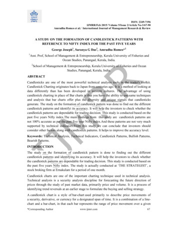
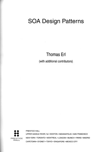
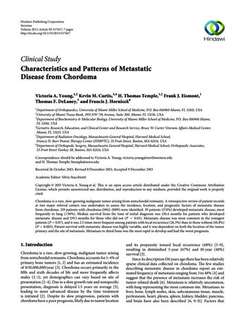
2Survival function1.00.8Cum survivalmay be predictive of metastasis include local recurrence,large tumor size, inadequate surgical margins, necrosis, longclinical duration, and exposure to high dose radiation [4, 9,11]. However, a recent study reported no significant differencein likelihood of metastasis based on age, gender, tumorlocation, or radiation therapy [9]. Earlier studies suggest arelationship between primary tumor location and metastaticdisease risk [4, 12, 13], though the strength of this correlationis uncertain as these reports contain conflicting data andrelatively few patients [6]. To better understand patterns ofmetastatic disease from chordoma, we conducted a retrospective review of 219 patients with primary chordoma, 39 ofwhom developed metastatic disease.Sarcoma0.60.40.20.002. Methods3. Results3.1. Patient Population and Disease Presentation. 219 patientswith chordoma presented to the Orthopaedics Services attwo major referral centers in a 46-year span (Table 1). There100 150 200 250 300 350Onset-death or endpoint (months)400450MetastasisYesNoFigure 1: Survival distribution stratified by presence of metastaticdisease. Diamonds represent censored data points.Survival functions1.00.8Cum survival2.1. Patient Data Collection. A retrospective review of allcases of chordoma diagnosed from November 1962 to October 2009 at two major US referral centers, Mass GeneralHospital, Boston, MA, and University of Miami, Miami, FL,was undertaken. Patient characteristics were recorded andcharts, radiographs, and histological slides were reviewed forsites of primary and metastatic lesions. Bone scans, MRI,chest radiographs, and/or CT scans were also reviewed. Thefollowing data were obtained or defined: patient age, gender,site of primary tumor, local recurrence, metastasis, site ofprimary metastasis, presence of multiple metastasis, site ofsecondary metastasis, and survival. Due to the rare incidenceof chordoma and lack of published large scale retrospectivestudies, we focused only on the prognostic factors of thedisease and did not include the effects of treatment or causeof patient death.Frequencies and descriptive statistics were obtained foranalysis; categorical variables were examined using chisquare and Fisher’s exact tests, where appropriate. KaplanMeier survival curves with log rank analysis were performedfor survival, time from onset to metastasis, and time frommetastasis to death. Survival was defined as time from diagnosis to date of death. Patients surviving beyond the studycutoff date were censored. There was sufficient data to analyse219 patients, yet only 177 cases of primary chordoma couldbe used for survival analysis (see Figure 1 and Table 2(a)).Excluded from the analysis were individuals known to bedeceased, but for which no date of death could be defined,and those lost to follow-up whose status (living or deceased)was unknown. Excluded from analyses involving primarytumor location were individuals with unknown, multiple, oratypical sites of presentation (see Figure 2 and Table 5(a)).Censored observations and data points are presented asvertical hashmarks in the survival curves. This study wasapproved by the institutional review boards of the respectiveinstitutions: UM/JMH IRB 20100483 and MGH IRB et-death or endpoint (months)Primary tumor locationCervicalLumbar350SacralThoracicFigure 2: Survival distribution stratified by primary tumor location.Diamonds represent censored data points.were 137 (63%) men and 81 (37%) women. Age at diagnosiswas divided into quartiles, with most individuals in the thirdquartile (median age at diagnosis 57, range: 3–88 years). Themost frequent site of presentation was sacral (60.7%). A fewprimary tumors were documented in the clivus/skull base(1.8%), and the remaining were observed in the mobile spine
Sarcoma3Table 1Patientcharacteristics𝑛 (%)Total number219GenderMaleFemale137 (63)81 (37)Age (years)MedianRange573–88Age quartilesQ1: (3–24 yrs)Q2: (25–46 yrs)Q3: (47–68 yrs)Q4: (69–88 yrs)9 (4.1)49 (22.4)116 (53)45 (20.5)ClivusCervical spineLumbar spineThoracic spineSacrumMultiple4 (1.8)22 (10.0)38 (17.4)16 (7.3)133 (60.7)4 (1.8)Primary only107 (47.4)Presenting siteExtension ofdiseaseRecurrent ( ) metastasisRecurrent ( ) metastasisTotal locally residual/recurrentdiseaseMetastasis ( ) local recurrenceMetastasis ( ) local recurrenceTotal metastatic732699 (45.2)261339 (17.8%)(34.7%), which is further subdivided into lumbar (17.4%),cervical (10.0%), and thoracic (7.3%).Chordoma location at initial presentation trended towarda dependence on patient age (𝑃 0.06). The youngestpatients (Q1: 3–24 yrs old) tended to have atypical primarytumor locations, with chordoma appearing in the cervicalspine (50%, 𝑛 4) more frequently than the sacrum (37.5%,𝑛 3) and clivus (12.5%, 𝑛 1). The remaining age quartiles(Q2–Q4: 25 yrs old) were more likely to have involvementin the typical/classical sacrococcygeal region (62.5–64.4%)than in the lumbar (12.5–21.4%), cervical (6.7–14.6%), thoracic spine (7.1–8.9%), and clivus (0.9–2.2%). Patients withyoungest age of presentation (Q1) were more likely to befemale (56% F : 44% M) as compared to older quartiles Q2–Q4 where males predominated (Q2–Q4 27–38% F : 60–73%M). Additionally, patients with primary tumors of the cervicalspine were more likely to be female as well (54.4% F : 45.5%M). Clival chordomas were found to occur equally among thegenders, with sacral, thoracic, and lumbar chordoma morefrequently occurring in males (64.4–68.8% M : 31.3–35.6% F).3.2. Overall Survival. Median overall survival was 140.5months (95% CI 115.7–165.3) (Table 2(c)). The duration ofsurvival differed based on site of presentation; individualswith primary tumors of the cervical and thoracic spine hadthe shortest median survival at 74.7 and 76.8 months, respectively. Those with primary tumors of the lumbar spine hadmedian survival of 126.7 months, while those with primarytumors of the sacrum had the longest median survival at 159.3months.While median survival did not differ based on presenceof local disease recurrence alone, median survival amongpatients with metastatic disease (Table 2(a)) was significantly(𝑃 0.05) (Table 2(b)) lower (130.4 months [95% CI 111.4–149.5]) than patients without metastatic disease (159.3 months[95% CI 123.9–194.6]) (Figure 1, Table 2(c)).3.3. Metastasis. Of the 39 (17.8%) patients who developed metastatic disease, lung was most frequently involved(53.8%). Other sites of primary metastasis include distalbone (20.5%), soft tissue (15.4%), and liver (7.7%) (Table 3).Metastatic disease was most frequent among the youngestpatients (Q1 44%, Q2 16%, Q3 20%, and Q4 9%) (𝑃 0.07)and 2.5 times more frequent among patients with local recurrence (26.3%) than without (10.8%) (𝑃 0.003). Tumorspresenting in the cervical spine rarely became metastatic(1 [4.5%] of 22), whereas tumors in the remainder of themobile spine demonstrated metastatic rates ranging from 17to 32% (Table 4). Throughout the course of their illness, 99patients (45.2%) developed locally recurrent disease (Table 1),with lumbar chordoma being the most likely to recur. Ifdisease was first locally recurrent, these lesions took longerto metastasize (67.2 months versus 20.5 months) (𝑃 0.001) and progressed more slowly from the diagnosis ofmetastasis to death (22.1 months) than disease that wasdirectly metastatic (4.5 months).The time from onset to metastasis was found to differsignificantly, depending on the site of presentation (𝑃 0.013). The shortest duration from onset to metastasis wasfound among patients with primary tumors located in thecervical spine (16.0 months). Time to metastasis of primarytumors located in the thoracic spine, lumbar spine, andsacrum was 22.0 months, 49.2 months, and 58.3 months,respectively. The longest time to metastasis was 120.1 months,which was observed in a patient with a primary clivalchordoma.3.4. Survival among Patients with Metastatic Disease. Amongpatients with metastasis (Table 5(a)), survival differed basedon primary tumor location (𝑃 0.05) (Table 5(b)). Patientswith metastatic tumors originating in the cervical spine hadthe shortest median survival (20.4 months, 𝑛 1), followedby tumors of the thoracic spine (70.1 months, 𝑛 3), lumbarspine (104.9 months, 𝑛 4), and sacrum (130.4 months, 𝑛 16) (Figure 2, Table 5(c)).Survival differed significantly based on site of metastasis(𝑃 0.01) (Tables 6(a) and 6(b)). Tumors that firstmetastasized to bone had the shortest median survival at46 months, followed by liver at 72 months (Table 6(c)).Sites of primary metastasis with the longest median survivalwere lung and soft tissue, at 130 months and 132 months,respectively (Figure 3, Table 6(c)).
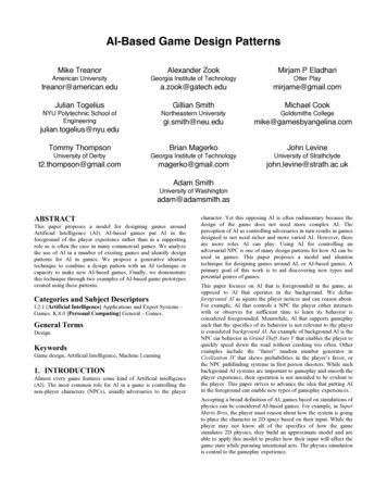
4SarcomaTable 2: Survival distribution stratified by presence of metastatic disease.(a) Case processing summary of patients included in analysisMetastasisNoYesOverallTotal 𝑁𝑁 of 8.5%52.0%(b) Overall statistical comparison and test of equality of survival distributionsChi-Square3.682Log rank (Mantel-Cox)df1Sig.0.055(c) Means and medians for survival time. Estimation is limited to the largest survival time if it is censoredMetastasisMean95% Conf. intervalLower bound Upper boundEstimateMedian95% Conf. intervalStd. error Lower bound Upper boundEstimateStd. 153.3202.2140.512.7115.7165.3Table 3: Primary metastasis.Total 00.8Cum survivalPrimary metastatic siteLungLiverBoneSternumSoft tissueSite not listedTotalSurvival functions0.60.44. Discussion4.1. Disease Presentation. In agreement with previous studies, individuals presenting with the typical sacrococcygealchordoma tended to be males in the 6th and 7th decades oflife, with a relatively good prognosis. Our data demonstrate,also in agreement with previous studies, that patients over 25years of age were more likely to be male and to present withchordoma in the sacral, thoracic, or lumbar spine [12, 14].The literature also describes a younger, atypical populationof chordoma patients with cranial primaries [3, 15], but ourdata suggest that this atypical, young female predominantgroup (under 25 yo.) has a predisposition toward aggressivechordomas of the cervical spine. And although this atypicalgroup of patients represents less than 5% of the chordomapopulation, cervical chordoma presentation has the lowestsurvival of 74.7 months. Thus, in concordance with priorstudy, our data reproduce two distinct groups: (1) an atypicalgroup of young female patients who predominantly presentwith a more aggressive form of cervical chordoma, and (2)a more common, typical group composed predominantlyof older male patients with sacral, thoracic, and lumbar0.20.0050100150200Onset-death or endpoint (months)Location of primary metastasisBoneLiver250LungSoft tissueFigure 3: Survival distribution among patients with metastasisstratified by location of metastatic disease. Diamonds representcensored data points.tumors. It is unclear from our data whether the relativelypoor prognosis of the atypical female cervical chordomas issecondary to more aggressive inherent tumor properties or tothe anatomical constraints imposed on surgical interventionin the cervical spine.
Sarcoma5Table 4: Patterns of primary metastasis.Primary tumor siteClivusCervical spineTotal (𝑛)422Metastatic (𝑛)11%25%4.5%Lumbar spine38718.4%Sacrum1332317.3%Thoracic spine16531.3%Multiple primary sitesPosterior mediastinumNot documentedTotal4112191013917.4%Site of primary metastasisSoft tissue [calf]LungSternum (2)Lung (3)Soft tissue [1: abdomen, 1: groin and psoas muscle]Lung (13)Liver (3)Bone [1: lumbar spine, 3: thoracic spine, 1: multiple bone sites] (5)Soft tissue (2)Lung (3)Bone [trochanter] (1)Soft tissue [abdominal wall] (1)LungLungTable 5: Survival distribution stratified by primary tumor location.(a) Case processing summary of patients included in analysisPrimary tumorlocationCensoredPercentTotal 𝑁𝑁 of 0%25%0%Overall2420416.7%𝑁(b) Overall statistical comparison and test of equality of survival distributionsChi-Square7.917Log rank (Mantel-Cox)df3Sig.0.048(c) Means and medians for survival time. Estimation is limited to the largest survival time if it is censoredPrimary tumorlocationMean95% Conf. intervalEstimate Std. error Lower bound Upper boundMedian95% Conf. intervalEstimate Std. error Lower bound Upper 2. Predicting Metastasis. In concordance with prior literature, nearly half of the individuals in the present study hadlocal recurrence during the course of their disease. As inthe literature, we found a significant correlation between thepresence of local recurrence and the subsequent developmentof metastasis [4, 6, 9]. Interestingly, in contrast to this generalrule, while lumbar chordoma was often recurrent, there wasa relative lack of metastatic disease originating from thissite. The strong correlation between local recurrence andsubsequent metastasis reinforces the importance of localtumor control, as our data suggest that chordomas mayacquire metastatic potential with uncontrolled growth at theprimary site.4.3. Patterns of Metastatic Disease and Prognosis. There isa paucity of data in the chordoma literature describing the
6SarcomaTable 6: Survival distribution among patients with metastasis stratified by location of metastatic disease.(a) Case processing summary of patients included in analysisLocation ofprimary 0%19313.6%Total 𝑁𝑁 of eventsBoneLiverLungSoft tissue32161Overall22(b) Overall statistical comparison and test of equality of survival distributionsChi-Square11.260Log rank (Mantel-Cox)df3Sig.0.010(c) Means and medians for survival time. Estimation is limited to the largest survival time if it is censoredLocation ofprimary metastasisMean95% Conf. intervalEstimate Std. error Lower bound Upper boundMedian95% Conf. intervalEstimate Std. error Lower bound Upper boundBoneLiverLungSoft d time to metastasis. One study addressed this issue,reporting times to metastasis ranging from 0.2 to 13.3 years[6]. In our large cohort, we found the median time from initialdiagnosis to metastasis to be 4.8 years (58.3 mo), with mostindividuals developing metastasis between 2.9 and 6.7 years.After metastatic disease is diagnosed, our data show survivalto be highly variable, dependent on both the location of thetumor primary and the site of metastasis, with a median of 1.7years between the diagnosis of metastatic disease and patientdeath, similar to one study by Bergh et. al. [6]. Metastasis todistal bone was the most rapid to develop and had the worstprognosis.Although initially contested, recent literature identifieslung to be the most common site for chordoma metastasis[9]. In our data, lung metastases account for more than 50% ofall metastatic disease, making lung metastases approximatelythree times more common than bone and soft tissue, whichare the second and third most frequent sites of metastaticdisease. And interestingly, metastatic disease to bone and softtissue almost always arises from locally recurrent chordoma.In contrast, metastasis to the lung was associated with priorlocal recurrence in only half of the cases.( 50%). Metastatic disease most commonly occurred in theyoungest patients ( 25 years old) (𝑃 0.07), and it was 2.5times more frequent among patients with local recurrence(26.3%) than in those without (10.8%) (𝑃 0.003). Patientsurvival with metastatic disease was highly variable, and itwas dependent on both the location of the tumor primarychordoma and the site of metastasis. But overall, metastaticdisease is a poor prognosticator. The median survival fromthe time of initial diagnosis is 130.4 months for patients whodeveloped metastatic disease and 159.3 months for those whodid not (𝑃 0.05). Metastasis to distal bone was the mostrapid to develop and had the worst prognosis.5. ConclusionAcknowledgmentsOur large retrospective study pooled data from two majorcancer centers to characterize the incidence, location, andprognostic factors of metastatic disease in patients withprimary chordoma. Lung is the favored site for metastasisPublication costs were provided from a National Institute ofArthritis and Musculoskeletal and Skin Diseases (NIAMS)of the National Institutes of Health-Postdoctoral Fellowship(F32); Award no. 5F32AT062990-04 (Kevin M. Curtis).Conflict of InterestsThe authors declare that there is no conflict of interestsregarding the publication of this paper.Authors’ ContributionVictoria A. Young and Kevin M. Curtis contributed equallyto this work.
SarcomaReferences[1] J. H. Healey and J. M. Lane, “Chordoma: a critical review ofdiagnosis and treatment,” Orthopedic Clinics of North America,vol. 20, no. 3, pp. 417–426, 1989.[2] D. M. Sciubba, J. J. Cheng, R. J. Petteys, K. L. Weber, D. A.Frassica, and Z. L. Gokaslan, “Chordoma of the sacrum andvertebral bodies,” The Journal of the American Academy ofOrthopaedic Surgeons, vol. 17, no. 11, pp. 708–717, 2009.[3] M. L. McMaster, A. M. Goldstein, C. M. Bromley, N. Ishibe, andD. M. Parry, “Chordoma: incidence and survival patterns in theUnited States, 1973–1995,” Cancer Causes & Control, vol. 12, no.1, pp. 1–11, 2001.[4] P. W. Chambers and C. P. Schwinn, “Chordoma. A clinicopathologic study of metastasis,” American Journal of ClinicalPathology, vol. 72, no. 5, pp. 765–776, 1979.[5] K.-S. Delank, J. Kriegsmann, P. Drees, A. Eckardt, and P. Eysel,“Metastasizing chordoma of the lumbar spine,” European SpineJournal, vol. 11, no. 2, pp. 167–171, 2002.[6] P. Bergh, L.-G. Kindblom, B. Gunterberg, F. Remotti, W. Ryd,and J. M. Meis-Kindblom, “Prognostic factors in chordoma ofthe sacrum and mobile spine: a study of 39 patients,” Cancer,vol. 88, no. 9, pp. 2122–2134, 2000.[7] N. Ashwood, P. J. Hoskin, and M. I. Saunders, “Metastaticchordoma: pattern of spread and response to chemotherapy,”Clinical Oncology, vol. 6, no. 5, pp. 341–342, 1994.[8] D. Baratti, A. Gronchi, E. Pennacchioli et al., “Chordoma:natural history and results in 28 patients treated at a singleinstitution,” Annals of Surgical Oncology, vol. 10, no. 3, pp. 291–296, 2003.[9] C. M. McPherson, D. Suki, I. E. McCutcheon, Z. L. Gokaslan,L. D. Rhines, and E. Mendel, “Metastatic disease from spinalchordoma: a 10-year experience,” Journal of Neurosurgery: Spine,vol. 5, no. 4, pp. 277–280, 2006.[10] G. Vergara, B. Belinchón, F. Valcárcel, M. Veiras, I. Zapata, andA. de la Torre, “Metastatic disease from chordoma,” Clinical &Translational Oncology, vol. 10, no. 8, pp. 517–521, 2008.[11] T. M. Markwalder, R. V. Markwalder, J. L. Robert, and A. Krneta,“Metastatic chordoma,” Surgical Neurology, vol. 12, no. 6, pp.473–478, 1979.[12] J. Bjornsson, L. E. Wold, M. J. Ebersold, and E. R. Laws, “Chordoma of the mobile spine: a clinicopathologic analysis of 40patients,” Cancer, vol. 71, no. 3, pp. 735–740, 1993.[13] N. L. Higinbotham, R. F. Phillips, H. W. Farr, and H. O. Hustu,“Chordoma. Thirty-five-year study at Memorial Hospital,” Cancer, vol. 20, no. 11, pp. 1841–1850, 1967.[14] S. Boriani, S. Bandiera, R. Biagini et al., “Chordoma of themobile spine: fifty years of experience,” Spine, vol. 31, no. 4, pp.493–503, 2006.[15] G. H. Choi, M.-S. Yang, D. H. Yoon et al., “Pediatric cervicalchordoma: report of two cases and a review of the currentliterature,” Child’s Nervous System, vol. 26, no. 6, pp. 835–840,2010.7
MEDIATORSofINFLAMMATIONThe ScientificWorld JournalHindawi Publishing Corporationhttp://www.hindawi.comVolume 2014GastroenterologyResearch and PracticeHindawi Publishing Corporationhttp://www.hindawi.comVolume 2014Journal ofHindawi Publishing Corporationhttp://www.hindawi.comDiabetes ResearchVolume 2014Hindawi Publishing Corporationhttp://www.hindawi.comVolume 2014Hindawi Publishing Corporationhttp://www.hindawi.comVolume 2014International Journal ofJournal ofEndocrinologyImmunology ResearchHindawi Publishing Corporationhttp://www.hindawi.comDisease MarkersHindawi Publishing Corporationhttp://www.hindawi.comVolume 2014Volume 2014Submit your manuscripts athttp://www.hindawi.comBioMedResearch InternationalPPAR ResearchHindawi Publishing Corporationhttp://www.hindawi.comHindawi Publishing Corporationhttp://www.hindawi.comVolume 2014Volume 2014Journal ofObesityJournal ofOphthalmologyHindawi Publishing Corporationhttp://www.hindawi.comVolume 2014Evidence-BasedComplementary andAlternative MedicineStem CellsInternationalHindawi Publishing Corporationhttp://www.hindawi.comVolume 2014Hindawi Publishing Corporationhttp://www.hindawi.comVolume 2014Journal ofOncologyHindawi Publishing Corporationhttp://www.hindawi.comVolume 2014Hindawi Publishing Corporationhttp://www.hindawi.comVolume 2014Parkinson’sDiseaseComputational andMathematical Methodsin MedicineHindawi Publishing Corporationhttp://www.hindawi.comVolume 2014AIDSBehaviouralNeurologyHindawi Publishing Corporationhttp://www.hindawi.comResearch and TreatmentVolume 2014Hindawi Publishing Corporationhttp://www.hindawi.comVolume 2014Hindawi Publishing Corporationhttp://www.hindawi.comVolume 2014Oxidative Medicine andCellular LongevityHindawi Publishing Corporationhttp://www.hindawi.comVolume 2014
University of Miami Tissue Bank, NW th Avenue, Suite , Miami, FL, USA Department of Biochemistry & Molecular Biology, University of Miami Miller School of Medicine, P.O. Box Miami, FL, USA Geriatric Research, Education, and Clinical Center and Research Service, Bruce W. Carter Veterans A airs Medical Center, Miami, FL, USA