Transcription
Proceedings of the International Conference on Non-Ionizing Radiation at UNITEN (ICNIR2003)Electromagnetic Fields and Our Health20th –22nd October 2003Magnetic Resonance Imaging: Health Effects and SafetyKwan-Hoong Ng, Azlan C Ahmad, MS Nizam, BJJ AbdullahDepartment of RadiologyUniversity of MalayaKuala LumpurMalaysiangkh@um.edu.myABSTRACTMagnetic Resonance Imaging (MRI) is one of the most rapidly advancing diagnostic imagingtools today. Hazards intrinsic to the MR environment must be understood, acknowledged andrespected. MRI is safe, but if something goes wrong, it can go very wrong. MRI-related accidentsdo happen, however they were not reported in most countries, including Malaysia. There are severalsafety issues to be considered by the radiologists, clinicians, radiographers, nurses andmedical physicists involving with MRI examinations. These include those related to themagnetic field, gradient magnetic fields, and radiofrequency (RF) magnetic fields. This paperreviews the health effects and current safety issues related to MRI environment for both thepatients as well as the staff members. Injuries from MRI accidents are occurring morefrequently now and there is an urgent need for MRI facilities to implement safety guidelines.We propose several safety recommendations for implementation by the MRI facilities and thehealth authority.INTRODUCTIONMagnetic resonance imaging (MRI) is one the most rapidly advancing imaging techniquesavailable today. It is normally used to produce detailed sectional images of the body in anyimaging plane. Compared to the x-ray based medical diagnostic techniques e.g. generalradiography, positron emission tomography (PET) and computed tomography (CT), MRIdoes not employ ionising radiation but uses radiofrequency (RF) fields. Therefore, themodality is considered to have less health effects than the ionising radiation-based imagingmodalities.As a start, let’s review your knowledge of MRI health effects and safety. How would youanswer the following questions?1. What advice would you give to a medical doctor about scanning a pregnant woman?2. Can a pregnant staff member enter the MRI scanning room?3. What procedures would you advise if a patient suffers from cardiac arrest in the MRIscanning room?If you are uncertain in answering these questions then you are advised to review yourunderstanding of MRI health effects and safety. There are many excellent books and reviewpapers on MRI health effects and safety [1-12].-1-
Proceedings of the International Conference on Non-Ionizing Radiation at UNITEN (ICNIR2003)Electromagnetic Fields and Our Health20th –22nd October 2003HEALTH EFFECTSThe potential benefits of MRI are numerous. However, there are hazards intrinsic to the MRenvironment which must be understood, acknowledged and respected. In general, during MRdiagnostic imaging and spectroscopy, patient being scanned and those individuals in theimmediate vicinity of the equipment can be exposed to three types of magnetic fieldssimultaneously:···The static (main) magnetic fieldsTime-varying magnetic field gradientsRadiofrequency (RF) magnetic fieldsThe hazards caused by these fields can affect patients, staff and other persons within themagnetic field environment.Several incidents involving MR have been reported in ECRI Health Device Alerts (HDA),U.S. Food and Drug Administration (FDA) Medical Device Reporting (MDR). Below aresome examples from the database:·“A 6-year-old boy died after undergoing an MRI exam at a New York-area hospitalwhen the machine's powerful magnetic field jerked a metal oxygen tank across theroom, crushing the child's head (2001)”·“A patient with an implanted cardiac pacemaker died during or shortly after an MRexam. The coroner determined that the death was due to the interruption of thepacemaker by the MR system (1989)”·“A patient with an implanted intracranial aneurysm clip died as a result of an attemptto scan her. The clip reportedly shifted when exposed to the magnetic field. The staffapparently had obtained information indicating that the material in this clip could bescanned safely (1992)”·“A patient complained of double vision after an MR exam. The MR exam as well asan x-ray revealed the presence of metal near the patient's eye. The patient was sedatedat the time of the exam and was not able to inform anyone of this condition (1993)”·“A patient received blistered burns on the finger where a pulse oximeter wasattached during MR scanning. A skin graft was required to treat the affected area(1995)”MRI is safe, but if something goes wrong, it can go very wrong. MRI-related accidents dohappen, however they were not reported in most countries, including Malaysia. Lack ofuniform safety rules and negligence in screening of the patients and staff contribute towardsthis unfortunate situation. Anecdotal accounts abound, only the fatal accidents make it to themass media and the professional journals.-2-
Proceedings of the International Conference on Non-Ionizing Radiation at UNITEN (ICNIR2003)Electromagnetic Fields and Our Health20th –22nd October 2003Effects of Static Magnetic Field (Bo)The static or main magnetic field is used to align the nuclei in patient’s body. This powerfulstatic magnetic field is always present even when the MR scanner is not imaging. Thestrengths of the static magnetic fields used in clinical and research MR systems for imagingand/or spectroscopy range from 0.012 T to more than 10 T compared to the 50 mT of theearth magnetic field. According to FDA, clinical basis for safety is for static magnetic fieldstrength of up to 2 T. However, above this level, evidence of safety must be provided by thesponsor or device manufacturer prior to routine clinical use [1].There are two major safety issues regarding the static magnetic fields used in MR: attractionof ferromagnetic material towards the magnet and biological changes.Devices made from ferromagnetic material such as surgical tools (e.g. aneurism clips scissorsand haemostats) and certain components of implantable medical devices (e.g. prostheses,pacemakers and neuro-stimulators) will be attracted to the core of the main magnet and thiseffect is known as projectiles. Anyone or anything in the direct path of the object may bestruck as the object moves toward the magnet. Individuals in or near the MR system could beseriously injured by the effect. In addition, the object, the MR system, or other equipment inthe room could be damaged. For some sensitive devices such as physiological monitoringinstruments and pacemaker, permanent damage may occur when exposed to certain level ofmagnetic fields. Furthermore, the implantable device such as pacemaker may experience atorque, which is sufficient to cause displacement in the body. Even devices such as sandbagthat might appear safe have become projectile in the MR environment. This is because somesandbag contains ferromagnetic pallet to add weight to the bag without increasing its size.The biological effects of static magnetic fields is one of the most controversial topics in thefield of MR safety. It have been reported in several literature that several structures withinhumans are affected by the static magnetic fields such as the retina, pineal gland, and somecells in the paranasal sinuses. However, the effects are not the same as harmful, orteratogenic/ carcinogenic. [2, 10]Effects of Time-varying Magnetic Field (dB/dt)Time-varying magnetic field gradients in MR system functions to provide position dependentvariation in magnetic field strength. The gradients are pulsed during and between RFexcitation pulses. The faster the sequence of imaging or spectroscopy, the greater the gradientfields change rate.The main safety concerns with the time-varying magnetic field gradients are biologicaleffects and acoustic noise. Subjecting the human body to time-varying magnetic fields leadsto induced electric fields and circulating currents in conductive tissues. At any particularlocation, the currents induced will be determined by the rate of change of the magnetic fieldand the local distribution of the body impedance, which is primarily resistive at frequenciesbelow about 1 MHz.-3-
Proceedings of the International Conference on Non-Ionizing Radiation at UNITEN (ICNIR2003)Electromagnetic Fields and Our Health20th –22nd October 2003At frequencies above 1 MHz, a reactive element begins to be significant and at frequenciesabove about 30 MHz, the wavelength begins to influence the electric field and currentdistribution. The time-varying field gradients employed in MR scanners are of relatively lowfrequency when compared, for example, to RF fields and microwaves.Time-varying magnetic fields induce electric currents that could be sufficiently large intissues to interfere with the normal function of nerve cells and muscle fibres. One of exampleof this is the sensation of flashes of light, caused by induced electric currents stimulating theretina. However, this effect has not been shown to be harmful. A more serious response toelectric currents flowing through the body is that of ventricular fibrillation.As the gradient coils are switched on and off during image acquisition, a significant amountof acoustic noise will be generated in the bore of the magnet. This noise occurs during rapidalterations of currents within the gradient coils. These currents, in the presence of a strongmagnetic field produce significant forces that act upon gradient coils and produce noise. Thenoise is manifested as loud tapping, knocking, or chirping sounds.Change in the gradient output (rise time or amplitude) caused by modifying the MR imagingparameters will cause the level of gradient-induced acoustic noise to vary. This noise isenhanced by decreases in section thickness, field of view (FOV), repetition time, and echotime. Temporary hearing loss has been reported using conventional sequences. [3]Effects of Radiofrequency FieldsRadiofrequency (RF) pulses are used in MR imaging for the excitation of the nuclei. RFfields may interact with both tissues and foreign bodies, such as metallic implants. During theimaging process, the majority of RF power is transformed into heat within the patient’stissue. Absorption of energy from RF fields by tissues results in generation of heat due toresistive losses. This local heating will be dissipated by the tissues. This ohmic heating oftissue is greatest at the surface and minimal at the centre of the patient’s body.The thermal characteristics of different organs and parts of organs are different. Limbs willdissipate thermal energy more rapidly than internal parts of the abdomen. The eyes are anexample of an organ that have very little blood flow, therefore, takes time to dissipate thermalenergy. The testes which is separated from the main volume of the body is regarded as heatsensitive.A rise of 1 C is generally acceptable to a normal healthy body. The actual temperature rise atany time depends on the balance between the energy absorbed and the energy transferredfrom the region of the body concerned. The ambient temperature and humidity play a majorrole in the rate of dissipation. The lower the ambient temperature and the lower the humiditythe greater the transfer.The rate of RF deposition is represented in terms of specific absorption ratio (SAR) which isnormally measure in W kg-1. The SAR actually is the mass normalized rate at which RFpower is coupled to biological tissues. For a given pulse sequence, the SAR is dependent-4-
Proceedings of the International Conference on Non-Ionizing Radiation at UNITEN (ICNIR2003)Electromagnetic Fields and Our Health20th –22nd October 2003upon the radiation, amplitude, and number of RF pulses during each repetition time of thesequence.A pulsed RF field can also induce a current in a coil made from loop of wire. The currentcould cause localized heating of the wire which may cause patient’s skin burns. These burnscould be caused by heating of the loop or by conductive effects between loop and thepatient’s skin.RF field may also cause athermal field-specific changes in biological systems that areproduced without increase elevation in temperature. However, these effects are not wellunderstood and have not been studied in association with the use of MR systems [4].SAFETY RECOMMENDATIONSMain Magnetic FieldsThere is no adverse effects have been found from whole-body exposure up to 2 T and of theextremities to 5 T. However for the patient scanned with static magnetic field greater than 2T, the International Radiation Protection Association (IRPA) has suggested that the patientsshould be monitored for symptoms referable to the nervous system [5].Although no deleterious biologic effects from the static magnetic fields used in MRI havebeen definitively associated with MRI’s static magnetic fields, there is no conclusive answeryet for this effect. Researches are still continued in this area using various animal models andat various magnetic field strengths.In order to prevent accidents caused by projectile, all equipment brought into the scanningroom such as anaesthetic trolleys, wheelchairs, and patient trolleys, must be ensured to benon-ferromagnetic. The installation of a metal detector which everybody has to pass throughbefore entering the MR suite is highly recommended [1].Some examples of medical devices and materials that could be dangerous in MR environmentare as follow:Examples of devices that may contain ferromagneticmaterials······Aneurism ClipsBiopsy Needles, markers,and devicesBreast Tissues expandersand implantsBone and spinal fusionstimulatorsCardiac pacemakersCochlear implants······-5-Carotid artery vascularclampsECG electrodesHearing aidsHeart valve prosthesisDental implants,devices and materialsTattoos and permanentmake-up
Proceedings of the International Conference on Non-Ionizing Radiation at UNITEN (ICNIR2003)Electromagnetic Fields and Our Health20th –22nd October 2003The constantly-updated pocket guide by Shellock [6] contains details on devices and theirsafety status.It is also advisable that all nursing, housekeeping, fire department, emergency and MRpersonnel be educated to the potential risks and hazards of the static magnetic field. Signsshould be posted to prevent possible entry into the scan room with ferromagnetic objects.Constant education of everybody involved is also vital. It is essential that every individualsinvolve with MR have a thorough understanding of the possibility of danger of the powerfulmagnetic fields of MRI.Time-varying Magnetic FieldsClinical experience indicates that no adverse effects are to be expected when the rate ofchange of magnetic flux density does not exceed 6 T s-1. However, patients with changes inthe electrocardiogram (ECG) indicative of abnormalities in conduction may particularlysusceptible to exposure to magnetic fields. Therefore, an assessment of cardiac functionshould be made when exposures above 6 T s-1 are contemplated, and the patient’s-1cardiovascular function should be monitored during exposure above6 T s . [5]In order to reduce the acoustic noise caused by the gradient field, the use of earplugs ofheadphones, ear defenders, or other means of hearing protection is highly recommended.A reduction of acoustic noise levels may also be achieved by decreasing the level of gradientpulsing in an imaging sequence.Recent advances in digital signal processing technology allow efficient active noise controlsystem. The system continuously sampling the sounds in the noise environment so that thegradient magnetic field-induced noise is attenuated. [3]Radiofrequency FieldsIn MR exposures up to 1 hour, the total body exposure should be limited to a total energydeposition of 120 W min kg-1 in order not to overload the thermoregulatory system. To avoidoverheating of any local area, the product of time and local SAR should not exceed:§ 60 W min kg-1 averaged over the head, or§ 120 W min kg-1 averaged over the trunk, or§ 180 W min kg-1 averaged over extremities, provided that the instantaneous SAR doesnot exceed:4 W kg-1 averaged over the head, or8 W kg-1 averaged over the trunk, or12 W kg-1 averaged over extremitiesTo protect poorly perfused tissues such as the eye, the tissues should not be exposed to a localSAR of more than 10 W kg-1, average over 0.01 kg for more than 10 min.-6-
Proceedings of the International Conference on Non-Ionizing Radiation at UNITEN (ICNIR2003)Electromagnetic Fields and Our Health20th –22nd October 2003For exposures of infants, pregnant women, or persons with cardio-circulatory and/or cerebralvascular impairment, a reduction of these values by a factor of two is recommended. For anRF-emitting surface coil, the average should be taken over the volume affected by the coil.[5]When utilizing surface coils, the operator must be careful to prevent any electricallyconductive material (i.e. cable of surface coil) form a "conductive loop" with itself or with thepatient. Tissue or clothing could potentially be ignited by uninsulated cables. Coupling of atransmitting coil to a receive coil may also cause severe thermal injury. [4]ANSWERS TO THE QUESTIONS POSED1. What advice would you give to a medical doctor about scanning a pregnant woman?If it is avoidable, don't scan her in the first trimester. This is precautionary advice only. Thereis no good evidence that the mammalian embryo or foetus suffers any detrimental effectwhen subjected to static magnetic fields of the MR scanner. There is some equivocal data thatsuggest that the developing chick embryo is sensitive to prolonged exposure to weak gradientmagnetic fields.There is still the acoustic noise question. The development of hearing in the foetus occursafter 24 weeks and the noise of the scans might pose risk to the development of the hearing ofthe child. The mothers heart sounds are actually very loud already for the foetus becausethere is very good propagation of sound from the heart to the womb.These guidelines are currently being reviewed by the MR community.2. Can a pregnant staff member enter the MRI scanning room?Yes. But they should be given the option to stay out of the inner Controlled Area during thefirst trimester. It is considered prudent to exclude all pregnant women from the scanningroom containing the magnet (Inner Controlled Area) during the first three months ofpregnancy. These guidelines are currently being reviewed by the MR community.3. What procedure would you advise if a patient suffers from cardiac arrest in the MRIscanning room?Begin resuscitation, call the resuscitation team and get the patient out of the inner controlledarea as soon as possible.The resuscitation equipment used in the event of cardiac arrest can be extremely dangerous inan MR environment, and should never be taken into the scanning room. In the event of acardiac arrest, resuscitation should begin immediately, but priority then moves to getting thepatient out of the scan room so that resuscitation can occur without risk of further disaster dueto unscreened medical personnel or equipment entering the scanning room. An MR-7-
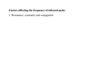
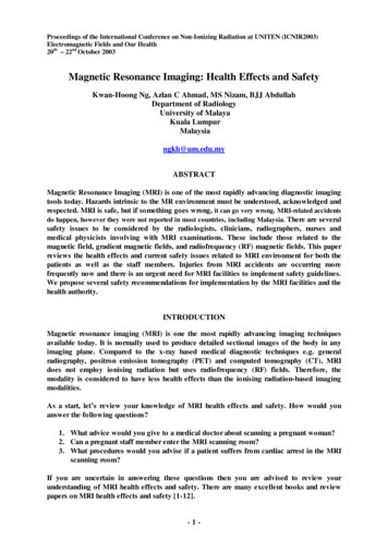
Proceedings of the International Conference on Non-Ionizing Radiation at UNITEN (ICNIR2003)Electromagnetic Fields and Our Health20th –22nd October 2003compatible patient trolley should be kept in the scanning room to enable swift evacuation ofthe patient.OUR RECOMMENDATIONS·Several incidents and accidents had already occurred in Malaysia, however these werenot documented and reported to the Ministry of Health. A standardised system ofincident and accident reporting should be formulated and implemented.·The authority must establish MRI safety guidelines for all MRI centres. Writtenprocedures for screening of patients must be available, read and understood by allauthorised persons. An example of MR procedure screening form for patients is givenin Appendix 1. [11]·MRI facilities should have their own set of Local Rules, which must be read by allstaff (both medical and supporting personnel) who have access to the department.These guidelines are department-specific information about the controlled areas, thedesignated Responsible Person, and Authorised Personnel. These should be reviewedand audited periodically. An example of Local Rules is shown in Appendix 2 [10].·Identify a safety officer responsible for ensuring that procedures are in effect andenforced to ensure safety in the MR environment.·An easily recognised and standardised warning sign is very important in the MRIenvironment. However, the information shown on the most signs is out-of-date,erroneous, or not displayed prominently enough. Appendix 3 shows two examples ofthe new proposed signs. [13]We must not be complacent!Develop a safety culture in MRI.REFERENCES[1]Shellock F.G., Kanal E. 1996. Magnetic resonance: Bioeffects, safety, and patientmanagement. 2nd Ed. New York: Lippincott-Raven Press[2]University of Pittsburgh Medical Centre Magnetic Resonance Safety Site. ologic effects.html[3]McJury M., Shellock F.G. 2000. Auditory Noise Associated With MR Procedures: AReview. Journal of Magnetic Resonance Imaging 12:37–45[4]Shellock F.G. 2000. Radiofrequency Energy-Induced Heating During MRProcedures: A Review, Journal of Magnetic Resonance Imaging 12:30–36-8-
Proceedings of the International Conference on Non-Ionizing Radiation at UNITEN (ICNIR2003)Electromagnetic Fields and Our Health20th –22nd October 2003[5]IRPA/INIRC Guidelines: Protection of the patient undergoing a Magnetic ResonanceExamination 1991. Health Physics 61: 923-928[6]Shellock F.G. 2003. Pocket guide to MR Procedures and Metallic objects: Update2003. Philadelphia: Lippincott Williams & Wilkins[7]Kanal E., Borgstede J.P., Barkovich A.J. et al. 2002. American College of RadiologyWhite Paper on MR Safety, Am Jour Roent: 178:1335-1347[8]Guidelines for Magnetic Resonance Diagnostic Equipment in Clinical Use withParticular Reference to Safety (2nd edition) 2002. Medical Devices Agency (MDA)[9]Principles for the protection of patients and volunteers during clinical magneticresonance diagnostic procedures 1991. National Radiological Protection Board(NRBP).Documents of the NRPB 2 (1).http://www.nrpb.org/publications/documents of nrpb/abstracts/absd2-1.htm[10]Martin C.J., Sutton D.G. 2002. Practical radiation protection in healthcare. Oxford:Oxford Univ. Press[11]Shellock F.G. 2002, Screening patients for MR procedures and individuals for the MRenvironment.Article excerpted from www.mrisafety.com with permission from Frank G.Shellock, Ph.D.[12]Abdullah B.J.J., Bux S.I., Chien D. 1997, Safety considerations in magneticresonance imaging (MRI). Medical Journal of Malaysia 52 (4): 445-53[13]Shellock F.G. 2003. Signs to help control accessto the magnetic resonanceenvironment.Article excerpted from DiagnosticImaging.com with permission of Frank G.Shellock, Ph.D.Some Useful WebsitesAmerican College of Radiology (ACR)http://www.acr.orgThe Emergency Care Research Institute (ECRI)http://www.ecri.orgFood and Drug Administration (FDA)http://www.fda.gov-9-
Proceedings of the International Conference on Non-Ionizing Radiation at UNITEN (ICNIR2003)Electromagnetic Fields and Our Health20th –22nd October 2003Institute for Magnetic Resonance Safety, Education, and Research (IMRSER)www.imrser.orgInternational Commission on Non-Ionizing Radiation Protection (ICNIRP)http://www.icnirp.deMRI Safetyhttp:// www.mrisafety.com- 10 -
Proceedings of the International Conference on Non-Ionizing Radiation at UNITEN (ICNIR2003)Electromagnetic Fields and Our Health20th –22nd October 2003Appendix 1MR Procedure Screening Form for Patients [11]- 11 -
Proceedings of the International Conference on Non-Ionizing Radiation at UNITEN (ICNIR2003)Electromagnetic Fields and Our Health20th –22nd October 2003- 12 -
Proceedings of the International Conference on Non-Ionizing Radiation at UNITEN (ICNIR2003)Electromagnetic Fields and Our Health21st -23rd October 2003, Malaysia- 13 -
Proceedings of the International Conference on Non-Ionizing Radiation at UNITEN (ICNIR2003)Electromagnetic Fields and Our Health21st -23rd October 2003, MalaysiaAppendix 2Basic Local Rules for Magnetic Resonance [From Martin and Sutton, Ref. 10]In view of the demanding electromagnetic environment associated with MR equipment, such equipment should belocated within a designated controlled area where free access is provided only to authorized staff.·It is recommended that the 0.5 mT contour is entirely within the controlled area.·Access to the controlled area should be through self-closing and self-locking doors.·All unauthorized persons must be medically screened and warned of potential hazards (projectile effect,malfunctioning of some devices in the presence of a magnetic field), before entering the controlled area.·Unauthorized persons include:§support staff (engineering staff, nurses, portering staff, cleaning staff, emergency servicesstaff) who should be aware of the potential hazards and should be appropriately trained§patients§volunteers for research projects who should be fully informed and have given consent§the general public (visitors, patients' relations or friends).·Patients, volunteers, and the general public must be supervised by authorized staff at all times within thecontrolled area.·Persons fitted with a heart pacemaker must not enter the controlled area.·It is also convenient to define an inner controlled area containing the 3 mT contour.·All persons entering this area should remove items such as watches, credit cards, and all ferromagneticobjects from their clothing and deposit them outside the area before entering.·No ferromagnetic object (tools, gas cylinders, trolleys, etc.) must be allowed in this inner area.·Persons with any metallic implant should be forbidden to enter the inner controlled area until the implant hasbeen declared safe by a suitably qualified person.·Persons with intracranial aneurysm clips or intraorbital metallic implants should not enter the innercontrolled area.·Persons with metallic implants such as artificial joints, surgical clips, or prosthetic cardiac valves neednot be excluded from MR procedures but care must be taken and the procedure terminated if discomfortor heating is experienced. A large database regarding MR compatibility and safety of implants has beencompiled.·It is prudent to exclude women in the first trimester of pregnancy from the inner controlled area.Procedures for dealing with emergency situations such as cardiac arrest should be defined. Resuscitation equipmentmust not be taken into the inner controlled area and support staff must be fully informed of such procedures. Careshould be taken in attaching physiological monitoring equipment to the patient. High-impedance leads should be usedand displays/recorders should be outside the 3 mT contour. Loops of cables should be avoided since these can lead tooverheating and local burning. Adverse incidents arising from the use of MR equipment should be reported to theMedical Devices Agency.- 14 -
Proceedings of the International Conference on Non-Ionizing Radiation at UNITEN (ICNIR2003)Electromagnetic Fields and Our Health21st -23rd October 2003, MalaysiaAppendix 3New MR safety warning sign designed to help control access to the MR environment. Thissign should be placed on the door to the MR system room. [13]15
to scan her. The clip reportedly shifted when exposed to the magnetic field. The staff apparently had obtained information indicating that the material in this clip could be scanned safely (1992)” · “A patient complained of double vision after an MR exam. The MR exam as well as an x-r