Transcription
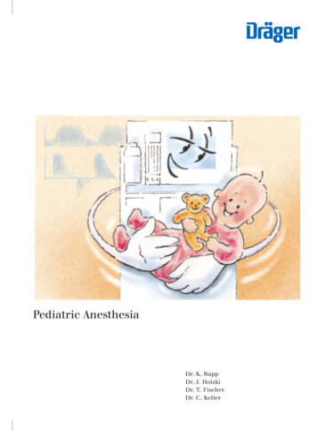
Pediatric Anesthesia-FiblePediatric AnesthesiaDr.Dr.Dr.Dr.K. RuppJ. HolzkiT. FischerC. Keller
2Author:Dr. Katrin RuppDräger Medizintechnik GmbHMoislinger Allee 53-55D-23542 LübeckWith special thanks to the following people for their kind assistance:Dr. J. HolzkiKinderklinik Köln (Children’s Hospital, Cologne)Amsterdamstraße 59D-50735 KölnDr. T. FischerDeutsches Herzzentrum Berlin(German Center for Cardiology, Berlin)Augustenbürgerplatz 1D-13353 BerlinDr. C. KellerKlinik für Anästhesie und Allgemeine Intensivmedizin(Clinic for Anesthesiology and General Intensive Care Medicine)Leopold-Franzens-Universität InnsbruckAnichstraße 35A-6020 InnsbruckDräger Medizintechnik GmbH reserves all rights to this publication,especially as pertaining to its duplication and distribution. No part ofthis publication may be reprinted or saved through any means,mechanical, electrical/electronic or photographic, without theexpressed written consent of the Dräger Medizintechnik GmbH.E-Mail: Katrin.rupp@draeger.comISBN 3-926762-48-9translated by: L.A. Weaver
3Pediatric Anesthesia
4Important Information:Medical knowledge is constantly changing as a result ofresearch and clinical testing. The editors and authors ofthis primer have taken great care to ensure that theinformation and therapeutic details contained hereincorrespond to the most up-to-date research results(especially as pertains to indications, dosages andundesirable side effects), they cannot, however, guaranteethis. Those persons using this book as a reference areadvised to carefully peruse all instructions included withmedications used and to make all decisions pertaining todosage or application at their own discretion.The EditorsOctober, 1999
5Table of Contents1. Introduction82. Special Anatomical and Physiological Features2.1 Breathing2.1.1 Anatomical Fundamentalsof the Respiratory Tract2.1.2 Controlling the Respiratory Process2.1.3 Respiratory Mechanics2.1.4 Pulmonary Volumes2.1.5 Surfactant2.1.6 Oxygen Requirements2.1.7 Extrapulmonary Oxygen Toxicity2.2 The Heart and Circulatory System2.2.1 The Fetal Circulatory System2.2.2 The Heart2.2.3 Blood Volume and Blood Pressure2.2.4 Hemoglobin Contents2.3 Temperature Regulation2.4 The Balance between Water and Electrolytes151719232426272729303132353. Anesthetic Agents3.1 Inhalation Anesthesia3.2 Interactions with Soda Lime3.3 Intravenous Anesthesia3.4 Muscle Relaxants37384549514. Anesthesia Accessories4.1 Masks4.2 Tubes4.3 Laryngeal Masks53545558121313
65. Ventilation in Pediatric Anesthesia5.1 Mechanical Modes of Ventilation5.1.1 The Ventilation Mode IPPV5.1.2 The Ventilation Mode SIMV5.1.3 The Ventilation Mode PCV5.2 Ventilation Parameters5.3 Breathing Systems5.3.1 Flow-controlledNon-rebreathing Systems5.3.2 Valve-controlledNon-rebreathing Systems5.3.3 Partial Rebreathing Systems(Semi-closed Breathing Systems)5.3.4 Complete Rebreathing Systems(Closed Systems)5.4 Characteristics of Ventilation forPediatric Anesthesia5.4.1 Manual and Mechanical Ventilation forNeonates and Infants5.4.2 Respirator Requirements for Children5.4.3 The Fresh-gas Decoupler5.4.4 Compliance Compensation5.4.5 Dead Space Volume5.5 Anesthetic Machines5.5.1 Cato/Cicero EM5.5.2 Julian5.5.3 PhysioFlex5.6 Monitoring5.6.1 The Stethoscope5.6.2 Pulse Oximetry5.6.3 Measuring Body Temperature5.6.4 Measuring Blood Pressure(NiBP/iBP)5.6.5 The 109110113115118
75.6.65.6.75.6.85.6.9CapnographyMeasuring Respiratory PressureVolumetryMeasuring the Concentrationof Anesthetic Agent5.6.10 Measuring the InspiratoryOxygen Concentration5.6.11 Oxygen Uptake5.6.12 Anesthetic Agent Uptake5.7 Low-flow Anesthesia5.7.1 The Wash-in/Wash-out Rateof Anesthetic Agent5.8 Breathing Gas Conditioningfor Anesthetic Ventilation Systems6. Bibliography1201251261271281291301321371421517. Practical Application1617.1 Anesthetic Ventilation for Infants161and Small ChildrenDr. J. HolzkiChildren’s Clinic, Cologne, Germany7.2 PCV during Pediatric Cardio-anesthesia176Dr. T. FischerGerman Heart Center, Berlin7.3 Using a Laryngeal Mask for Pediatric Anesthesia 185Dr. C. KellerLeopold-Franzens University, Innsbruck8. Abbreviations1939. Index196
81. IntroductionChildrenpose many questionsfor anesthetists.
9IntroductionVentilating Children under AnesthesiaAn anesthetic workstation to be used for pediatricanesthesia has to meet numerous requirements and musttake into consideration the special physiological aspects ofthe various age groups of children, from premature babiesto school children. Children are not simply to beconsidered “little adults”. They differ from adultsanatomically, physiologically, psychologically, and biochemically. These differences are especially marked whencomparing premature infants and neonates to adults, andthey only begin to recede around a child’s tenth year.newborns1 to 28 daysinfantsup to end of 1st yearsmall children2 to 5 yearsschool-aged children6 to 14 yearsMany anesthetists who do not care for or administer toinfants or small children on a daily basis are somewhatinsecure with pediatric patients. A large number ofanesthetists were part of a systematic survey whichgathered questions on the general topic of pediatricanesthesia and included other specialized topics, forexample Dräger products used for pediatric anesthesia.The goal of this primer is, on the one hand, to provideanswers for those questions most often asked and, on theother hand, to consolidate, in compact form, the mostimportant basic knowledge in the field of pediatricanesthesia.Figure 1: Age groups
10IntroductionIt is not the intent of this primer to replace other textbooks,but rather to supplement known textbooks by providinginformation on practical applications for pediatricanesthesia. Concrete examples for application topics likeanesthetic ventilation for small children, pressurecontrolled ventilation (PCV), and laryngeal mask usage inpediatric anesthesia, help complete this primer.The following complex questions on the topics of anestheticmachines, accessories, special physiological featuresand anesthetics shall be addressed: Are the latest anesthetic machines able to ventilateusing a pressure-controlled ventilation mode (PCV)?(p. 64) What are the advantages of pressure-controlledventilation over volume-controlled ventilation? (p. 64) Which ventilation parameter settings should be selectedfor which age group? (p. 73) Does it make sense to use PEEP, on principle, for eachand every case of newborn ventilation? (p. 75) What usage problems are associated with nonrebreathing systems? (p. 79) What features does an efficient pediatric respiratorhave? (p. 92) How does dead space volume affect ventilation? (p. 98) How low is the minimum tidal volume which ananesthetic machine can apply? (p. 99) What are the advantages of closed-system ventilation forpediatric anesthesia? (p. 104) What kind of monitoring is used for pediatric anesthesiaand what special features are available for this agegroup? (p. 108)
Introduction How much is volume-controlled or, respectively,pressure-controlled ventilation affected by a siphoninggas measurement procedure during side-streamcapnography? (p. 123) Can low-flow anesthesia be carried out for pediatricanesthesia? (p. 132) How quickly does the system react to changes inconcentration (wash-in/wash-out rate)? (p. 137) Why is it necessary to condition breathing gases forchildren (p. 143), and which types of breathing gashumidification should be used for which age group?(p. 144) Which oxygen concentration is dangerous for neonates?(p. 26) How high are the anesthetic MAC values for children?(p. 39) How do inhalation anesthetics interact with soda lime(p. 46) and how can this be prevented? (p. 48) When would one use intravenous anesthesia on childreninstead of inhalation anesthesia? (p. 50) When is intubation preferred to mask anesthesia forpediatric anesthesia? (p. 54) What are the advantages and disadvantages of laryngealmask usage? (p. 59)These questions shall appear on those pages, indicated bythe blue italicized page numbers behind the questions.11
122. Special Anatomical andPhysiological FeaturesThe “little” patientsshould not be considered“miniature adults”.
Special Anatomical and Physiological Features2.1 1.1 Breathing2.1.1 Anatomical Fundamentals of the Respiratory TractKnowing the differences between the respiratory tract of achild and that of an adult is essential for anesthetists inorder for them to safely administer anesthesia. A child’s nostrils, oropharynx and trachea are relativelynarrow (see Figure 2). Breathing can be hindered byirritation of the mucous membrane due to edema buildup in this area. The trachea is short – it only measures approximately4 cm from the larynx to the carina – and has a narrowdiameter of 6 mm. The tongue is relatively large and tends to fall backwardsunder anesthesia. Neonates, infants and small children have a very softthorax compared to their lungs. The thorax is relativelyshort. The ribs run horizontally and not diagonally, as isthe case with adults. The intercostal muscles areimmature. The salivary secretions of children are more pronouncedthan those of adults. The larynx of a child is more ventrally located and onlevel with the third to fourth (neck) vertebrae, thusabout a whole vertebrae higher than that of an adult.Until the age of 8 to 10 years, the most narrow point is avery sensitive mucous membrane on level with thelarynx cartilage and not, as is the case with adults, withthe glottis. The epiglottis is relatively large and shaped like a U. The size of the tonsils and the adenoid in children cancomplicate the intubation process.13
14Special Anatomical and Physiological Features Infants breath through their nose until they reach anage of 5 months. Inserting a stomach probe through thenose can be a massive respiratory hindrance.Figure 2: Sagittal tomogramof a newborn and an adultadultinfantnasal cavityupperairwayoral cavityepiglottislowerairwaywindpipe
Special Anatomical and Physiological Features2.1.2 Controlling the Respiratory ProcessThe respiratory process of both premature newborns andneonates, like that of adults, is essentially controlled bychanges in paCO2, paO2 and pH (see Figure 3). The hypoxiabreathing regulation of newborns is not, however, fullydeveloped; right after birth, the oxygen receptors and theirfunctions are immature. The paCO2 and paO2 values ofnewborns and infants are lower than those of adults untilthe end of their first year.Premature infants often experience respiratory arrest(apnea) either at regular (periodical breathing) orirregular intervals. Periodical breathing is considered anepisode of 3 or more respiratory pauses of at least 3seconds. Normal breathing periods of less than 20 secondsaccompany these. Apnea phases can be due to a centralproblem (no physical breathing exertion) or, less often,caused by an obstruction (no flow despite physicalbreathing exertion). In addition, there are mixed forms ofboth. Nevertheless, these breathing abnormalities are notusually dangerous. Respiratory arrest can, however, lead todecreased O2 partial pressure in the blood and can causebradycardia if the length of such a phase is longer than 30seconds.Infants react to hypoxia biphasically. First there is a 30second increase in minute volume, followed by hypoventilation or apnea. If hypothermia or hypoglycemiaoccurs simultaneously, hypoventilation is the immediateresult. An adequate reaction to lack of oxygen on the part ofthe child can only be observed 2 to 4 weeks after birth.15
16Special Anatomical and Physiological FeaturesThe breathing regulation of premature newborns andneonates continues to be influenced by pulmonarycompliance. Compliance triggers breathing reflexes via themechanoreceptors. The most well-known reflex is theinspiratory repressive Hering-Breuer reflex which isespecially noticeable in premature babies (born betweenthe thirty-second and thirty-eight week of pregnancy) withlittle pulmonary compliance. This effect decreases as theneonate matures. Ventilating with high tidal volume, whichmay cause the lungs to overinflate, leads to an inhibition ofreflexes in the central breathing system’s inspiratoryneurons and, thus, to an interruption and extension of theexpiratory phase.It is thought that this reflex protects the system fromrespiratory fatigue caused by ineffective muscle work andfrom volutrauma [Davis, 1987; Mortola, 1998].Figure 3: Children’srespiratory regulationmain IIR Hering omplianceIIR Inspiration Inhibiting ReflexEPR Exspiration Prolonging Reflex
Special Anatomical and Physiological Features172.1.3 Respiratory MechanicsA newborn’s pulmonary compliance (or elasticity) is verylow and does not differ greatly from the total compliance(i.e. compliance of the lungs and thorax). A newborn’sthorax is substantially more elastic than an adult’s andoffers little resistance to e.g. overinflation. As a child growsolder, the total compliance increases (see Table lchildrenschool-agedchildren1 to 28 days up to1 year 2 to 5 years 6 to14 years2.5 to 5 kg55 to 10 kg 10 to 20 kg10 to 2020 to 40 20 kg100In newborns and infants, the diaphragm does almost allthe work expended for breathing. Abdomen hindrances, forexample due to intra-abdominal pressure, can lead toinsufficient spontaneous breathing.Table 1: The relationshipbetween age and compliance
18Special Anatomical and Physiological FeaturesPulmonary compliance can be reduced due to manycauses, for example:parenchyma changes bronchopneumonia pulmonary edema ARDS fibrosisfunctional surfactant disorders alveolar pulmonary edema atelectasis aspiration RDS/ARDSreduced volume pneumothorax raised diaphragm[Oczenski, 1997]
Special Anatomical and Physiological Features192.1.4 Pulmonary VolumesRelative to its size, the volume of a child’s lung isequivalent to that of an adult’s. An infant born after a fullterm pregnancy has a total lung capacity of approximately160 ml, a functional residual capacity of 80 ml and a tidalvolume of approximately 16 ml. One-third of the tidalvolume is equal to the dead space volume. The proportion,dead space volume to tidal volume, remains constant forspontaneously breathing children, it can, however,increase during controlled ventilation. In order to keeptotal dead space volume to a minimum, accessories of theanesthetic systems should be operated using the smallestpossible dead space available, especially when ventilatingan infant with a dead space volume of only 5 ml (seeChapter 5.4.5, Dead Space Volume).Figure 4: Static tidal volumes[Lotz, 1984]inspiratoryinspiratorycapacity reserve nalresidualcapacityexpiratoryreserve volumeresidualvolume
20Special Anatomical and Physiological FeaturesThe following static tidal volumes can be distinguished: total capacity (TC): includes total air volume in thelungs after a maximal inspiratory phase (TC FRC IC) vital capacity (VC): the maximum volume which can beexhaled after a maximal inspiratory phase(VC ERV IC) residual volume (RV): the volume remaining in thelungs after a maximal expiratory phase functional residual capacity (FRC): the volumeremaining in the lungs after a normal expiratory phase(FRC RV ERV) expiratory reserve volume (ERV): additional volumewhich can be exhaled after a normal expiratory phase tidal volume (VT): the volume normally inspired andexpired inspiratory capacity (IC): the maximum volume whichcan be inspired (IC VT IRV) inspiratory reserve volume (IRV): additional volumewhich can be inspired after a normal inspiratory phaseIn addition to the above tidal volumes, closing volume isimportant (see Table 2). Although all respiratory paths areopen in a completely filled lung, decreasing expiratoryvolume may cause peripheral paths to become blocked.The closing volume of infants and small children is ratherlarge compared to that of adults. It could exceed thefunctional residual capacity and, during normalventilation, impair tidal volume.
Special Anatomical and Physiological FeaturesIntubation eliminates physiologically intrinsic PEEP in thelarynx area, which counteracts peripheral respiratory pathblockage. Autogenic PEEP can be compensated for in mostanesthetic machines by using a slightly extrinsic PEEP ofabout 3 mbar, or by implementing the system ALICE [automatic lung inflation control effect] (see page 104).The alveolar ventilation (ventilation of the alveoli for thepurpose of pulmonary/blood gas exchanges) of a child, 100to 150 ml/kg/min, is twice as high as that of an adult. Thisis achieved mainly through an increased respiratory rate,not through increased tidal volume (VT). The ratio ofalveolar ventilation to functional residual capacity is 5:1 forinfants and 1.5:1 for adults. As a result, the functional residual capacity of an infant is only a slight “buffer” againstfluctuations of breathing gas and anesthetic agentconcentrations so that changes in anesthetic agentconcentrations are reflected very quickly in the arterialblood gas values.Any reduction in functional residual capacity, e.g. byanesthetics, can lead to blockage in the smaller paths ofthe respiratory tract, uneven breathing gas distribution,and hypoxemia.21
22Table 2: Average respiratoryrates, tidal volumes andresistance values accordingto ageSpecial Anatomical and Physiological Featuresnewbornssmallchildrenschool-agedchildren2 to 5 years6 to 14 years5 to 10 kg10 to 20 kg 20 kgrespiratory rate(min-1)40 to 6030 to 6030 to 4012 to 20tidalvolume(ml kg -1)8 to 108 to 108 to 108 to 104020 to 30201 to 2ageweightresistance(mbar L -1 s)infants1 to 28 days up to 1 year2.5 to 5 kgRespiratory resistance in newborns is high. This is due tothe smallness of the inside diameter of the nostrils, theminute diameter of the smaller bronchi and low tidalvolume. During an operation, significant fluctuations inrespiratory resistance can occur. For example, bronchialdilatatorical effects from volatile anesthetics reducerespiratory resistance while even the slightest swelling oraccumulation of fluids in the respiratory tract, or thetiniest obstruction in the tube area can increase total flowresistance.
Special Anatomical and Physiological Features2.1.5 SurfactantSurfactant is a surface-active substance found on thealveolar surface which is synthesized by type 2 alveolar cellsand released during inspiration. Surfactant mainly consistsof lipoproteins made of lecithin. It stabilizes the alveoli andprevents them from collapsing during the expiratory phase.In addition to this, surfactant reduces the risk of too littleventilation and atelectasis build-up. Premature newbornssuffer from respiratory distress syndrome (RDS) becausetheir surfactant is not yet mature; surfactant first maturesin the thirty-fifth and thirty-sixth weeks of pregnancy.Although hypo- and hyperoxia, acidosis and hyperthermiacan affect surfactant development, the anesthetic agents ofinhalation anesthesia seem to have little influence onsurfactant production. Those newborns with insufficientsurfactant will have a considerable amount of trouble withpulmonary gas exchange. These children usually requireventilation.When premature newborns are treated with surfactant,functional residual capacity (FRC) builds up during thefirst 6 to 12 hours after application. The dynamiccompliance of the lungs can only increase after this timelapse has occurred. It is important to monitor tidal volumeand reduce pressure simultaneous to applying surfactantin order to prevent potential pneumothorax build-up. Anysurfactant already generated can be destroyed again bydelivering 100 % oxygen, by aspiration pneumonia or byventilating too aggressively, e.g. by applying a tidal volumewhich is too high.23
24Special Anatomical and Physiological Features2.1.6 Oxygen RequirementsAt 7 ml/kg x min, the oxygen needs of a neonate or apremature newborn are about twice as high as that of anadult (see Table 3). At the same time, the level of oxygenconsumption depends on the child’s state of health, bodilymaturity, and stress due to cold. For example, post-operativehypothermia causes an increase of 15 to 16 ml/kg x min,whereas the oxygen required under anesthesia decreases asthe body temperature sinks.Table 3: Interdependency ofoxygen consumption andfunctional residual capacity tobody weightoxygen consumptionfunctionalresidual capacity5 kg9 ml/kg x min10 ml/kg10 kg7 ml/kg x min15 ml/kg20 kg6 ml/kg x min30 ml/kgadults3.5 ml/kg x minNewborns and infants are significantly more susceptible tohypoxia than adults because of higher oxygen consumptionlevels, higher alveolar ventilation (twice that of adults),and little functional residual capacity; the latter is reducedeven more after anesthetics have been administered (60 %vs. 30 %). The small oxygen reserve available to a newbornor small child is quickly used up; hypoxia can occur within10 to 20 seconds after an apnea. For comparison, an adulthas a length of tolerance of 2 to 3 minutes (see Figure 5)[Frei, 1994].
Special Anatomical and Physiological Featuresadultinfant25/min12/min2 ml2 ml dead space4 ml4 ml alveolar100 ml/min(- 60 %)12 mlreserved for 18 s7 ml/min48 ml/min alveolar ventilation(- 30 %)32 ml FRC(FO2 30 %)reserved for 165 s3.5 ml/min O2 consumptionPremature newborns 1500 g do not react to a lack ofoxygen, like an adult, with a tachycardia, but rather with areduced heart rate (bradycardia). This state is notremedied by administering medication, like Atropine, butby sufficiently ventilating the patient and increasing theoxygen supply [Jöhr, 1998].25Figure 5: Comparing infantsand adults (standardizedweight in kg); an infant cango into a hypoxic state within10 to 20 seconds after anapnea because it has lessoxygen reserve than andtwice as much oxygenconsumption as an adult
26Special Anatomical and Physiological FeaturesWhich oxygen concentrationis dangerous for prematureinfants?2.1.7 Extrapulmonary Oxygen ToxicityTheir immature state and contact with partial oxygen pressures which are too high put premature infants at risk ofgoing blind (retinopathy of prematurity). This risk increases with the degree of immaturity, the duration of oxygenapplication and the height of the partial pressures.Premature infants born before the forty-forth week ofpregnancy and exposed to a paO2 of more than 80 mmHgfor more than 3 hours, or a paO2 of more than 150 mmHgfor more than 2 hours, are in acute danger [Shann, 1988].For this age group, an oxygen saturation level of 90 to 95 %should be targeted using pulse oximetry in order to avoidpaO2 values of 70 mmHg (see the oxygen disassociationcurve for premature infants on page 111).The risk of damaging the retina by excessive partialpressure depends on age and is almost non-existent whenpremature infants reach the infant stage.
Special Anatomical and Physiological Features2.2 The Heart and Circulatory System2.2.1 The Fetal Circulatory SystemThe lungs of a fetus are not ventilated before birth and areonly supplied with very little blood. Oxygen-enriched bloodis carried from the placenta of the mother through thevena cava inferior to the right atrium. The main portion ofthis blood flows through the foramen ovale directly into theleft atrium without being mixed with other blood. It istransported to the brain through the aorta ascendens andthe carotid artery while blood pushed from the rightventricle is pumped into the aorta through the ductusarteriosus. All this causes high pulmonary tissue resistanceto be present, with a right-to-left shunt via the ductusarteriosus and the foramen ovale (see Figure 6).Lung tissue resistance decreases shortly after birth as aresult of lung expansion and an increased paO2 value in thealveoli. The result is functional blockage of the ductus andthe foramen ovale. After only 6 to 18 weeks, this blockagebecomes permanent. Before this, however, the ductus of anewborn can be reopened by acidosis and/or hypoxia.27
28Special Anatomical and Physiological nary eriorvenacavasuperioraortapulmonary arteryforamen ovale(closed)pulmonaryveinrightventricleductus levenacavainferiorFigure 6: Circulatory system of a fetus and a newbornpulmonaryveinleftventriclerightventricle
29Special Anatomical and Physiological Features2.2.2 The HeartThe heart of a newborn has fewer contractile elementsthan that of an adult. Because the metabolic rate is raised,cardiac output is relatively high: infants 200 ml/kg/min;adults 70 ml/kg/min. Cardiac output can only beincreased by increasing the heart rate. Systolic dischargecannot be increased significantly.The normal resting heart rate of a newborn isapproximately 120 beats per minute, but the breadth forvariation is wide (see Table 4). Heart rhythm disorders areoften observed in infants. Extra ventricular systoles occurquite often during anesthesia induction, especially whenthe depth of anesthesia is insufficient. These disorders can,however, be removed by deepening anesthesia. However, inthis case there must be no risk of hypoxemia andhypercapnia.heart 1 to 12 months120801602 years110801304 years100801206 years100751158 years907011010 years9070110Table 4: Heart ratesaccording to age[Kretz, 1998]
30Special Anatomical and Physiological Features2.2.3 Blood Volume and Blood PressureA child’s blood pressure is lower than an adult’s blood pressure, and it increases in proportion to age (see Table 5).The rule of thumb for premature infants is: mean arterialpressure (MAP): 1 mmHg per week of pregnancy. Bloodpressure sinks under anesthesia; it should not drop below50 mmHg for infants and 30 mmHg for prematurenewborns.Table 5: Average systolic anddiastolic blood pressurevalues according to age[Versmold, 1981]agesystolic(mmHg)diastolic(mmHg)newborns75 to 8540 to 502 weeks to 4 years85606 years90608 years956210 years1006515 years11572The blood volume of children is, when compared to theirweight, larger than that of adults and decreases with age(see Table 6). The total amount of blood is, however, low;the slightest loss of blood can lead to lack of volume andanemia.Table 6: Average bloodvolume values according toage [Jöhr, 1998]premature nadults8070
31Special Anatomical and Physiological Features2.2.4 Hemoglobin ContentsThe hemoglobin of an infant consists of fetal (HbF) andadult (HbA) hemoglobin and often varies during the first 6months (see Figure 7). Compared to HbA, HbF has higheroxygen affinity, making it more difficult for the oxygen tobe transferred to body tissues. Increased hemoglobin atbirth, about 200 g/l blood, compensates for this deficiency.This value decreases greatly, as low as 100 g/l blooddepending on the individual, within the first 3 months asfetal hemoglobin is replaced with adult hemoglobin. Afterthis phase, it increases again (see Table 7). In the case ofpremature newborns, the hemoglobin value decreasesmore rapidly and reaches lower points.hemoglobin(g/l)1 to 7 Tage160 to 2001 to 4 weeks110 to 1602 to 3 months 100 to 1201 year100 to 1205 years110 to 130Table 7: Hemoglobin valuesaccording to ageFigure 7: Portions of fetal andadult hemoglobin in the blood(WP: week of pregnancy)portion in %10080agefetalhemoglobinadulthemoglobin6040203 WP6 WPbirth3 months 6 months
32Special Anatomical and Physiological Features2.3 Temperature RegulationOne of the anesthetist’s main tasks during anesthesia ismaintaining the patient’s body temperature. Infants andsmall children are especially susceptible to hypothermiaboth during and after an operation. Children absorb andgive off heat more quickly than adults because they have arelatively large body surface compared to their size andbecause their skin is thin and has little subcutaneous fat.Infants and small children are not able to produce heat byshivering. Postoperative shivering appears only at about sixyears. Unil then, heat is produced by increasingmetabolism, for which so-called “brown adipose tissue” isbroken down.During an operation, thermal regulatory responses to hypothermia only begin if the body temperature deviates greatlyfrom the usual ( 35.5 C), and heat is transferred fromthe core of the body to the periphery. Figure 8 comparesthe slightness of a neonate’s core temperature constant tothat of an adult’s when ambient air temperature changes.Anesthetic agents like halothane or isoflurane (see Chapter 3)lead to an even greater loss of heat due to peripheryvasodilation. If hypothermia occurs during anesthesia, therecovery time of the child could be delayed. Respiration,heart rate, blood pressure, and/or cardiac output coulddecrease, and the effectiveness of non-depolarized musclerelaxants could increase. The need for oxygen decreases ifthe body temperature sinks sharply, and the metabolic ratedecreases by 6 to 7 % per degree Celsius.
33Special Anatomical and Physiological FeaturesFigure 8: Changes in coretemperature for prematureinfants and adultsThe Core Temperature of an Adultcore temperature C3730hypothermiathermoneutral zonehyperthermia Cambient temperatureThe Core Temperatue of a Premature Infantcore temperature C3730hyperthermia Chypothermiathermoneutral zoneambient temperature
34Special Anatomical and Physiological FeaturesHypothermia can also lead to hypoxia, acidosis, and hypoglycemia.These are the reasons why measures must be taken to preserve the body heat and protect the patient from hypothermia. Such measures are many-sided (see Table 8) anddepend upon the length of the operation to be undertaken,as well as the age of
Those persons using this book as a reference are . Ventilation in Pediatric Anesthesia 60 5.1 Mechanical Modes of Ventilation 61 5.1.1 The Ventilation Mode IPPV 61 5.1.2 The Ventila
