Transcription
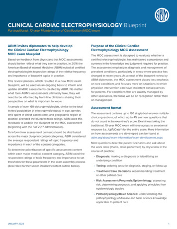
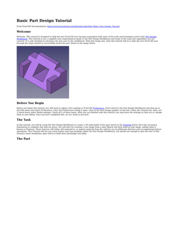
Basic Electrophysiology Part 1Directions for nursing CEU credit: Review the material in this module. Download the test form.Take the test. Fax or send the answer sheet with a form of payment. Passing score is 75%. Youhave 3 chances to take the test. Upon completion you will receive a CEU certificate. Critical CareED is an approved provider of continuing education in nursing by the California Board of Nursing.Module Objectives: Upon completion of this module the learner will be ableto:State in correct order the movement of the electrical impulse through the conductionsystem.Describe the anatomy of the AV junction.Name two types of accessory AV connections.Define automaticity, excitability, conductivity, refractoriness, and contractility.Differentiate depolarization from repolarization.Discuss the refractory periods in ventricular conduction.Describe the ventricular action potential.Describe two types of arrhythmia mechanisms.State the inherent or intrinsic rate of the various parts of the conduction system.I. Specialized Conduction System of the HeartThe heart has a specialized conduction or electrical system. The conduction system isresponsible for the movement of an electrical impulse from the top to the bottom of the heart,stimulating the heart so that it squeezes or contracts. Electrical impulses are normally initiated bythe sinus or sinoatrial (SA) node. The SA node is located in the right atrium next to the superiorvena cava (SVC). The SA node is a sizable structure with a crescent shape. The SA node isactually a band of tissue that lies between the spot where the SVC attaches to the RA and aportion of the RA called the RA appendage. It is more an epicardial structure than endocardial.There are no documented specialized connections linking the SA node to the AV node. The SAnode is often referred to as the pacemaker of the heart because in the normal, healthy heart theSA node will “fire” automatically more quickly than any other part of the conduction system, ata rate between 60 – 100 beats per minute (though some texts state that 50 is the low value fornormal and 110 is the upper limit).The SA node is richly innervated by the sympathetic and parasympathetic (vagus) nervoussystem. The effect of sympathetic stimulation would cause the SA rate to increase, while theparasympathetic effect would cause the rate to slow.The following figure shows the location of the SA node from a frontal view. It should be notedthat the SA node is structurally a band of tissue. A rhythm disorder called SA node reentry canoccur in which an impulse recirculates through portions of the SA node and causes a “sudden1
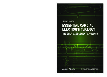
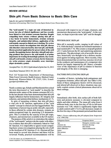
onset” sinus tachycardia at a rate of 100 – 150. The treatment for this rhythm problem may be an“SA node modification” via radiofrequency (RF) catheter ablation.SA node from the frontal viewAtrial Floor PlanThe routes of impulse conduction throughout the right and left atria are largely determined by theregions of ordinary atrial myocardium left over when you exclude the spaces taken up by holes(orifices of great vessels emptying into the atrium) and nonconducting borders (the annuli).Atrial Floor Plan2
The above diagram is a rough illustration of the atrial floor plan as if the atria were flattened intoan equal area map and is intended to show how we might envision the “preferential” routes ofconduction from sinus to AV node. In general, these routes contain muscle fibers which maintaina reasonably parallel alignment for a considerable distance, thereby contributing to the appearanceof “preferential” conduction through these routes.The actual area of contact between the right and left atrium is relatively small. The majorconnection to the left atrium is via the coronary sinus os and Bachmann’s bundle, a rather bulkyappearing muscular band coursing over the roof of the of the right atrium, connecting to the leftabove the interatrial septum.Connections to the left atriumThe coronary sinus is the other pathway an impulse could follow from the right to the left atrium.The figure above, which looks down into the atria, highlights the Bachmann’s bundle connectionand the pathway an impulse could follow from the right to the left atrium along the coronary sinus.Historically drawings of the conduction system portrayed internodal tracts, displayed in such amanner as to suggest that they bear some resemblance to the bundle branches/Purkinje system.Such anatomic suggestions, including the inaccurate representation of the AV junction, are stillfound in current textbooks.The AV JunctionThe AV junction could be the most interesting structure within the heart. As the sole normalconnection between atria and ventricles, it is responsible for introducing complexity to the cardiacrhythm. Without it, the classification of arrhythmias would be rudimentary, including onlytachycardia, bradycardia and fibrillation -with emphasis on the ventricles since the atria would3
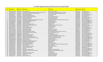
assume a secondary importance. Because of the AV junction, there are a wide variety of AV andVA interactions of single beats or rhythms arising from the sinus node, atria and ventricles thatimpart complexity to rhythm analysis. The AV junction is located at the anterosuperior portion ofthe interatrial septum near the tricuspid annulus. The inherent rate for the AV junction to pace theheart is 40-60 beats per minute (slower than the sinus rate of 60-100). The AV junction is capableof being a ‘backup’ pacemaker should the SA node fail. The AV junction is innervated by bothsympathetic and parasympathetic fibers, as the SA node is. Electrical impulses which arrive in theAV junction are slowed briefly to allow atrial contents to empty into the ventricles prior toventricular contraction.The AV junction can be considered as having three component parts:1. the compact AV node2. the anterosuperior approaches to the node located in the anterosuperior interatrialseptum (fast AV nodal pathway)3. the posteroinferior approaches extending from the compact node to the coronary sinusos (slow AV nodal pathway)AV nodal anatomy as seen from the RAO viewTerminology varies depending on who you read, but the term “junction” is best because it allowsdistinction between the compact AV node and its atrial approaches while at the same timeminimizes confusion about what is meant by the “node”. Some authors may be referring to theentire AV junction as the AV node while others may infer only parts.The fast and slow pathways converge onto the compact AV node in a way that promotes properorientation for conduction into and through the node. The compact node is the primary site ofconduction delay, and is responsible for the PR interval. Normal AV conduction proceeds via thefast pathway, termed fast because nodal conduction is quicker than when it conducts through the4
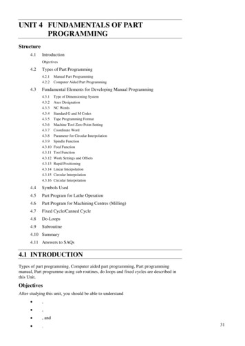
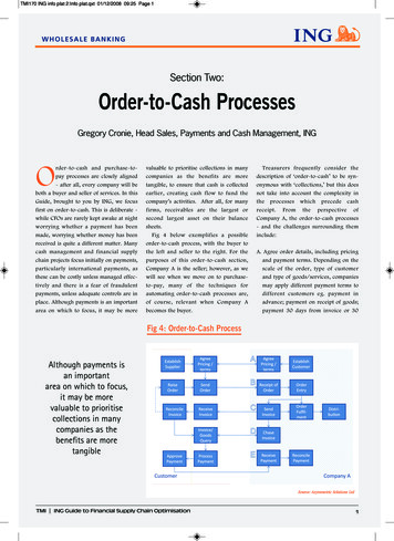
node via the slow pathway. Thus, the normal PR interval of 0.12-0.20 sec reflects preferentialconduction over this faster route.Although the spectrum of AV conduction behavior in the majority of patients suggests twopathways, hence the well-known reference to “dual physiology”, many variations on the themeexist. At one extreme, there may be no detectable evidence for a slow pathway (fast pathwaydominance) while at the other extreme multiple slow pathways can be found. Fast pathwaydominance is unusual, accounting for less than 5-10% of patients. An arrhythmia called AV nodereentry (AVNRT) can occur when an impulse travels down one pathway and up the other in acircle causing a ventricular rate of about 150-250 per minute.His-Purkinje SystemThe bundle of His represents the beginning of the conduction system in the ventricles. Thebundle of His divides into two main branches, the right bundle branch and the left bundlebranch. The right bundle branch is long and slender and delivers the electrical impulse to theright ventricle. A blockage of impulse through this structure is called a right bundle branchblock (RBBB). During RBBB an impulse must travel down the left bundle and cross theventricular septum to provide stimulation to the right side so that the right ventricle will contract.The left bundle branch (LBB) is broad and fanlike. The LBB delivers the electrical impulse tothe ventricles. In most individuals there are three divisions (fascicles) to the LBB. Thesedivisions are the posteroinferior, the anterosuperior and the septal. There are identifiableEKG criteria for a blockage of the anterosuperior or posteroinferior divisions. Blockages ofthese divisions are called left anterior hemiblock and left posterior hemiblock. The followingfigure shows the location of this left bundle branch toward the upper right from the AV nodalarea and the right bundle branch below. The LAD is the left anterior descending coronary arteryand the PDA is the posterior descending artery.The bundle branches5
The last stop for the electrical impulse in the ventricle is the Purkinje fibers. The ventricles arethen stimulated sufficiently to contract. The ventricles may also act as a backup pacemaker forthe heart if the areas above fail; however the rates would be in the range of 10-40, depending onwhich site served as the backup.Self-Study Questions:1. Describe the shape of the SA node.2. What is SA node reentry?3. What is the basic or inherent rate for the SA node?4. What is Bachmann’s Bundle?5. How does the electrical impulse travel?6. What is the inherent rate for the AV junction?7. Describe the fast and slow AV nodal pathways.8. How does AV node reentry occur?9. Describe the conduction system components that are found in the ventricles.10. What is right bundle branch block?11. What is the most common treatment for AV node reentry?II. Accessory Atrioventricular Connections(Pathways, Bypass Tracts)Accessory atrioventricular connections (also known as pathways or bypass tracts) areconnections between the atria and ventricles which are not present in everyone. They provide analternative route for the travel of the electrical impulse instead of the AV junction. Thus thesebypass tracts may be responsible for alterations in QRS width or changed P wave/QRSrelationships.Rapidly Conduction Accessory AV PathwaysRapidly conducting accessory AV pathways are the pathways which connect the atria andventricle in the Wolff-Parkinson-White (WPW) Syndrome. These pathways exist because ofan abnormality in development of the AV groove of the heart in utero. The AV groove separatesthe atria from the ventricle electrically, except for the AV node, thus making impulse travel fromthe atria to ventricle orderly.With WPW, there are stray pieces of muscle which still connect the atria to the ventricle, thusallowing an alternative route for the electrical impulse. This may result in the triad of short PRinterval, wide QRS and a delta wave (gradual incline at the onset of the QRS), which ‘preexcites’the ventricle. Or its presence may provide the pathway for an impulse to circulate down the AVnode and up the pathway, or down the pathway and up the AV node. This is called a reentranttachycardia (AV reentry tachycardia, AVRT, or circus movement tachycardia).6
Rapidly conducting accessory pathways exist on either the left or right side of the heart. Theyare usually capable of antegrade and retrograde conduction. About 75% of patients exhibitpreexcitation on a 12 lead ECG during normal sinus rhythm. About 25% will conductretrograde only (concealed accessory pathways) and less than 2-4% conduct antegrade only.Characteristics of rapidly conducting pathwaysRapidly conducting accessory pathways (WPW) are right or left-sided.Slowly Conducting Accessory AV PathwaysSlowly conducting accessory AV pathways exist only on the right side of the heart. Those thatare capable of only antegrade conduction are called Mahaim fibers. Preexcitation will beevident on the 12 lead ECG only during tachycardia. The clinical syndrome is known asMahaim PSVT.The slowly conducting accessory AV pathways capable of only retrograde conduction areresponsible for a permanent form of junctional reciprocating tachycardia (PJRT). With thispathway, preexcitation is never evident on the 12 lead ECG in normal sinus rhythm or duringPSVT. The slowly conducting accessory AV pathways are diagramed below.7
Slowly conducting accessory pathways (Mahaim, PJRT) are only right-sidedSummary of characteristics for Mahaim fibersSummary of characteristics for PJRT pathwaySelf-Study Questions1. What is an accessory pathway?2. What are rapidly conducting accessory pathways called?3. Describe the aberrant anatomy that is known as WPW.4. What are the three abnormal findings that may be present on the EKGcomplex in WPW during normal sinus rhythm?8
5. On what side(s) (L or R) of the heart will WPW pathways be found?6. Describe antegrade and retrograde conduction.7. Name two slowly conducting pathways.8. Describe preexcitation.III. Properties of Cardiac FunctionFive properties of cardiac function should be reviewed to adequately understand electrical andmechanical events. The first of these is automaticity. Automaticity is the ability of certain cellsin the heart to initiate electrical impulses spontaneously. This property exists in all cells knownas pacemaker cells. Areas of the conduction system thus capable of initiating impulses includesthe SA node and atrium, the AV junction and the His-Purkinje system. Normal automaticity foreach of these regions would thus elicit the following rates: SA node 60-100 bpm; AV junction40-60 bpm; and His-Purkinje system 10-40 bpm. Altered automaticity occurs when these areasperform more slowly (depressed automaticity) or more rapidly (enhanced automaticity). Theseabnormal situations may be the cause for some of the arrhythmias that we see.Excitability is the ability of cardiac cells to respond to stimulation. A cell must be properlyoxygenated and have electrolytes and ph within normal range to respond in a normal fashion.When an impulse makes its way to an individual cell, the impulse must be strong enough tochange a cell from predominately electrically negative to electrically positive. The success ofthe impulse to achieve this is also determined by its ability to measure up to the “threshold level”for that cell. The threshold level is analogous to a bar over which the impulse must jump. If thethreshold level or bar is lowered, weaker impulses may be able to affect the cell and depolarizeit. A lowering of the bar occurs with low serum potassium levels and low tissue oxygen levels.Thus during these situations arrhythmias are allowed to occur which one normally would not see.A raising of the bar on the other hand, may prohibit even the normal impulse from the SA nodefrom causing the cell to depolarize. An example of this would be the situation caused by highserum potassium levels, which in the extreme could cause asystole.9
Conductivity is the ability to transmit an impulse through the specialized conduction system andthe atrial and ventricular musculature. Conductivity is analogous to traveling or the taking of atrip. We measure the ‘trip’ of an impulse in three different parts of the ECG complex. The first‘trip’ we measure is that taken from the SA node to the ventricles, and the time that trip takes isnormally 0.12 to 0.20 seconds and is known as the PR interval. The second trip is that takenthrough the ventricles, and corresponds with the QRS width. That trip is usually less than 0.12seconds. The third trip that we measure is the combined trip of ventricular depolarization andrepolarization, and this is known as the QT interval. The QT interval normal varies for men andchildren versus women. A general yardstick for measuring however, is that normal is probablyhalf of an individual’s R to R interval.Some conditions that may alter conductivity include increased sympathetic or parasympatheticinfluence, disease related factors such as ischemia, cardiomegaly, calcification, congenitaldisorders, fibrosis, neurological disorders, tumors, metabolic disorders, myocarditis, and aging.Medications such as digoxin and antiarrhythmics may also cause conduction problems.Refractoriness is the inability of a cell or tissue to undergo repeat stimulation until after acertain period of time has elapsed. This means that once a cell is stimulated and responds to animpulse, the cell must recover before it can be stimulated again. State of refractoriness is amoving target, changing from moment to moment. We ask where in the cycle we are at anygiven point.Finally, contractility is the ability of cardiac muscle fibers to shorten when they are stimulated,resulting in muscle contraction (pump action). Cardiac muscle cells must be healthy with normalelasticity to make pumping efficient. Muscle cells that are dilated and that have experiencedexcessive stretch will not contract in a normal fashion and this pump action will be decreased.This is the situation we see with heart failure.We can use drugs or devices to alter contractility. Bi-ventricular pacing is an effort to enhancecontractility. Giving inotropic drugs enhances contractility. Sometimes we give drugs to depresscontractility. For example, in intractable angina we may give beta blockers or calcium channelblockers to decrease contractility and therefore reduce pain. Also, for those with hypertrophiccardiomyopathy beta blockers and calcium channel blockers reduce contractility, causing the leftventricular muscle to relax a bit and decrease aortic valve obstruction, thus allowing more bloodto be ejected with each contraction.Self-Study Questions1. Describe the principle of automaticity and its relationship to the SA node.2. In order for a cell to be excitable, what must be true?3. Name the three parts of the EKG complex in which conductivity may bemeasured.4. Define refractoriness.5. Name a diagnosis in which contractility is impaired.10
IV. Depolarization and RepolarizationCardiac cells can exist in three major states electrically. The first is that of the polarized cell.This is the cell at rest. The interior of the cell is negative with respect to the outside. Potassiumand sodium are maintained with normal distribution.The next cell state is depolarization. With depolarization the electrical impulse arrives at thecardiac cell and the cell is stimulated. Stimulation will occur appropriately if tissue oxygenationand electrolytes are at normal levels. With depolarization, sodium enters the cell, producing aninward electrical current. Calcium enters the cells soon after. This process proceeds from theinner muscle layer, the endocardium, to the outer layer, epicardium, and begins with theventricular septum. The impulse moves from cell to cell as depolarization occurs successfully.The third cell state is repolarization. With this process the cell returns to the resting state.Potassium leaves the cell and an outward electrical current is produced. Eventually the sodiumpotassium pump plays a roll and the polarized state is again reached.The three electrical states of the cellNotice that we are speaking of sodium, potassium, and calcium. Three of the major classes ofantiarrhythmic drugs act by blocking these very electrolytes. The drug actions occur at thecellular level. These drugs therefore alter depolarization and repolarization in different ways.The next figure further explains the principles of depolarization and repolarization. In theDomino Analogy one starts with a group of dominos stood on end, representing polarized cells.The depolarization or activation man arrives and knocks over the first domino which in turnknocks down the second, etc. The recovery or repolarization man arrives and begins to stand thedominos up again. The key point to understand here is that activation or depolarization cannotoccur again until recovery has taken place.11
The domino analogyThe other analogy for depolarization and repolarization that has been made is that of flushing atoilet (this one you’ll remember!!). Flushing corresponds with depolarization. The filling of thetank corresponds with repolarization. One cannot flush the toilet again until an adequate amountof water has filled the tank.V. Refractory PeriodsRefractoriness refers to the inability of cells to undergo repeat stimulation until after a certainperiod of time has elapsed. With this elapsing time are certain further periods to note. These canbe seen in the next figure.The absolute refractory period (ARP) is a relatively long period of time following excitationduring which cells cannot respond to another stimulus, regardless of its strength. Oneremembers this through the idea that ‘absolutely nothing else’ can happen here. The ARP periodroughly corresponds to the duration of systole.The relative refractory period (RRP) is a narrow window of time near the beginning of theARP during which stimulus strength must be above normal to cause depolarization and theresponse elicited will be less than normal (not all the dominos are back up).The vulnerable period is not a refractory period, but is a window of time during the refractoryphase in which the heart is prone to develop fibrillation in response to a premature beat deliveredat that time. This roughly corresponds to the top of the T wave. When a PVC hits on the top ofthe T and causes ventricular fibrillation, this is called R or T phenomenon. Another situationwhich may also cause this is when a pacemaker is improperly sensing and delivers a pacingstimulus on a T wave and causes fibrillation. A third way in which this may occur would be inthe setting of a cardioversion in which the synchronized button is not pressed and insteaddefibrillation occurs on a T wave, thus sending a patient with an atrial flutter or atrial fibrillationinto a ventricular fibrillation. Synchronized cardioversion ensures that electricity is deliveredonly within the period of the QRS, thus protecting against the accidental delivery of current on12
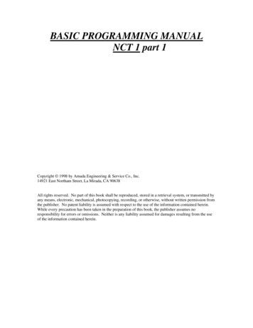
the T wave. With cardioversion you also need to check the height of the T wave on your monitorscreen before delivering the shock, because if the T wave is as tall as the QRS, a cardioversionshock could be delivered on the T wave. If this equal height exists, you need to put the patient ina lead where the QRS is the tallest complex.Finally, the supernormal period (which is really not a refractory period), is an interval of timeduring the latter portion of the T wave when it may be possible for a subthreshold stimulus(premature beat) to conduct.Self-Study Questions1. Describe the polarized cell.2. What happens during depolarization?3. Define repolarization.4. Name electrolytes involved with depolarization and repolarization.5. Will the ventricles respond to another impulse during the absoluterefractory period?6. Why may the vulnerable period be a time for concern?7. What is the super-normal period?VI. Action Potential 101The next figure demonstrates the activities within a single ventricular cell during depolarizationand repolarization. The upper figure shows the various numbered phases of electrical activitywithin the cell. The lower figure corresponds with the phases and tells the story of electrolytemovement within these phases. It should be noted that potassium appears to be quite activethroughout these phases. This is because there are at least seven different potassium currentswhich have been identified, each behaving in a slightly different manner.The upper figure (the action potential diagram) contains a horizontal line called the thresholdpotential. This line defines the strength an impulse must possess in order to successfullydepolarize a cell. A weak impulse will not be able to ‘jump’ this high. The height of the line13
may be lowered with low serum potassium level or low tissue oxygenation levels, allowing weakimpulses to cause ectopic beats or rhythms. A high serum potassium will ‘raise the bar’ andpossibly prevent even a normal SA node impulse from depolarizing the cell, leading to asystole.The baseline level of the action potential curve is -90mV until depolarization occurs, at whichtime the cell becomes electrically positive. The various numbered phases which will bedescribed below are referred to in discussion of antiarrhythmic drugs. Sodium channel blockersalter phase 1 of the action potential. Beta blockers and calcium channel blockers have an effecton phase two of SA node and AV node action potentials. Potassium channel blockers exertchanges on the various potassium current activities.Phase 0 of the action potential is the phase of rapid depolarization. During this phase the cell isstimulated and the cell membrane becomes more permeable to sodium ions. Fast sodiumchannels open and sodium rushes into the cell.In phase 1, known as the period of initial repolarization, there is a brief very rapid attempt toreturn to resting membrane potential. During this time sodium influx ends in fast channels andpotassium begins to flow out of the cell.Phase 2 is called the plateau phase. During this phase there is an influx of calcium and sodiumthrough the slow channels (though this sometimes also occurs during phases 0 and 1). Thisinflux is responsible for the refractory period during which the depolarized state is maintained.Calcium also plays a role in maintenance of normal heart muscle contractibility.Phase 3 is known as the phase of rapid repolarization. Membrane potential is driven to thenegative level due to the increased loss of intracellular potassium.Action potential of a ventricular cellPhase 4, the quiescent period, is the time when the cell returns to the resting membrane potentiallevel (-90 mV). During this period, normal distribution of sodium and potassium are restored.14
The sodium potassium pump plays a roll during these latter phases. ATP is required to fuel thispump and an adequate level of serum magnesium insures that the pump performs properly.Repolarization will be prolonged if the pump malfunctions, thus prolonging the QT interval onthe ECG. This may lead to a potentially lethal arrhythmia, torsade de pointes, a polymorphicventricular tachycardia. Treatment for this arrhythmia is intravenous magnesium.Local Variations in Action PotentialsThe next figure contrasts the action potential curves for each type of pacemaker cell in the heart.Notice that each curve has a somewhat different contour. The phases 0-4 occur with each ofthese. The SA node and atrial cells depolarize early and repolarize early, thus making thesefaster areas normally in control of the depolarization impulse, with a rate of 60-100 bpm.Local variations in action potentials15
The table below lists the various currents or channels operational during the cardiac cycle.Notice the various potassium related currents and the timing of their efflux. I current I current Ina(slow, medium, fast) Ina -Inward(slow,currentmedium, fast)-Inwardcurrent-Present in atrial, HPS, ventricular cells-Presentatrial,HPS,ventricular cells-TargetedinbyClassI drugs-Targetedby Class I drugs Slow conduction Slowconduction Prolong refractory period Prolong refractory period Ica Ica -Inward current, two types (L and T)-Inwardcurrent,intwo(L andT) cells-L-type presentSAtypesand AVnodal-L-type presentand AV nodal cells-Targetby ClassinIVSAdrugs-Target by Class IV drugs If funny current – automaticity If funny current – automaticity Ik Ik -Outward current-Outward-Present incurrentatrial, AV nodal, HPS and ventricular cells-Presentin atrial,nodal, HPS and ventricular cells-Manytypesof K AVcurrent-ManytypesofK current Transient outward Ito Transientrectifieroutward(existIto during the plateau) Delayed Delayed rectifier (exist during the plateau)-Ikr (rapid) seen in slower heart rate-Iseen inin fasterslowerheartheartratesrate-Ikr(slow) seenks (rapid)-Iks (slow) seen in faster heart rates Ik1 dominant background K outward current Ik1 dominant background K outward currentCurrents operational during the cardiac cycleSelf-Study Questions1. Describe the threshold potential and its importance.2. What is the role of potassium during the ventricular action potential?3. What occurs during phase 0 of the action potential?4. What is the baseline mV level of the ventricular action potential?5. What occurs during phase 1 of the action potential?6. What electrolyte is instrumental in causing the activity during phase 2 ofthe action potential?16
7. Describe the activities that occur during phase 4 of the ventricular actionpotential.8. Discuss the implications of the difference in action potential timing andshape among the various parts of the conduction system.VII. Mechanisms of ArrhythmiasThere are two major categories of rhythm problems:those that are fastthose that are slowSlowThe slow rhythm problems are called bradyarrhythmias or brachycardias. Two types ofmechanisms are said to be responsible for causing bradycardias:Failure of impulse formation (eg. The SA node fails or automaticity is depressed).These situations may lead to an asystolic situation, presence of a backup rhythm, slowerrhythms from the AV junction or ventricles, or the presence of sinus bradycardia.Failure of impulse propagation. With this problem, the impulse does not conductnormally to the ventricles. Example: AV block, when the impulse is prevented fromsuccessfully traveling through the AV node and ventricular rate slows.FastRhythm abnormalities that are fast are called tachycardias. There are three mechanismsresponsible for tachycardias: altered automaticity, reentry, and triggered activity.These mechanisms are not mutually exclusive: eg. triggered beats can:Engage a pre-existing reentrant circuitCreate arcs of functional block, generating substrate for reentryAutomatic RhythmsAutomaticity is the ability of cells to ‘automatically’ initiate an impulse. With alteredautomaticity there is a gradual increase of rate which takes place (a warming up), and then agradual decrease or ‘cooling down’. There may be an ‘enhanced normal’ as in sinus tachycardia,or an enhanced abnormal, as in atrial tachycardia. If there ap
Basic Electrophysiology Part 1 Directions for nursing CEU credit: Review the material in this module. Download the test form. Take the test. Fax or send the answer sheet with a form of payment. Passing score is 75%. You have 3 chances to take the test. Upon com