Transcription
Acquire. Process. Visualize.Analyze. Correlate. Store.ZEN Microscopy SoftwareYour Complete Solution from Sample to Knowledgezeiss.com/zen
Experience the End-To-EndMicroscopy Software›In Brief›The Advantages›The Applications›The System›Technology and Details› ServiceZEISS Efficient Navigation – ZEN, for short – is the universal user interface youwill see on every imaging system from ZEISS. No matter what microscopy taskyou have, you will find intuitive tools and modules to assist you: Acquire images using smart automationProcess images with scientifically proven algorithmsVisualize big data by GPU powered 3D engineAnalyze images via Machine Learning-based toolsCorrelate between light-light or light-electron microscopesStore raw data in a secure format locally or in the cloudDesigned to balance simplicity and flexibility for microscopy usersFor simple and routine works, ZEN leads you straight to result, simply and quickly.You will always see which options are available and which step is most appro priate to take next. The universal user interface lets you operate any ZEISS microscopes with the same graphical elements and workflows. As a result, you’ll savetime and reduce costs for training and support. For complex research experiments, ZEN offers the full flexibility to design multi dimensional workflows the way you wanted. The expert mode options openendless possibilities. Wanting even more customization? Try the Python-basedprogramming with an interface to compatible third-party software. Feelingoverwhelmed with the settings? The “Optimal” buttons let you quickly start withdefault parameters. You’ll never run out of ideas when designing experiments.2
Acquire. Process. Visualize.›In Brief›The Advantages›The Applications›The System›Technology and Details› ServiceSmart AcquisitionQuantitative ProcessingPowerful VisualizationMake your most complex experiments a success.Obtain reliable and reproducible results.Interact with your big data confidently.ZEN controls all ZEISS imaging systems so you canAcquiring stunning microscopy images is just theAdvances in microscopy automation, cameraoperate every one of your devices with the samebeginning of the experimental journey. More thantechnology, and light-sheet microscopy have fu-convenient interface. As a researcher, you might180 image processing tools, using transparent andeled the exponential growth of data size and thenot be familiar with all microscope components,scientifically proven algorithms, help you transformchallenge of big data visualization. Using ZEISSbut you surely know your sample and the dyesand manage your data. Simply search the keywordAxioscan 7 slide scanner, you can quickly gener-used. With “Smart Setup”, the intelligent controlof your intended method, e.g., kymograph orate a huge 2D tile scan with multi-channels atcenter, simply search and select the dyes and ZENdeconvolution, ZEN will lead you straight to it. Nothigh magnification. The powerful ZEN pyramidwill automatically generate the settings for you,familiar with those processing settings? ZEN willdata structure enables you to smoothly zoom inregardless of the microscope types. Aiming forread the metadata of the input image, then displayand out of such 2D data with a simple mousereproducible experiments? So long as you haveonly the logical processing steps, and optimize thescroll, just like using a map application on yourone raw image from a previous acquisition, adefault parameters automatically. You can evensmartphone. Having 200 GB of cleared mousesimple click lets you replicate the exact experi-process images from other platforms using ZENbrain data from ZEISS Lightsheet 7? Just load it inment precisely. And that’s just the start of ZEN’sthird-party import tools. With a dedicated work-ZEN – you will be amazed by the speed and claritysmart acquisition. You will find plenty of otherspace, you can also batch process multiple imagesof the various 3D rendering methods. Poweredsmart automation, some guide you for rare eventswith ease for quantitative and unbiased results.by arivis ImageCore technology and efficient usedetection, and others assist you with hardwareWith many of those processing tools in our freeof system resources, you can view your large 3Dcalibration.version, ZEN lite, you can comfortably carry on theimages even on regular consumer hardware.works on your laptops at home.3
Analyze. Correlate. Store.›In Brief›The Advantages›The Applications›The System›Technology and Details› ServiceEffortless AnalysisUnique Correlative MicroscopySecure StorageGain insight into your research efficiently.Combine perspectives across scales and modalities.Stay confident when dealing with complex data.Image analysis is essential to extract meaningful in-The fascinating variety of microscopy imagingData security gets top priority as ZEN stores eachformation from your microscopy images, via digitaltechnologies allows observing the same speci-of your valuable experiments with all its metadatatools of segmentation and registration. However,men faster, deeper, longer, and at a greater level(476 entries and counting). The proprietary .czibuilding a specific image analysis workflow thatof detail from different perspectives. Imagine thefile format is comprehensive, easy-to-work-with,adapts to one’s application is not an easy task. Itpossibilities for answering your scientific questionsbig data-supported, yet open. It is Bio-Formatsrequires knowledge of image processing, and thewhen you combine these images from multiplecompatible and can be read directly by ImageJ,ability to assemble a series of operations. ZEN ad-instruments, ZEISS or third-party. ZEN Connect isand many other third-party software. With adresses this imbalance with the simple and modu-your unique correlative microscopy solution thatsingle click, you can also export them into OME-lar BioApps modules. Each module is optimizedoverlays, navigates, and organizes your multimodalTIFF, the image format specification of the Openfor one type of application, e.g., cell counting ordata. Spending days painfully locating synapsesMicroscopy Environment, to further facilitateconfluency measurement, with tailored segmenta-between two special neurons for the ultrastructuralcross-platform image data exchange. Managingtion settings and streamlined data presentation.information? You can substantially improve yourmassive amounts of data in your facility? The ZENNeeding to build a special workflow? The wizard-efficiency by combining the large field of view ofData Storage module provides you a server-basedbased ZEN Image Analysis module guides you stepa widefield fluorescent microscope, and the highcentral platform to store and organize your rawby step to create your unique measurements. Youresolving power of a field emission electron micro-data and manage user access right; while the ZENcan also integrate our machine learning-basedscope. ZEN Connect lets you quickly overviewingData Explorer module grants user browser-basedsegmentation tool, Intellesis, into the workflow forthe whole brain slice with fluorescent markers,access from anywhere.the easy and accurate analysis of noisy images.identifying and relocating ROIs for EM imaging.4
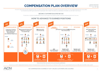
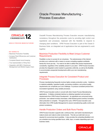
ZEN at Work›In Brief›The Advantages›The Applications›The System›Technology and DetailsTime Lapse Imaging› Service20 µmClick here to view this videoClick here to view this videoLiving cell with mitochondria in green and microtubule tips in red (EB3). ZEN allows to gain insightsLateral line primordium migration and deposition of immature neuromasts in a Zebrafish embryointo fast dynamic cellular processes while providing you with tools to process your data in the same(Danio rerio). Maximum intensity projection of 155 z-planes, acquired with Airyscan 2. Mebranes infamiliar user interface.green, actin in violet.ZEN allows you to observe your specimen longer and under more natural conditions than everbefore, as it precisely controls acquisition in widefield or LSM modes. No photons are wasted andimages are processed and restored to yield the highest signal to noise ratio.Sample courtesy of J. Hartmann and D. Gilmour, EMBL, Heidelberg, Germany5
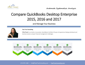
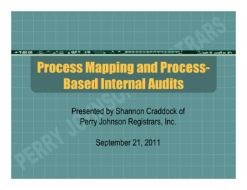
ZEN at Work›In Brief›The Advantages›The Applications›The System›Technology and DetailsLarge Area imaging› Service1000 µmTiled image of a mouse kidney section with four labels. ZEN provides you with the best strategyto optimize sample focus across large areas at high resolution. You get better images in shorter time.Brightfield images of a tissue section acquired using Live Panorama. ZEN automatically takes imagesand stitches them, while you manually navigate the area of interest on your sample.6
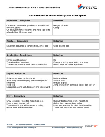
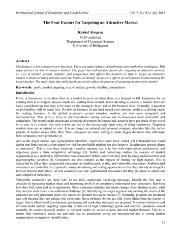
ZEN at Work›In Brief›The Advantages›The Applications›The System›Technology and Details3D Imaging› ServiceOrtho view of mouse brain slice, acquired with LSM 900.3D rendering of a Zebrafish embryo. Deconvolved Apotome Z stack.Z stack of the hippocampus area of a brain slide with neurons (green) and nuclei (red)With its powerful viewing and processing options ZEN gives you the insights into your specimen thatZEN assists you in finding the perfect spot in your sample and effortlessly handles large files so thatyou need to draw conclusions and to plan further experiments.you can always concentrate on examining your data.7
ZEN at Work›In Brief›The Advantages›The Applications›The System›Technology and DetailsImage Analysis› ServiceA 129 slice Z stack of cell nuclei, fully segmented and quantified, using APEER on-site in ZEN.Counting of DAB-positive (brown with green outline) cells in tissue sections and calculation of percentage of total cells (blue and brown): 36 %.8
ZEN Software Packages›In Brief›The Advantages›The Applications›The System›Technology and DetailsZEN systemZEN proZEN liteZEN deskSoftware package for all imaging systems,Software package for all non-laser basedFree basic version of ZEN with AxiocamOffline analyses, processing and visualization.including laser-based 3D imaging systems.standalone widefield systems.control that can be further extended with› Servicespecific modules.ZEN SlidescanZEN CelldiscovererZEN Lattice LightsheetZEN SEMTailored for ZEISS Axioscan 7 systemsTailored for ZEISS Celldiscoverer 7 systemsTailored for ZEISS Lattice Lightsheet 7Basic software for correlative modules thatsystems.allows SEM image acquisition.9
Your Flexible Choice of Modules›In Brief›The Advantages›The Applications›The System›Technology and DetailsGeneral Features of ZENGraphical user interface can be switched between bright and dark design to adapt to ambient brightness.User interface offers step-less scaling and zooming for optimal adjustment to the screen size.All functional elements can be displayed either in reduced- or full-sized mode.Full integration with ZEISS microscopes that can be configured in MicroToolbox, Axiocam cameras and additional componentsInteractive and automatic control of the individual motorized microscope componentsTransfer of information from the encoded component into the softwareComplex acquisition experiments can be configured, saved and reloaded.› ServiceMovie Recorder enables films to be acquired very simply with Start and Stop (interval setting and duration setting are not possible).Sequence of acquisition dimensions can be selected (depending on active dimensions).Hardware settings can be created with the help of a graphical light path.Sequences of commands can be easily combined to create hardware settings: Contains the Smart Setup function for the fully automatic creation of experiments to acquire multichannel fluorescence and transmitted light images using motorized systems. Image acquisition with b/w, color, high-resolution and high-sensitivity cameras, b/w images with up to 16 bits, color images with up to 3 16 bits.Display parameters can be adjusted without changing the pixel values.Assignment of geometric scaling is fully automatic when acquiring an image (depending on the microscope configuration).Acquisition history is recorded and saved as metadata in CZI image format. This format has been developed in consideration of the standards of the OME-TIFF and OME-XML format of theOpen Microscopy Environment. This allows far-reaching compatibility with the Bio-Formats Reader of the Open Microscopy Environment.Acquired images are automatically saved in CZI or other image formats (including metadata). Saving in CZI format is also possible in compressed form.Full integration into the Windows multi-user functionality (separation of user data and program installation, user-specific configurations, etc.)Configuration options for the graphical user interface enable creation of menu bars, saving of workplace configurations and definition of properties of standard graphic elements.Export into OME-TIFF (image format specification of the Open Microscopy Environment which enables the exchange of microscopic image data).Export into ZVI, BMP, GIF, JPG, PNG, TIFF, HDP image formats and export into AVI and Windows Media video formats. Batch export of images and videos.Image import (LSM, ZVI, BMP, TIFF, JPG, GIF, PNG) and function to convert images (TIFF, JPG, BMP) into CZI formatNavigator windowInteractive measurement: length, contour-based measurement data (area, box, perimeter, gray values), angleZEN Connect workspace with Project-based file architecture: Zoom in from the full macroscopic view of your sample down to nanoscale details. Combine data from any image source and view multiple layers withadjustable transparency. Manual alignment of images allows correction of xy-shift, rotation, re-scaling, shearing and mirroring.10
Your Flexible Choice of Modules›In BriefOptions/ModulesZEN liteBasisZENDevice controlControl of Axiocams›The Advantages›The Applications›The SystemControl of Apotome›Technology and DetailsControl of PMTs› ServiceZEN deskZEN proZEN systemControl of other camerasControl of motorized microscopesControl of 3rd party accessoriesControl of laser modulesAcquisitionMovie recorderManual extended focusPanoramaFast acquisitionDual cameraMulti channelTime lapseZ stackSoftware autofocusTile & PositionsExperiment designerGuided acquisitionCorrelative Array TomographyPhoton countingFCSAiryscanConfocal HDRConnectConnect basicConnectConnect 2D Add-onConnect 3D Add-onIncludedOptional11
Your Flexible Choice of Modules›In BriefOptions/ModulesProcessing›The Advantages›The Applications›The System›Technology and DetailsZEN liteZEN deskZEN proZEN systemDeconvolution basicSpectral unmixingDeconvolutionColocalizationExtended focusDirect processingAdvanced processing & analysisMacro environment› ServiceEM processing toolboxThird party importAiryscan processingLightsheet processingVisualization3D rendering basic3Dxl, powered by arivis3Dxl plus, powered by arivisAnalysisMeasurementBioApps (4x)Image analysisIntellesisIntellesis object classificationImage analysis IntellesisPhysiologyFRAP*FRETAPEER on-site basicAPEER on-site advancedStorageIncludedZEN Data Storage ClientOptional*available in image processing mode only12
Your Flexible Choice of Modules›In Brief›The Advantages›The Applications›The System›Technology and DetailsOptions/ModulesZEN SlidescanBasisZENDevice controlControl of AxiocamsZEN CelldiscovererZEN Lattice LightsheetZEN SEMControl of other camerasControl of motorized microscopesControl of 3rd party accessoriesControl of PMTs› ServiceControl of laser modulesAcquisitionFast acquisitionMulti channelTime lapseZ stackSoftware autofocusTile & PositionsExperiment designerGuided acquisitionCorrelative Array TomographyAutomated photomanipulationAiryscanConnectConnect basicConnectConnect 2D Add-onConnect 3D Add-onIncludedOptional13
Your Flexible Choice of Modules›In BriefOptions/ModulesProcessing›The Advantages›The Applications›The System›Technology and DetailsZEN SlidescanZEN CelldiscovererZEN Lattice LightsheetZEN SEMDeconvolution basicSpectral unmixingDeconvolutionColocalizationExtended focusDirect processingAdvanced processing & analysisMacro environment› ServiceEM processing toolboxThird party importAiryscan processingVisualization3D rendering basic3Dxl, powered by arivis3Dxl plus, powered by arivisAnalysisMeasurementBioApps (4x)Image analysisIntellesisIntellesis object classificationImage analysis IntellesisPhysiologyFRAPFRETAPEER on-site basicAPEER on-site advancedStorageIncludedZEN Data Storage ClientOptional14
Your Flexible Choice of Modules›In Brief›The Advantages›The Applications›The System›Technology and Details› hannelZ stackTime LapseAcquire fluorescence and transmitted light imagesAcquire Z stacks with the help of a motorizedAcquire images over a period of time:in independent channels:focus drive: Images acquired at maximum possible speed. Technically unlimited number of independent Definition of the Z stack in first and last or Definition of intervals between images, totalchannels for reflected light and transmittedlight techniques. Fully automatic generation of the required microscope setting for a channel with possi bility of adjusting the setting manuallyfor the channel. Independent exposure times and shading- corrections for each channel. Supports simultaneous acquisition of twocenter mode. Z stack limited only by the travel range of thesystem and minimum increments. Optimum Distance button sets the correctincrement to satisfy the Nyquist criterion. Integrated Z-drive backlash compensation formaximum precision. Z stack can be acquired at relative or absolutefocus positions in the experiment.acquisition duration and number of time points. Acquisition can be interrupted to analyze images already acquired or change the experiment parameters. Experiment size is limited only by free space onthe hard drive. Time series can be started and stopped manually, at fixed times, after a waiting period or byinput (trigger) signal. channels using two synchronized cameras.Click here to view this video15
Your Flexible Choice of Modules›In Brief›The Advantages›The Applications›The System›Technology and Details› ServiceACQUISITION:ACQUISITION:ACQUISITION:Tiles & PositionsManual Extended FocusSoftware AutofocusGenerate precise, high-resolution images throughGenerate images manually with no limit on depthDetermine the optimum focus position of theautomatic scanning of predefined regions andof field:specimen:positions of a sample: Extract sharp details from individual images at Works in transmitted light, reflected light and Regions of tile images and individual positionscan be combined freely. A motorized stage allows automatic scanningof specimens. Overlapping individual images can be combined into an overview image using “stitching”algorithms. Select from predefined or generate your ownvarious focus positions and combine them intoan image with extended depth of field. Works with images acquired interactively. Images can be added to the intermediate resultvia a time interval function or key function. Wavelet algorithm allows use in transmittedlight, reflected light and fluorescence.fluorescence. Calibration-free operation for all objectives andfilter sets. Options for adjusting the quality, search areaand sampling rate of the autofocus to the application. Autofocus can be activated automatically during the experiment at defined time intervals andmultiwell plates, multi-chamber slides, slideschannels, and at predefined tile positions orand dishes.individual positions. Intuitive focus strategy wizard to guide you tothe best focus.16
Your Flexible Choice of Modules›In Brief›The Advantages›The Applications›The System›Technology and Details› aExperiment DesignerGuided Acquisition*Generate precise, high-resolution overview imag-Configure inhomogeneous acquisition experi-Perform fully automated targeted acquisitiones from manually-acquired 2D individual images:ments:of objects of interest: Acquire overlapping individual multi channel Support for all experiment dimensions: time Save time and storage space by focusingimages interactively and combine them to forma panorama image on microscopes with anencoded or motorized stage. 3D panorama images can be acquired onstands with a motorized Z-drive. Live Panorama: ZEN automatically takes imagesand stitches them during sample navigation.series, Z stacks, tile images and channels. Operation via a simple graphical interface usingfour different types of experiment blocks alongthe image acquisition on objects of interest(e.g., rare events) only. Automate your workflow comprising ofa timeline: Acquisition, Execute, Pause andoverview scan, object detection via auto-Interaction blocks.mated image analysis, and high-resolution, Synchronous or asynchronous control of hardware actions during the experiment. Definition of a number of iteration loops. Set of powerful processing functions to extractor fuse multiblock images.multi- dimensional image acquisition for eachdetected object. Customize focusing strategies for both overview scan and detailed acquisition. Automatically save all images, tables and settings in one folder for easy access and reuse.* requires ZEN module Image Analysis17
Your Flexible Choice of Modules›In Brief›The Advantages›The Applications›The System›Technology and Details› ve Array Tomography (CAT)Third Party ImportDirect ProcessingImage ultrathin serial sections automatically inImport third-party microscopy images into ZEN:Perform time-consuming image processing taskswidefield and scanning electron microscopes: Imports third-party images in native formatsimultaneously during image acquisition: Regions of interest defined manually in oneincluding extraction of relevant metadata.section will be automatically propagated to allfollowing sections. Selected regions of interest can be imaged inlight and electron microscopes. The 2D image sequences are aligned into a 3DZ stack, resulting in a correlative data set combining information from the light and electronmicroscopes into one image volume. Supports metadata extraction depending onthe original format (powered by Bio-Formats). Deblurring for fast and easy 2D backgroundremoval with truly quantitative output. Supports a selection of processing methods,such as deconvolution, Airyscan processing,raw convert, denoising or unsharp mask. Employs pipeline to set up a sequence of imageprocessing functions. Remote processing to maximize computationalresources during acquisition. Instantaneous side-by-side comparison of rawand processed data.Click here to view this video18
Your Flexible Choice of Modules›In Brief›The Advantages›The Applications›The System›Technology and Details› onExtended FocusEM Processing ToolboxUse 3D deconvolution algorithms to enhance yourGenerate images with no limitation of depth of fieldSelect from a list of processing tools to improve3D image stacks: Extraction of the sharp details from individualyour EM datasets: Efficient multi-CPU based processing Additional speed gain via GPU acceleration withdedicated CUDA-compatible graphics card. Improvements in resolution down to 120 nm(depending on imaging system). Compatible with conventional widefield, Apotome, Lightsheet 7, confocal or multi photon microscopes. Choice of four primary methods, plus more than15 published methods (e.g., Richardson-Lucy)can be employed by changing the parameters.images at various focus positions and combination into an image with high depth of field. Processes Z stacks that have already beenacquired. Wavelet algorithm allows use in transmittedlight, reflected light and fluorescence Easily import EM images acquired by SmartSEMadditionally to the well-known ZEN ModuleThird Party Import. Remove artifacts such as noise and stripes Reconstruct a 3D dataset out of subsequent2D images: Equalize and align your datasets bythe automatic z-alignment tool. Replace individual slices of poor qualitywithin the 3D Z stack. Cut out free-form 3D regions of interestin order to remove unwanted areas fromthe EM dataset for a more customized3D v isualization.19
Your Flexible Choice of Modules›In Brief›The Advantages›The Applications›The System›Technology and Details› ServicePROCESSING:PROCESSING:PROCESSING:Advanced Processing & Analysis*APEER On-siteMacro EnvironmentExtend the processing functions and performUse APEER modules fully integrated in ZENCustomize and automate ZEN using powerfulfeedback experiments Use public and private APEER modules to en-Python scripts: Add more collections of image processingfunctions: Edges, Arithmetics, Morphology,Segmentation and Binary. Experiment feedback allows to adapt and modify running experiments using Pythonscripts. Access results from the online image analysisable additional processing and analysis featuresand workflows in ZEN incl. Python scripting. Package your own tools into an APEER moduleand use them inside ZEN. Remote execution within your local IT infrastructure is supported. Execute customized and open-source imageduring runtime of the experiment as well asanalysis functions in ZEN (on-site), providedthe current system status.via APEER, the cloud-based image and data Data Logging or starting of external applica- Integrated script editor with debugging, recording and code completion. Integration of APEER modules and externalsoftware packages like Python, MATLAB or Fijiin an automated workflow is easily possible. Uses IronPython in order to integrate.NET-based functions.processing platform.**tions like Python, Fiji or MATLAB directly fromwithin the imaging experiment.* requires the ZEN Module Image AnalysisContact us: apeer-solutions@zeiss.comImageJFIJIMATLABPython** If you need support developing customized solutions, we havea team of data scientists to rapidly develop applications usingtraditional and machine learning tools.20
Your Flexible Choice of Modules›In Brief›The Advantages›The Applications›The System›Technology and Details› Dxl Plus*MeasurementsVisualize 3D/4D image stacks:This module introduces the Tomo3D viewer:Perform interactive measurements Display 3D volume models using efficient ray Render up to three 2D and one 3D view panes Measure morphological parameters of interac-tracing technology, even for large data. Display up to 6 channels and time series. Choose from five rendering methods: Transparency, Volume, Max Intensity Projection,Surface, mixed and with up to three clippingplanes. Improved transparency mode for better visualization of dense structures, such as EM,XRM and dense fluorescent data. Bridge functionality: Send to arivis vision4Dwith saved settings and sample pipelines fortogether. 3D view features ray-casting-based volumetively defined contours: area, orientation angle,perimeter, diameter, center of gravity, radius ofrendering with transparency, volume and maxi-circle with equal area, shape factor, boundingmum intensity modes.box, projections, etc. Flexible channel-wise adjustment of 3D view,background color and lighting. The position of the three orthogonal 2D viewpanes are synchronized with the 3D view. Navigation through the sample and projectionviews via colored cut lines. Measure intensity values for rectangles andcontours. Free configuration of all interactive measurement tools displays desired parameters intables, lists or graphs. Option for interactive measurement in onlineimages.fast and easy 3D analysis. Generate animations.* powered by arivis21
Your Flexible Choice of Modules›In Brief›The Advantages›The Applications›The System›Technology and Details› ServiceANALYSIS:ANALYSIS:ANALYSIS:Image AnalysisCell CountingAutomated Spot DetectionCreate an automated measurement programFrom our BioApps portfolio delivering out-of-From our BioApps portfolio delivering out-of-guided by an intuitive software wizard:the-box image analysis, tailored result presen-the-box image analysis, tailored result presen- Flexible class and subclass definitiontation with interactive measurement tables,tation with interactive measurement tables, Segment objects either by selecting a fewheatmaps and plots.heatmaps and plots. Automatically detect fluorescently labelled cell Automatically quantify spots in the cell nuclei.reference objects, by automatic thresholding orbased on machine learning. Refine results with additional object separationand filters. Measure geometric and intensity features ofindividual objects or in the entire image. Examine results at one glance using interactiveplotting (line chart, histogram, scatter plot,nuclei in biological samples. Suitable for measuring proliferation or survival. Measure cell counts, nuclear intensities, meanintensities and mean areas. Optimized for measurements in screening applications with multi-well setups. Applicable for FISH applications, telomere / centromere analysis or focus counting. Measure the total number of spots, the averagenumber of spots per cell or the mean intensityof spots. Optimized for measurements in screening applications with multi-well setups.heatmap), with a direct link between images,tables and graphs. Export results in tables or lists for furtheranalysis.22
Your Flexible Choice of Modules›In Brief›The Advantages›The Applications›The System›Technology and Details› ServiceANALYSIS:ANALYSIS:ANALYSIS:ConfluencyGene and Protein ExpressionIntellesisFrom our BioApps portfolio delivering out-of-From our BioApps portfolio delivering out-of-Enable machine-learning algorithms to segmentthe-box image analysis, tailored result presen-the-box image analysis, tailored result presen-images:tation with interactive measurement tables,tation with interactive measurement tables, Train a simple image segmentation modelheatmaps and plots.heatmaps and plots.labelli
For complex research experiments, ZEN offers the full flexibility to design multi-dimensional workflows the way you wanted. The expert mode options open endless possibilities. Wanting even more customization? Try the Python-based programming with an interface to compatible thi