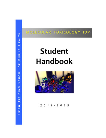
Transcription
16The MolecularBasis of Inheritanceyou inherited from your parents. DNA, the substance of inheritance, is the most celebrated molecule of our time.Of all nature’s molecules, nucleic acids are unique in theirability to direct their own replication from monomers. Indeed, the resemblance of offspring to their parents has itsbasis in the precise replication of DNA and its transmissionfrom one generation to the next. Hereditary information isencoded in the chemical language of DNA and reproduced inall the cells of your body. It is this DNA program that directsthe development of your biochemical, anatomical, physiological, and, to some extent, behavioral traits. In this chapter,you will discover how biologists deduced that DNA is the genetic material and how Watson and Crick worked out itsstructure. You will also learn about DNA replication, theprocess by which a DNA molecule is copied, and how cells repair their DNA. Finally, you will explore how a molecule ofDNA is packaged together with proteins in a chromosome.CONCEPT16.1DNA is the genetic materialToday, even schoolchildren have heard of DNA, and scientists routinely manipulate DNA in the laboratory, often tochange the heritable traits of cells in their experiments. Earlyin the 20th century, however, identifying the molecules ofinheritance loomed as a major challenge to biologists.The Search for the Genetic Material:Scientific Inquiry䉱 Figure 16.1 How was the structure ofDNA determined?KEY CONCEPTS16.1 DNA is the genetic material16.2 Many proteins work together in DNA replicationand repair16.3 A chromosome consists of a DNA moleculepacked together with proteinsOVERVIEWLife’s Operating InstructionsIn April 1953, James Watson and Francis Crick shook the scientific world with an elegant double-helical model for thestructure of deoxyribonucleic acid, or DNA. Figure 16.1 showsWatson (left) and Crick admiring their DNA model, whichthey built from tin and wire. Over the past 60 years or so,their model has evolved from a novel proposition to an iconof modern biology. Mendel’s heritable factors and Morgan’sgenes on chromosomes are, in fact, composed of DNA.Chemically speaking, your genetic endowment is the DNAOnce T. H. Morgan’s group showed that genes exist as parts ofchromosomes (described in Chapter 15), the two chemicalcomponents of chromosomes—DNA and protein—becamethe candidates for the genetic material. Until the 1940s, thecase for proteins seemed stronger, especially since biochemistshad identified them as a class of macromolecules with greatheterogeneity and specificity of function, essential requirements for the hereditary material. Moreover, little was knownabout nucleic acids, whose physical and chemical propertiesseemed far too uniform to account for the multitude ofspecific inherited traits exhibited by every organism. This viewgradually changed as experiments with microorganismsyielded unexpected results. As with the work of Mendel andMorgan, a key factor in determining the identity of the geneticmaterial was the choice of appropriate experimental organisms. The role of DNA in heredity was first worked out whilestudying bacteria and the viruses that infect them, which arefar simpler than pea plants, fruit flies, or humans. In this section, we will trace the search for the genetic material in somedetail as a case study in scientific inquiry.Evidence That DNA Can Transform BacteriaThe discovery of the genetic role of DNA dates back to 1928.While attempting to develop a vaccine against pneumonia,CHAPTER 16The Molecular Basis of Inheritance305
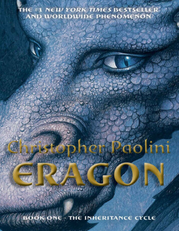
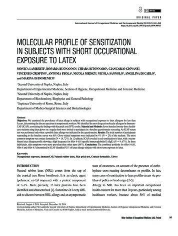
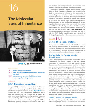
a British medical officer named Frederick Griffith was studying Streptococcus pneumoniae, a bacterium that causes pneumonia in mammals. Griffith had two strains (varieties) of thebacterium, one pathogenic (disease-causing) and one nonpathogenic (harmless). He was surprised to find that when hekilled the pathogenic bacteria with heat and then mixed thecell remains with living bacteria of the nonpathogenic strain,some of the living cells became pathogenic (Figure 16.2). Furthermore, this newly acquired trait of pathogenicity wasINQUIRY䉲 Figure 16.2Can a genetic trait be transferred betweendifferent bacterial strains?EXPERIMENT Frederick Griffith studied two strains of the bacteriumStreptococcus pneumoniae. Bacteria of the S (smooth) strain can causepneumonia in mice; they are pathogenic because they have an outer capsule that protects them from an animal’s defense system. Bacteria of the R(rough) strain lack a capsule and are nonpathogenic. To test for the trait ofpathogenicity, Griffith injected mice with the two strains:Living S cells(pathogeniccontrol)Heat-killedLiving R cellsS cells(nonpathogenic (nonpathogeniccontrol)control)Mixture ofheat-killedS cells andliving R cellsinherited by all the descendants of the transformed bacteria.Clearly, some chemical component of the dead pathogeniccells caused this heritable change, although the identity of thesubstance was not known. Griffith called the phenomenontransformation, now defined as a change in genotype andphenotype due to the assimilation of external DNA by a cell.(This use of the word transformation should not be confusedwith the conversion of a normal animal cell to a cancerousone, discussed near the end of Concept 12.3)Griffith’s work set the stage for a 14-year effort by American bacteriologist Oswald Avery to identify the transformingsubstance. Avery focused on three main candidates: DNA,RNA (the other nucleic acid in cells), and protein. Averybroke open the heat-killed pathogenic bacteria and extractedthe cellular contents. He treated each of three samples withan agent that inactivated one type of molecule, then testedthe sample for its ability to transform live nonpathogenicbacteria. Only when DNA was allowed to remain active didtransformation occur. In 1944, Avery and his colleaguesMaclyn McCarty and Colin MacLeod announced that thetransforming agent was DNA. Their discovery was greetedwith interest but considerable skepticism, in part because ofthe lingering belief that proteins were better candidates forthe genetic material. Moreover, many biologists were notconvinced that the genes of bacteria would be similar incomposition and function to those of more complex organisms. But the major reason for the continued doubt was thatso little was known about DNA.Evidence That Viral DNA Can Program CellsAdditional evidence for DNA as the genetic material camefrom studies of viruses that infect bacteria (Figure 16.3).These viruses are called bacteriophages (meaning “bacteriaeaters”), or phages for short. Viruses are much simpler thanRESULTSMouse diesMouse healthy Mouse healthyMouse diesPhageheadIn blood sample,living S cells arefound that canreproduce,yielding moreS cells.TailsheathTail fibertransformed into pathogenic S bacteria by an unknown, heritable substance from the dead S cells that allowed the R cells to make capsules.SOURCE F. Griffith, The significance of pneumococcal types, Journal ofHygiene 27:113–159 (1928).WHAT IF? How did this experiment rule out the possibility that theR cells could have simply used the capsules of the dead S cells to become pathogenic?306UNIT THREEGeneticsBacterialcell100 nmDNACONCLUSION Griffith concluded that the living R bacteria had been䉱 Figure 16.3 Viruses infecting a bacterial cell. Phages calledT2 attach to the host cell and inject their genetic material through theplasma membrane while the head and tail parts remain on the outerbacterial surface (colorized TEM).
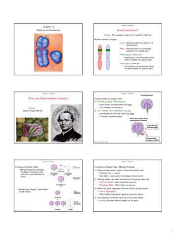
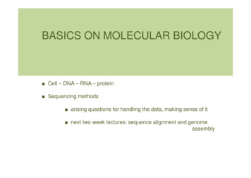
cells. A virus is little more than DNA (or sometimes RNA)enclosed by a protective coat, which is often simply protein.To produce more viruses, a virus must infect a cell and takeover the cell’s metabolic machinery.Phages have been widely used as tools by researchers in molecular genetics. In 1952, Alfred Hershey and Martha Chaseperformed experiments showing that DNA is the genetic material of a phage known as T2. This is one of many phages thatinfect Escherichia coli (E. coli), a bacterium that normally livesin the intestines of mammals and is a model organism for molecular biologists. At that time, biologists already knew that T2,like many other phages, was composed almost entirely of DNAand protein. They also knew that the T2 phage could quicklyturn an E. coli cell into a T2-producing factory that releasedmany copies when the cell ruptured. Somehow, T2 couldreprogram its host cell to produce viruses. But which viralcomponent—protein or DNA—was responsible?Hershey and Chase answered this question by devising anexperiment showing that only one of the two components ofT2 actually enters the E. coli cell during infection (Figure 16.4).In their experiment, they used a radioactive isotope of sulfur totag protein in one batch of T2 and a radioactive isotope ofINQUIRY䉲 Figure 16.4Is protein or DNA the genetic material of phage T2?EXPERIMENT Alfred Hershey and Martha Chase used radioactive sulfur and phosphorus to tracethe fates of protein and DNA, respectively, of T2 phages that infected bacterial cells. They wanted tosee which of these molecules entered the cells and could reprogram them to make more phages.1 Mixed radioactivelylabeled phages withbacteria. The phagesinfected the bacterial cells.Phage3 Centrifuged the mixture 4 Measured theradioactivity inso that bacteria formed athe pellet andpellet at the bottom ofthe liquid.the test tube; free phagesand phage parts, whichare lighter, remainedRadioactivitysuspended in the liquid.(phage protein)in liquid2 Agitated the mixture ina blender to free phageparts outside thebacteria from the cells.RadioactiveproteinEmptyprotein shellBacterial cellBatch 1: Phages weregrown with radioactivesulfur (35S), which wasincorporated into phageprotein (pink).DNAPhageDNACentrifugePellet (bacterialcells and contents)RadioactiveDNABatch 2: Phages weregrown with radioactivephosphorus (32P), whichwas incorporated intophage DNA (blue).CentrifugePelletRESULTS When proteins were labeled (batch 1), radioactivity remained outside the cells; but whenRadioactivity(phage DNA)in pelletDNA was labeled (batch 2), radioactivity was found inside the cells. Bacterial cells with radioactivephage DNA released new phages with some radioactive phosphorus.CONCLUSION Phage DNA entered bacterial cells, but phage proteins did not. Hershey and Chaseconcluded that DNA, not protein, functions as the genetic material of phage T2.SOURCE A. D. Hershey and M. Chase, Independent functions of viral protein and nucleic acid ingrowth of bacteriophage, Journal of General Physiology 36:39–56 (1952).WHAT IF?How would the results have differed if proteins carried the genetic information?CHAPTER 16The Molecular Basis of Inheritance307
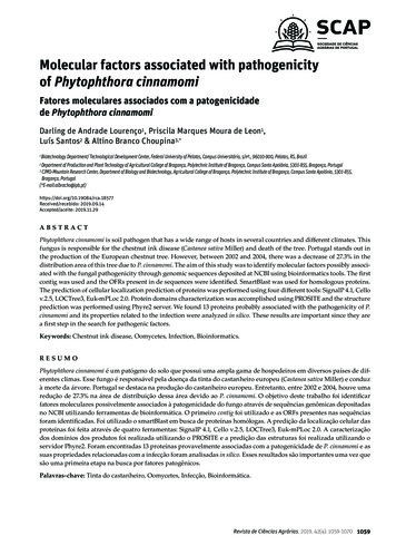
phosphorus to tag DNA in a second batch. Because protein,but not DNA, contains sulfur, radioactive sulfur atoms wereincorporated only into the protein of the phage. In a similarway, the atoms of radioactive phosphorus labeled only theDNA, not the protein, because nearly all the phage’s phosphorus is in its DNA. In the experiment, separate samples of nonradioactive E. coli cells were allowed to be infected by theprotein-labeled and DNA-labeled batches of T2. The researchers then tested the two samples shortly after the onsetof infection to see which type of molecule—protein or DNA—had entered the bacterial cells and would therefore be capableof reprogramming them.Hershey and Chase found that the phage DNA entered thehost cells but the phage protein did not. Moreover, when thesebacteria were returned to a culture medium, the infection ranits course, and the E. coli released phages that contained someradioactive phosphorus, further showing that the DNA insidethe cell played an ongoing role during the infection process.Hershey and Chase concluded that the DNA injected by thephage must be the molecule carrying the genetic informationthat makes the cells produce new viral DNA and proteins. TheHershey-Chase experiment was a landmark study because itprovided powerful evidence that nucleic acids, rather thanproteins, are the hereditary material, at least for viruses.5‘ endOO–P–OCH3O4‘HUNIT NAnucleotideH2‘NNOCH2HOPPhosphateCytosine (C)NHHO–OHHHAdenine (A)HOCH2HNHPOHNOCH2–ONOOONH3‘OAdditional Evidence That DNA Is the Genetic Material3081‘HP–OThymine (T)OHOOH5‘CH2OHFurther evidence that DNA is the genetic material came fromthe laboratory of biochemist Erwin Chargaff. It was alreadyknown that DNA is a polymer of nucleotides, each consistingof three components: a nitrogenous (nitrogen-containing)base, a pentose sugar called deoxyribose, and a phosphategroup (Figure 16.5). The base can be adenine (A), thymine(T), guanine (G), or cytosine (C). Chargaff analyzed the basecomposition of DNA from a number of different organisms.In 1950, he reported that the base composition of DNA variesfrom one species to another. For example, 30.3% of humanDNA nucleotides have the base A, whereas DNA from thebacterium E. coli has only 26.0% A. This evidence of molecular diversity among species, which had been presumed absent from DNA, made DNA a more credible candidate for thegenetic material.Chargaff also noticed a peculiar regularity in the ratios ofnucleotide bases. In the DNA of each species he studied, thenumber of adenines approximately equaled the number ofthymines, and the number of guanines approximatelyequaled the number of cytosines. In human DNA, for example, the four bases are present in these percentages: A 30.3% and T 30.3%; G 19.5% and C 19.9%.These two findings became known as Chargaff’s rules: (1) thebase composition varies between species, and (2) within aspecies, the number of A and T bases are equal and the number of G and C bases are equal. The basis for these rules remained unexplained until the discovery of the double helix.Nitrogenous basesSugar–phosphate backbone1‘ONHHSugar(deoxyribose)Guanine (G)NHNNHHNitrogenous base3‘ end䉱 Figure 16.5 The structure of a DNA strand. Each DNAnucleotide monomer consists of a nitrogenous base (T, A, C, or G), thesugar deoxyribose (blue), and a phosphate group (yellow). The phosphategroup of one nucleotide is attached to the sugar of the next, forming a“backbone” of alternating phosphates and sugars from which the basesproject. The polynucleotide strand has directionality, from the 5 end (withthe phosphate group) to the 3 end (with the —OH group of the sugar).5 and 3 refer to the numbers assigned to the carbons in the sugar ring.Building a Structural Model of DNA:Scientific InquiryOnce most biologists were convinced that DNA was the geneticmaterial, the challenge was to determine how the structure ofDNA could account for its role in inheritance. By the early1950s, the arrangement of covalent bonds in a nucleic acidpolymer was well established (see Figure 16.5), and researchersfocused on discovering the three-dimensional structure ofDNA. Among the scientists working on the problem were LinusPauling, at the California Institute of Technology, and MauriceWilkins and Rosalind Franklin, at King’s College in London.First to come up with the correct answer, however, were twoscientists who were relatively unknown at the time—the American James Watson and the Englishman Francis Crick.
The brief but celebrated partnership that solved the puzzle ofDNA structure began soon after Watson journeyed to CambridgeUniversity, where Crick was studying protein structure with atechnique called X-ray crystallography (see Figure 5.24). Whilevisiting the laboratory of Maurice Wilkins, Watson saw an X-raydiffraction image of DNA produced by Wilkins’s accomplishedcolleague Rosalind Franklin (Figure 16.6a). Images produced byX-ray crystallography are not actually pictures of molecules. Thespots and smudges in Figure 16.6b were produced by X-rays thatwere diffracted (deflected) as they passed through aligned fibersof purified DNA. Watson was familiar with the type of X-ray diffraction pattern that helical molecules produce, and an examination of the photo that Wilkins showed him confirmed thatDNA was helical in shape. It also augmented earlier data obtained by Franklin and others suggesting the width of the helixand the spacing of the nitrogenous bases along it. The pattern inthis photo implied that the helix was made up of two strands,contrary to a three-stranded model that Linus Pauling had proposed a short time earlier. The presence of two strands accountsfor the now-familiar term double helix (Figure 16.7).Watson and Crick began building models of a double helixthat would conform to the X-ray measurements and what wasthen known about the chemistry of DNA, including O0.34 nmOPO3‘ endAOGOTOPOOPOHACH2OH2CGOCOOGGOPOO1 nmCCH2OP-OAAOH2CTTOOACOHOH2CG3‘ endOP-OCCrule of base equivalences. Having also read an unpublishedannual report summarizing Franklin’s work, they knew shehad concluded that the sugar-phosphate backbones were onthe outside of the DNA molecule, contrary to their workingmodel. Franklin’s arrangement was appealing because it put theHydrogen bondH2C3.4 nmG䉱 Figure 16.6 Rosalind Franklin and her X-ray diffractionphoto of DNA. Franklin, a very accomplished X-ray crystallographer,conducted critical experiments resulting in the photograph that allowedWatson and Crick to deduce the double-helical structure of DNA.OCCT(b) Franklin’s X-ray diffractionphotograph of DNA5‘ endGGG(a) Rosalind FranklinOPO5‘ end(a) Key features of DNA structure. The(b) Partial chemical structure. For clarity, the two DNA strands“ribbons” in this diagram represent theare shown untwisted in this partial chemical structure. Strongcovalent bonds link the units of each strand, while weakersugar-phosphate backbones of the twoDNA strands. The helix is “right-handed,”hydrogen bonds hold one strand to the other. Notice that thestrands are antiparallel, meaning that they are oriented incurving up to the right. The two strandsare held together by hydrogen bondsopposite directions.(dotted lines) between the nitrogenousbases, which are paired in the interior ofthe double helix.(c) Space-filling model. Thetight stacking of the basepairs is clear in this computermodel. Van der Waalsinteractions between thestacked pairs play a majorrole in holding the moleculetogether (see Chapter 2).䉱 Figure 16.7 The double helix.CHAPTER 16The Molecular Basis of Inheritance309
relatively hydrophobic nitrogenous bases in the molecule’s interior, away from the surrounding aqueous solution, and thenegatively charged phosphate groups wouldn’t be forced together in the interior. Watson constructed a model with thenitrogenous bases facing the interior of the double helix. In thismodel, the two sugar-phosphate backbones are antiparallel—that is, their subunits run in opposite directions (seeFigure 16.7). You can imagine the overall arrangement as a ropeladder with rigid rungs. The side ropes represent the sugarphosphate backbones, and the rungs represent pairs of nitrogenous bases. Now imagine holding one end of the ladder andtwisting the other end, forming a spiral. Franklin’s X-ray data indicated that the helix makes one full turn every 3.4 nm along itslength. With the bases stacked just 0.34 nm apart, there are ten layers of base pairs, or rungs of the ladder, in each full turn of the helix.The nitrogenous bases of the double helix are paired in specific combinations: adenine (A) with thymine (T), and guanine(G) with cytosine (C). It was mainly by trial and error thatWatson and Crick arrived at this key feature of DNA. At first,Watson imagined that the bases paired like with like—for example, A with A and C with C. But this model did not fit theX-ray data, which suggested that the double helix had a uniform diameter. Why is this requirement inconsistent with likewith-like pairing of bases? Adenine and guanine are purines,nitrogenous bases with two organic rings, while cytosine andthymine are nitrogenous bases called pyrimidines, which havea single ring. Thus, purines (A and G) are about twice as wideas pyrimidines (C and T). A purine-purine pair is too wide anda pyrimidine-pyrimidine pair too narrow to account for the2-nm diameter of the double helix. Always pairing a purinewith a pyrimidine, however, results in a uniform diameter:Purine purine: too wideHN310UNIT THREEGeneticsNHNNOSugarThymine (T)Adenine (A)HNSugarNHONNHNNHONNSugarHGuanine (G)Cytosine (C)䉱 Figure 16.8 Base pairing in DNA. The pairs of nitrogenousbases in a DNA double helix are held together by hydrogen bonds,shown here as black dotted lines.adenine equals the amount of thymine, and the amount ofguanine equals the amount of cytosine. Although the basepairing rules dictate the combinations of nitrogenous basesthat form the “rungs” of the double helix, they do not restrictthe sequence of nucleotides along each DNA strand. The linearsequence of the four bases can be varied in countless ways, andeach gene has a unique order, or base sequence.In April 1953, Watson and Crick surprised the scientific worldwith a succinct, one-page paper in the journal Nature.* Thepaper reported their molecular model for DNA: the double helix,which has since become the symbol of molecular biology. Watson and Crick, along with Maurice Wilkins, were awarded theNobel Prize in 1962 for this work. (Sadly, Rosalind Franklin diedat the age of 38, in 1958, and was thus ineligible for the prize.)The beauty of the double helix model was that the structure ofDNA suggested the basic mechanism of its replication.CONCEPT CHECKWatson and Crick reasoned that there must be additionalspecificity of pairing dictated by the structure of the bases.Each base has chemical side groups that can form hydrogenbonds with its appropriate partner: Adenine can form two hydrogen bonds with thymine and only thymine; guanine formsthree hydrogen bonds with cytosine and only cytosine. Inshorthand, A pairs with T, and G pairs with C (Figure 16.8).The Watson-Crick model took into account Chargaff’s ratios and ultimately explained them. Wherever one strand of aDNA molecule has an A, the partner strand has a T. And a G inone strand is always paired with a C in the complementarystrand. Therefore, in the DNA of any organism, the amount ofCH3OHNSugarPyrimidine pyrimidine: too narrowPurine pyrimidine: widthconsistent with X-ray dataNN16.11. A fly has the following percentages of nucleotides inits DNA: 27.3% A, 27.6% T, 22.5% G, and 22.5% C.How do these numbers demonstrate Chargaff’s ruleabout base ratios?2. Given a polynucleotide sequence such as GAATTC,can you tell which is the 5 end? If not, what furtherinformation do you need to identify the ends? (SeeFigure 16.5.)3. WHAT IF? Griffith did not expect transformation tooccur in his experiment. What results was he expecting? Explain.For suggested answers, see Appendix A.*J. D. Watson and F. H. C. Crick, Molecular structure of nucleic acids: a structure for deoxyribose nucleic acids, Nature 171:737–738 (1953).
CONCEPT16.2Figure 16.9 illustrates Watson and Crick’s basic idea. ToMany proteins work togetherin DNA replication and repairThe relationship between structure and function is manifestin the double helix. The idea that there is specific pairing ofnitrogenous bases in DNA was the flash of inspiration thatled Watson and Crick to the double helix. At the same time,they saw the functional significance of the base-pairing rules.They ended their classic paper with this wry statement: “Ithas not escaped our notice that the specific pairing we havepostulated immediately suggests a possible copying mechanism for the genetic material.” In this section, you will learnabout the basic principle of DNA replication, as well as someimportant details of the process.The Basic Principle:Base Pairing to a Template StrandIn a second paper, Watson and Crick stated their hypothesisfor how DNA replicates:Now our model for deoxyribonucleic acid is, in effect, a pair oftemplates, each of which is complementary to the other. Weimagine that prior to duplication the hydrogen bonds are broken, and the two chains unwind and separate. Each chainthen acts as a template for the formation onto itself of a newcompanion chain, so that eventually we shall have two pairsof chains, where we only had one before. Moreover, the sequence of the pairs of bases will have been duplicated exactly.*make it easier to follow, we show only a short section of double helix in untwisted form. Notice that if you cover one of thetwo DNA strands of Figure 16.9a, you can still determine itslinear sequence of nucleotides by referring to the uncoveredstrand and applying the base-pairing rules. The two strands arecomplementary; each stores the information necessary to reconstruct the other. When a cell copies a DNA molecule, eachstrand serves as a template for ordering nucleotides into anew, complementary strand. Nucleotides line up along thetemplate strand according to the base-pairing rules and arelinked to form the new strands. Where there was one doublestranded DNA molecule at the beginning of the process, thereare soon two, each an exact replica of the “parental” molecule.The copying mechanism is analogous to using a photographicnegative to make a positive image, which can in turn be usedto make another negative, and so on.This model of DNA replication remained untested for several years following publication of the DNA structure. Therequisite experiments were simple in concept but difficult toperform. Watson and Crick’s model predicts that when adouble helix replicates, each of the two daughter moleculeswill have one old strand, from the parental molecule, andone newly made strand. This semiconservative modelcan be distinguished from a conservative model of replication, in which the two parental strands somehow come backtogether after the process (that is, the parental molecule isconserved). In yet a third model, called the dispersive model,all four strands of DNA following replication have a mixtureof old and new DNA. These three models are shown in*F. H. C. Crick and J. D. Watson, The complementary structure of deoxyribonucleic acid, Proceedings of the Royal Society of London A 223:80 (1954).ATATATATCGCGCGCGTATATATAATATATATGCGCGCGC(a) The parental molecule has two complementary strands of DNA. Each base is paired byhydrogen bonding with its specific partner,A with T and G with C.(b) The first step in replication is separation ofthe two DNA strands. Each parental strandcan now serve as a template thatdetermines the order of nucleotides alonga new, complementary strand.(c) The complementary nucleotides line up andare connected to form the sugar-phosphatebackbones of the new strands. Each“daughter” DNA molecule consists of oneparental strand (dark blue) and one newstrand (light blue).䉱 Figure 16.9 A model for DNA replication: the basic concept. In this simplifiedillustration, a short segment of DNA has been untwisted into a structure that resembles a ladder. Theside rails of the ladder are the sugar-phosphate backbones of the two DNA strands; the rungs arethe pairs of nitrogenous bases. Simple shapes symbolize the four kinds of bases. Dark blue representsDNA strands present in the parental molecule; light blue represents newly synthesized DNA.CHAPTER 16The Molecular Basis of Inheritance311
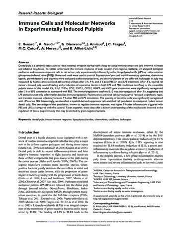
Parent cellFirstreplicationSecondreplication(a) Conservativemodel. The twoparental strandsreassociate afteracting astemplates fornew strands,thus restoringthe parentaldouble helix.Does DNA replication follow the conservative,semiconservative, or dispersive model?EXPERIMENT At the California Institute of Technology, MatthewMeselson and Franklin Stahl cultured E. coli for several generations in amedium containing nucleotide precursors labeled with a heavy isotopeof nitrogen, 15N. They then transferred the bacteria to a medium withonly 14N, a lighter isotope. A sample was taken after DNA replicatedonce; another sample was taken after DNA replicated again. They extracted DNA from the bacteria in the samples and then centrifugedeach DNA sample to separate DNA of different densities.1 Bacteriacultured inmediumwith15N (heavyisotope)(b) Semiconservative model.The two strandsof the parentalmoleculeseparate, andeach functionsas a templatefor synthesis ofa new, complementary strand.3 DNA samplecentrifugedafter firstreplication4 DNA samplecentrifugedafter secondreplicationLessdenseMoredenseCONCLUSION Meselson and Stahl compared their results to those pre-dicted by each of the three models in Figure 16.10, as shown below.The first replication in the 14N medium produced a band of hybrid (15N14N) DNA. This result eliminated the conservative model. The secondreplication produced both light and hybrid DNA, a result that refutedthe dispersive model and supported the semiconservative model. Theytherefore concluded that DNA replication is semiconservative.Predictions:䉱 Figure 16.10 Three alternative models of DNAreplication. Each short segment of double helix symbolizes the DNAwithin a cell. Beginning with a parent cell, we follow the DNA for twomore generations of cells—two rounds of DNA replication. Newlymade DNA is light blue.persive DNA replication are not easy to devise, these modelsremained possibilities until they could be ruled out. Aftertwo years of preliminary work in the late 1950s, MatthewMeselson and Franklin Stahl devised a clever experimentthat distinguished between the three models, described indetail in Figure 16.11. Their experiment supported the semiconservative model of DNA replication, as predicted byWatson and Crick, and is widely acknowledged among biologists to be a classic example of elegant experimental design.The basic principle of DNA replication is conceptuallysimple. However, the actual process involves some complicated biochemical gymnastics, as we will now see.DNA Replication: A Closer LookThe bacterium E. coli has a single chromosome of about 4.6 million nucleotide pairs. In a favorable environment, an E. coli cellGeneticsFirst replicationSecond igure 16.10. Although mechanism
inheritance loomed as a major challenge to biologists. The Search for the Genetic Material: Scientific Inquiry Once T. H. Morgan’s group showed that genes exist as parts of chromosomes (described in Chapter 15), the two chemical components of chromosomes—DNA and protein—became the