Transcription
JOURNAL OF VIROLOGY, Feb. 2007, p. 1261–12730022-538X/07/ 08.00 0 doi:10.1128/JVI.01510-06Copyright 2007, American Society for Microbiology. All Rights Reserved.Vol. 81, No. 3Mutational Analysis of Aminopeptidase N, a Receptor for SeveralGroup 1 Coronaviruses, Identifies Key Determinants ofViral Host Range䌤Sonia M. Tusell,1 Stephanie A. Schittone,2 and Kathryn V. Holmes1,2*Molecular Biology Program1 and Microbiology Department,2 University of Colorado at Denver andHealth Sciences Center, Aurora, Colorado 80045Feline coronavirus (FCoV), porcine transmissible gastroenteritis coronavirus (TGEV), canine coronavirus(CCoV), and human coronavirus HCoV-229E, which belong to the group 1 coronavirus, use aminopeptidase N(APN) of their natural host and feline APN (fAPN) as receptors. Using mouse-feline APN chimeras, weidentified three small, discontinuous regions, amino acids (aa) 288 to 290, aa 732 to 746 (called R1), and aa764 to 788 (called R2) in fAPN that determined the host ranges of these coronaviruses. Blockade of infectionwith anti-fAPN monoclonal antibody RG4 suggested that these three regions lie close together on the fAPNsurface. Different residues in fAPN were required for infection with each coronavirus. HCoV-229E infection wasblocked by an N-glycosylation sequon present between aa 288 to 290 in murine APN. TGEV required R1 offAPN, while FCoV and CCoV required both R1 and R2 for entry. N740 and T742 in fAPN and the homologousR741 in human APN (hAPN) were key determinants of host range for FCoV, TGEV, and CCoV. Residue N740in fAPN was essential only for CCoV receptor activity. A conservative T742V substitution or a T742R substitution in fAPN destroyed receptor activity for the pig, dog, and cat coronaviruses, while a T742S substitutionretained these receptor activities. Thus, the hydroxyl on T742 is required for the coronavirus receptor activityof fAPN. In hAPN an R741T substitution caused a gain of receptor activity for TGEV but not for FCoV orCCoV. Therefore, entry and host range of these group 1 coronaviruses depend on the ability of the viral spikeglycoproteins to recognize small, species-specific amino acid differences in the APN proteins of differentspecies.with another coronavirus that naturally infects the new hostspecies.An important determinant of coronavirus host range is theinteraction of the 200-kDa viral spike (S) glycoprotein with areceptor glycoprotein on the surface of susceptible cells (18,30, 41, 49). Several coronavirus receptors have been identified.Mouse hepatitis virus, in phylogenetic group 2a, uses murinecarcinoembryonic cell adhesion molecule 1a as a receptor (14,15, 62). Human angiotensin-converting enzyme 2 (hACE-2) isa receptor for severe acute respiratory syndrome (SARS)-CoVin phylogenetic group 2b and HCoV-NL63 in group 1 (21, 36,52). HCoV-229E, TGEV, FCoV, and CCoV in group 1 useaminopeptidase N (APN) of their natural host species to entercells (12, 28, 54, 65). In cell culture, human APN (hAPN) is areceptor for only HCoV-229E, and porcine APN (pAPN) is areceptor for only TGEV (12, 65). However, feline APN(fAPN) is a receptor for not only FCoV but also HCoV-229E,TGEV, and CCoV (54). The purpose of this study was toidentify key regions and residues in fAPN that determine thehost range of these group 1 coronaviruses.APN (CD13) is a 150- to 160-kDa type II transmembraneglycoprotein expressed as a homodimer on the apical membranes of epithelial cells in the respiratory and enteric tracts,endothelial cells, and kidney cells; at synaptic junctions; and oncells of the immune system (monocytes, dendritic cells, andgranulocytes) (34). APN is a zinc-dependent protease thatcleaves N-terminal amino acids from biologically active peptides (34). The APN proteins from human, mouse, rat, rabbit,Coronaviruses are important respiratory and enteric pathogens of humans and many animal species (32, 40, 58). Coronavirus phylogenetic groups 1, 2a, 2b, and 3 differ in host rangeand pathogenicity (17, 32, 40, 58). Group 1 contains two human coronaviruses, HCoV-229E and HCoV-NL63, that causeacute respiratory tract infections (4, 6, 56, 60) and severalimportant veterinary viruses: in pigs, porcine epidemic diarrhea virus and transmissible gastroenteritis coronavirus(TGEV) cause enteric disease, and porcine respiratory coronavirus causes respiratory disease (25, 26); feline coronavirus(FCoV) causes enteric or systemic disease in cats (10, 27, 42,57); and canine coronavirus (CCoV) causes enteric disease indogs (44).Human, feline, canine, and porcine group 1 coronavirusescause transmissible disease within a single host species. However, experimental inoculation of several other species withthese coronaviruses can result in viral replication, seroconversion, and, in some cases, nontransmissible disease (2, 3, 63, 64).For serial transmission to occur in a new host species, the spikeglycoproteins of group 1 coronaviruses need to adapt to theirreceptor in the new host species by mutation or recombination* Corresponding author. Mailing address: University of ColoradoHealth Sciences Center, Microbiology Department, Mail Stop 8333,12800 E. 19th Avenue, P.O. Box 6511, Aurora, CO 80045. Phone:(303) 724-4231. Fax: (303) 724-4226. E-mail: kathryn.holmes@uchsc.edu.䌤Published ahead of print on 8 November 2006.1261Downloaded from http://jvi.asm.org/ on April 13, 2015 by UCSF Library & CKMReceived 14 July 2006/Accepted 31 October 2006
1262TUSELL ET AL.MATERIALS AND METHODSCell lines and viruses. Felis catus whole fetus (Fcwf) cells (provided by NielsPedersen, University of California at Davis, Davis, CA), a hamster kidney (BHK)fibroblast cell line, a swine testicle cell line (provided by David Brian, Universityof Tennessee, Knoxville, TN), canine tumor cell line A-72 (provided by LeonardBinn, Walter Reed Army Institute for Research, Silver Spring, MD), and humanlung fibroblast cell line MRC5 (ATCC CCL-171, Manassas, VA) were grown aspreviously described (54, 59). FCoV genotype II strain 79-1146, CCoV genotypeII strain 1-71 (provided by Leonard Binn, Walter Reed Army Institute forResearch), TGEV clone E (provided by David Brian, University of Tennessee),and HCoV-229E (ATCC VR-740) were propagated, and titers of infectiousviruses were determined by plaque assay as previously described (54, 59).Chimeric and mutant APN plasmids. All mouse-feline APN (m/fAPN) chimeric and mutant APN cDNA sequences were constructed by standard fusionPCR techniques (24) or by site-directed mutagenesis as described previously (59)using Pfu Turbo polymerase (Stratagene, La Jolla, CA). All cDNAs encodingchimeric m/fAPN, mutant, and wild-type fAPN (accession number NM001009252) (54) and mAPN (accession number NM 008486; provided by LindaShapiro, University of Connecticut Health Center, Farmington, CT) were clonedinto the pcDNA3.1D/V5-His-TOPO mammalian expression vector (Invitrogen,Carlsbad, CA) in frame with the C terminal V5 and six-His tags, according to themanufacturer’s instructions. The expression plasmid containing hAPN cDNA (59)was used to introduce mutations into the hAPN DNA sequence. All DNA constructswere sequenced by the University of Colorado Cancer Center DNA Sequencing andAnalysis Core Facility.Transient transfections. BHK cells were transfected with plasmids containingcDNA encoding fAPN, mAPN, hAPN, and mutant or chimeric APN proteinsusing Lipofectamine 2000 (Invitrogen) according to the manufacturer’s instructions. Twenty-four hours after transfection, cells were seeded on glass coverslips,and 48 h after transfection, they were used for virus inoculation or for detectionof APN protein expression by immunofluorescence or flow cytometry as described below.Generation of cells stably expressing fAPN. A plasmid construct containingthe fAPN cDNA in the pCiNeo mammalian expression vector (Promega Corp.,Madison, WI), fAPN-pCiNeo, was generated by subcloning the fAPN cDNAfragment from the pCR3 expression plasmid (Invitrogen) described by Tresnanet al. (54) into the pCiNeo expression vector using EcoRI and NotI. BHK cellswere transfected with the fAPN-pCiNeo construct using Lipofectamine 2000(Invitrogen) according to the manufacturer’s instructions, and 48 h after transfection cells were placed under G418 selection (GIBCO BRL, Grand Island,NY). Two weeks later, cells expressing fAPN were selected by fluorescenceactivated cell sorting with a mouse anti-fAPN RG4 monoclonal antibody (MAb)from a hybridoma cell line (23) kindly provided by Tsutomu Hohdatsu (KitasatoUniversity, Japan). Cell sorting was done at the University of Colorado CancerCenter Flow Cytometry Core Facility.Virus inoculation. BHK cells transfected with cDNAs encoding wild-type,chimeric, or mutant APN proteins were inoculated 48 h after transfection withHCoV-229E, FCoV, TGEV, or CCoV diluted in Dulbecco’s modified Eagle’sminimal essential medium (GIBCO BRL) supplemented with heat-inactivated10% fetal bovine serum (HyClone, Logan, UT) and 2% antibiotic-antimycotic(GIBCO BLR) at a multiplicity of infection (MOI) of 0.3 to 0.8. After 1 h ofadsorption, the inocula were removed and replaced with fresh medium. Inoculated cells were fixed with methanol:acetic acid (3:1) for detection of viralantigens as described below.Immunofluorescence assay to detect expression of APN proteins or viralantigens. To detect APN expression, transfected cells on coverslips were fixedwith 1.6% paraformaldehyde (Ted Pella, Inc., Redding, CA), and an immunofluorescence assay (IFA) was performed with purified, fluorescein-conjugated ratanti-mAPN R3-242 MAb (BD Biosciences Pharmingen, San Jose, CA), purifiedRG4 MAb, or anti-hAPN DW1 MAb. Incubation with DW1 or RG4 MAbs wasfollowed by incubation with fluorescein-conjugated goat anti-mouse immunoglobulin G (IgG; Jackson Immunoresearch, West Grove, PA). FCoV, TGEV,and CCoV antigens were detected with a polyclonal fluorescein-conjugated feline anti-FCoV serum (FITC-FIP; VMRD, Inc., Pullman, WA) that cross-reactswith TGEV and CCoV. HCoV-229E antigens were detected with a polyclonalgoat anti-HCoV-229E serum (59). Immunolabeled cells were analyzed using aZeiss Axioplan 2 microscope (Carl Zeiss, Inc., New York, NY).Flow cytometry. Surface expression of APN protein was detected with antifAPN RG4 MAb, anti-mAPN R3-242 MAb, or anti-hAPN DW1 MAb. Cellswere washed and incubated with phycoerythrin-conjugated goat anti-mouse IgG(Jackson Immunoresearch) or fluorescein-conjugated goat anti-rat IgG (JacksonImmunoresearch) and fixed with 1.6% paraformaldehyde. Cells were analyzed atthe University of Colorado Cancer Center Flow Cytometry Core Facility.Blockade of coronavirus infection with anti-fAPN RG4 MAb. BHK cells stablyexpressing fAPN were preincubated with 4.8 g of total protein of purified RG4MAb or a control mouse MAb (control MAb) against an irrelevant antigen inmedium for 45 min at 4 C and then inoculated with FCoV, TGEV, CCoV, orHCoV-229E at an MOI of 0.1. After virus adsorption for 1 h, cells were washed,and fresh medium was added containing 16 ng/ l of RG4 or control MAb. Cellswere fixed with methanol:acetic acid (3:1) 16 to 18 h after inoculation, and viralantigens were detected by IFA. In two experiments, BHK cells transiently transfected with plasmids encoding fAPN or m/fAPN chimeric proteins were preincubated with 14.4 g or 4.8 g of total protein of RG4 MAb or control MAb andthen inoculated with FCoV or HCoV-229E at an MOI of 0.6 or 0.3. Viralantigens were detected as described above. Cells expressing viral antigens werecounted in five fields at a magnification of 40 for all APN constructs except form/fAPN containing aa 582 to 967 of fAPN in the mAPN backbone (m/fAPN582–967),for which positive cells were counted in five fields at a magnification of 10.For each wild-type or chimeric APN protein, the number of cells positive for viralantigens in samples treated with the control MAb was set as 100%. SamplesDownloaded from http://jvi.asm.org/ on April 13, 2015 by UCSF Library & CKMpig, cow, cat, dog, and chicken are highly conserved at theamino acid level (70 to 80% amino acid identity). Secondarystructure predictions and biochemical studies suggest thatAPN consists of seven domains (51). Domain I, at the Nterminus, is a short cytoplasmic tail; domain II is the transmembrane domain; and domain III (amino acids [aa] 40 to 70of hAPN) is the “stalk region.” In hAPN, domain IV includesaa 70 to 252. Domains V and VI (aa 253 to 580 of hAPN)contain the active site of the enzyme and a conserved zincbinding motif (HELAH). Domain VII (aa 581 to 967) at the Cterminus of hAPN is predicted to be mainly -helical (51).Until the crystal structures for APN and group 1 coronavirusS glycoproteins are determined, the identification of receptordeterminants on APN that affect coronavirus host range depends on the use of mutant and chimeric APN proteins. Several regions of APN that are important for entry of group 1coronaviruses were previously identified using chimeras between APN proteins of different species (13, 28). Human-pigAPN chimeras showed that aa 717 to 813 in domain VII ofpAPN are essential for TGEV receptor activity (11), while aa288 to 295 in domain V of hAPN are necessary for HCoV229E receptor activity (29). The introduction into hAPN of asequon encoding a potential N-glycosylation sequon at aa 291,as found in pAPN, abrogates HCoV-229E receptor activity(59). Regions in fAPN that are important for HCoV-229E,TGEV, and FCoV receptor activity were identified using pigfeline and human-feline APN chimeras. Residues 670 to 840 indomain VII of fAPN are required for TGEV and FCoV receptor activity, and aa 135 to 297 in domain V of fAPN arerequired for HCoV-229E receptor activity (19, 29). In addition, aa 643 to 841 in domain VII of canine APN in an hAPNbackbone can mediate entry of CCoV, TGEV, and FCoV (5).In this study, chimeras between fAPN and mouse APN(mAPN), which lacks coronavirus receptor activity, were usedto identify three small, discontinuous regions in fAPN betweenaa 288 to 290 in domain V and aa 732 to 746 (called R1) andaa 764 to 788 (called R2) in domain VII that were criticaldeterminants of coronavirus entry and host range. Using mutant APN proteins, we identified single residues in fAPN andhAPN that are critical determinants of group 1 coronavirushost range and infection in vitro. These results provide a modelfor the evolution and emergence of coronaviruses as theyadapt to recognize species-specific differences in the APN proteins of different host species.J. VIROL.
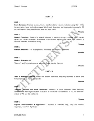
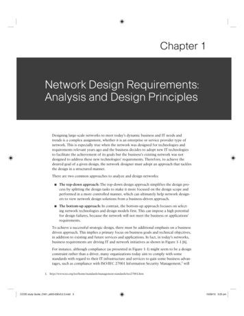
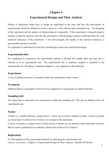
VOL. 81, 2007CORONAVIRUS HOST RANGE DETERMINANTS ON APN1263treated with the RG4 MAb were scored as positive for receptor blockade if thepercentage of infected cells was 5% of control MAb-treated samples andscored as negative for receptor blockade if the percentage of infected cells wasⱖ90% of control MAb-treated samples. The Fab fragments of purified RG4MAb and a control MAb were prepared using the ImmunoPure Fab preparationkit (Pierce, Rockford, IL) according to the manufacturer’s instructions. BHKcells transiently expressing wild-type fAPN were preincubated with 20 g of totalprotein of RG4 Fab or control Fab, inoculated with HCoV-229E or FCoV, andthen processed as described above.RESULTSHCoV-229E and the animal coronaviruses FCoV, CCoV,and TGEV require different regions of fAPN for entry. BHKcells transfected with plasmids encoding fAPN or mAPN expressed the APN protein on the cell surface 48 h after transfection (Fig. 1). Cells expressing fAPN were susceptible toinfection with HCoV-229E, FCoV, TGEV, and CCoV as previously reported (54), while cells expressing mAPN were resistant to infection with all four of these group 1 coronaviruses(Fig. 1).To identify fAPN regions required for coronavirus receptoractivity, BHK cells were transfected with chimeric m/fAPNproteins containing different regions of fAPN in an mAPNbackbone. All chimeric m/fAPN proteins were expressed at thecell surface 48 h after transfection (Fig. 2). Two m/fAPN chimeras, m/fAPN1–582 and m/fAPN251–582, had receptor activityfor HCoV-229E but not for FCoV or TGEV (Fig. 2). In contrast, m/fAPN582–967, m/fAPN704–967, and m/fAPN704–831 hadreceptor activity for FCoV and TGEV but not for HCoV-229E(Fig. 2). Chimera m/fAPN704–831 also had receptor activity forCCoV (data not shown). In contrast, m/fAPN831–967 had noreceptor activity for the human or animal group 1 coronaviruses (Fig. 2). Thus, the region between aa 251 to 582 of fAPNwas required for HCoV-229E entry, while the region betweenaa 704 to 831 of fAPN was required for entry of the animalcoronaviruses tested.The anti-fAPN RG4 MAb binds to an epitope between aa251 to 582 of fAPN and blocks fAPN-mediated infection ofHCoV-229E, FCoV, CCoV, and TGEV. Immunolabeling andflow cytometry were used to test the ability of the anti-fAPNRG4 and anti-mAPN R3-242 MAbs to recognize fAPN,mAPN, and chimeric m/fAPN proteins expressed on BHK cells.The RG4 MAb recognized fAPN, as previously reported (23), butnot mAPN. The anti-mAPN MAb recognized mAPN but notfAPN (Table 1 and Fig. 1). Two chimeras, m/fAPN251–967 andm/fAPN251–582, were recognized by both MAbs. In contrast,m/fAPN582–967 was recognized by only the anti-mAPN MAb,and m/fAPN1–582 was recognized by only the anti-fAPN MAb(Table 1 and Fig. 2). Thus, the anti-mAPN MAb R3-242 boundto an epitope on mAPN between aa 1 to 251. The anti-fAPNRG4 MAb bound to an epitope on fAPN that lies between aa251 to 582 and includes domain V that is required for HCoV229E entry, but anti-fAPN RG4 did not bind to aa 582 to 967in domain VII of fAPN that is required for entry of FCoV,CCoV, and TGEV.Anti-fAPN RG4 MAb was previously shown to block FCoV,TGEV, and CCoV infection of feline cells expressing fAPN(23). Infection of BHK cells stably expressing fAPN by thesethree animal coronaviruses and by HCoV-299E was blocked bytreatment of the cells with RG4 MAb, but virus infection wasnot blocked in cells treated with a control MAb (data notshown). Thus, the RG4 MAb blocked infection by the threeanimal coronaviruses and HCoV-299E even though these viruses required different domains of fAPN for entry. Treatmentof cells transiently expressing fAPN or m/fAPN chimeric proteins with a control MAb did not block FCoV or HCoV-229Einfection, but treatment with anti-fAPN RG4 MAb blockedHCoV-229E infection of BHK cells expressing fAPN,m/fAPN251–967, or m/fAPN251–582 (Table 2). RG4 MAb alsoblocked FCoV infection of cells expressing fAPN, orm/fAPN251–967, but RG4 MAb did not block FCoV infection ofcells expressing m/fAPN582–967 (Table 2). In addition, the small( 50 kDa) Fab protein of RG4 MAb, but not the Fab fragment of a control MAb, blocked both FCoV and HCoV-229Einfection of cells expressing fAPN (data not shown).The recognition of m/fAPN proteins (Table 1) and the inhibition of coronavirus infection (Table 2) by the RG4 MAbDownloaded from http://jvi.asm.org/ on April 13, 2015 by UCSF Library & CKMFIG. 1. BHK cells transfected with fAPN but not mAPN are susceptible to infection with FCoV, TGEV, CCoV, and HCoV-229E. Surfaceexpression of fAPN or mAPN proteins on paraformaldehyde-fixed BHK cells transiently transfected with plasmids encoding fAPN or mAPN wasdetected by immunolabeling with anti-fAPN RG4 or anti-mAPN R3-242 MAb, respectively. Viral antigens in cells inoculated with FCoV, TGEV,CCoV, or HCoV-229E were detected after 24 h by immunolabeling with antiviral antibodies.
1264TUSELL ET AL.J. VIROL.Downloaded from http://jvi.asm.org/ on April 13, 2015 by UCSF Library & CKMFIG. 2. HCoV-229E and the animal coronaviruses FCoV and TGEV require different regions of fAPN for receptor activity. BHK cells weretransiently transfected with plasmids encoding m/fAPN chimeric proteins. The numbers in parentheses indicate fAPN residues present in them/fAPN protein. At 48 h posttransfection, the surface expression of the chimeric m/fAPN1–582 and m/fAPN251–582 proteins was detected withanti-fAPN RG4 MAb, and the surface expression of all other m/fAPN chimeric proteins was detect
001009252) (54) and mAPN (accession number NM 008486; provided by Linda Shapiro, University of Connecticut Health Center, Farmington, CT) were cloned into the pcDNA