Transcription
Ruan et al. BMC Infectious Diseases (2017) 17:369DOI 10.1186/s12879-017-2459-9CASE REPORTOpen AccessDisseminated cryptococcosis with recurrentmultiple abscesses in an immunocompetentpatient: a case report and literature reviewQiaoling Ruan†, Yimin Zhu†, Shu Chen, Liping Zhu, Shu Zhang and Wenhong Zhang*AbstractBackground: Cryptococcus neoformans is frequently present as an opportunistic pathogen mainly affectingimmunocompromised populations. Disseminated C. neoformans infection in immunocompetent population is rareand usually involves lung and central nerve system. Cryptococcus from biologic samples can easily grow on routinefungal and bacterial culture media. Besides, cryptococcal latex agglutination test has been established as a reliablediagnostic tool with overall sensitivities of 93–100%.Case presentation: We report a rare disseminated cryptococcosis case which presented with chronic recurrentmultiple abscess in an immunocompetent male involving skin, lung, spine and iliac fossa without evidence ofcentral nerve system involving. The results of serum cryptococcal latex agglutination tests and standard microbialcultures were negative. The patient underwent empirical anti-bacterial and anti-tuberculosis therapy which turnedout to be effectless. Finally, bedside inoculation of the pus was carried out and revealed Cryptococcus neoformans,which was confirmed by polymerase chain reaction. After the administration of anti-fungal drugs includingliposomal amphotericin B, the patient recovered from fever and paraplegia.Conclusions: This case reveals an uncommon pattern of disseminated C. neoformans infection in immunocompetentpopulation presented with chronic multiple abscess and without central nerve system involving. Negativeroutine microbial cultures may not necessarily rule out cryptococcosis, especially in early stage. Besides,cryptococcal latex agglutination test does have a chance of false negative, which might be related with“capsule-deficiency”. Moreover, this phenomenon could be related with low-grade virulence and relative longillness duration.Keywords: Cryptococcosis, Immunocompetent, AbscessBackgroundCryptococcus neoformans is generally considered as anopportunistic pathogen because of its tendency to causeinfection in immunocompromised individuals, particularly individuals with HIV infection. The lungs arepredominantly the primary locus of infection, with extrapulmonary dissemination through hematogenous routeaffecting the meninges and, less commonly, the skin,bones, prostate, and other organs. The present report* Correspondence: zhangwenhong@fudan.edu.cnYimin Zhu as co-first†Equal contributorsDepartment of infectious disease, Huashan Hospital of Fudan University,Shanghai, Chinadescribes a rare case of disseminated cryptococcosis whichpresented with recurrent multiple abscesses involvingskin, lung, spine and iliac fossa in a patient with no detectable immune deficiencies and negative results of serumcryptococcal latex agglutination tests (SCLATs) and routine microbial cultures.Case presentationA 68-year-old man presented with a 7-month history ofprogressive multiple abscesses, followed by fever, lowerextremity weakness and urinary retention. Seven monthsbefore, he occasionally found a bean-size lump withslight tenderness on the left side of the lower back. Onemonth later, the lump increased to the size of an egg The Author(s). 2017 Open Access This article is distributed under the terms of the Creative Commons Attribution 4.0International License (http://creativecommons.org/licenses/by/4.0/), which permits unrestricted use, distribution, andreproduction in any medium, provided you give appropriate credit to the original author(s) and the source, provide a link tothe Creative Commons license, and indicate if changes were made. The Creative Commons Public Domain Dedication o/1.0/) applies to the data made available in this article, unless otherwise stated.
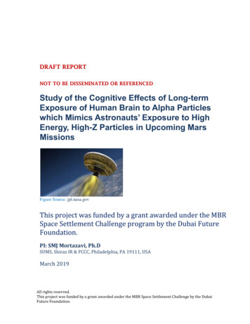
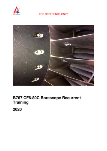
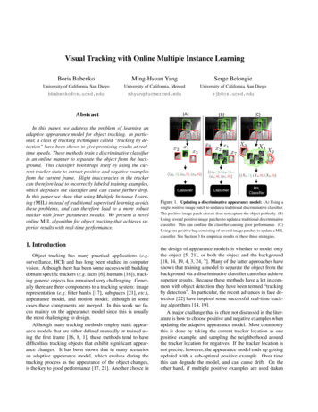
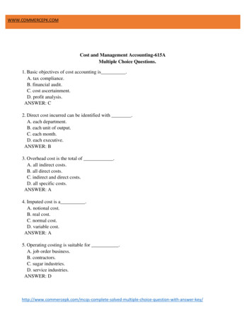
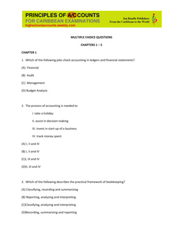
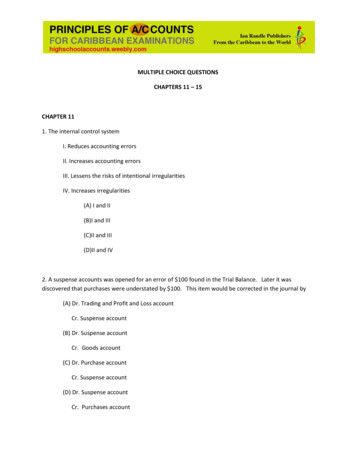
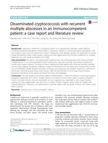
Ruan et al. BMC Infectious Diseases (2017) 17:369Fig. 1 The lump on the back: There was a bean-size lump withslight tenderness on the left side of the lower backFig. 2 Abscess in lung and fascia: Chest CT scan revealed pulmonaryabscess formation (above) and an abscess inside the fascia (below)Page 2 of 6(Fig. 1). Computed tomography (CT, for short) scan revealed an abscess inside the fascia. Chest CT scanshowed pulmonary abscess formation (Fig. 2). Repeatedcultures of blood and percutaneous aspiration wereconstantly negative. Abscess incision and drainage wereperformed and empirical antibiotic treatment was given.Three months later, chest CT revealed the unresolvedpulmonary abscess and additional multiple destructionsof vertebral bodies, especially T5 and T11 (Fig. 3).Empirical anti-tuberculosis drugs were administrated for2 months. Instead of improvement, the patient becamefebrile to 38.4 C, and developed weakness and numbness in both legs and urinary retention. The patient reported no significant past medical history, and he deniedany exposure to bird droppings.On examination, physician identified a skin lesion withdraining sinus on the left side of the lower back. He hadgrade 0 muscle strength of both legs and slightlyFig. 3 CT scan revealed progressing infection: Chest CT scanrevealed the unresolved pulmonary abscess (above) compared with2 months before (Fig. 2). Moreover, there were additional multipledestructions of vertebral bodies, especially T5 in the figure below
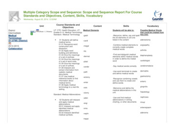
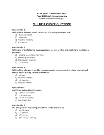
Ruan et al. BMC Infectious Diseases (2017) 17:369Fig. 4 Bone destructions and compressed spine: These two sagittal viewsof spinal MRI T2-weighted sequence revealed multiple bone destructionsof thoracic vertebrae, with spine compressed at T4 and T5 levelincreased muscle tone. There was slight tenderness onthe T4 and T5 vertebrae bodies, with hypoesthesia belowT4 level. Laboratory testing revealed a leucocyte countof 16.55 103 cells/μL, a C-reactive protein concentration of 128 mg/L, an erythrocyte sediment rate of120 mm/h and a negative result of HIV serology test.CD4 T cell count was normal and the levels of serumglobulins including IgG, IgA, IgM and total IgE werePage 3 of 6within normal range. T-SPOT. TB was negative. The(1, 3)-β-D-glucan test (or G test) and repeated SCLATs wereall negative. Other laboratory results were unremarkable.Spinal magnetic resonance imaging (MRI, for short) andsubsequent CT revealed multiple bone destruction of thoracic and lumber vertebrae and ribs, with spine compressedat T4 and T5 level (Fig. 4). Surgical excision of paravertebral abscess at T4-T5 level was performed and pathologicalexamination of the excisional biopsy specimen foundnothing but inflammatory changes. Specimen cultureswere negative for bacteria, tuberculosis and fungi. Antimycobacterial treatment was ceased and the antibioticschanged to intravenous sulfamethoxazole-trimethoprimcompound and oral doxycycline. Gradually, his temperaturereturned to normal and abscesses regressed. The patientwas discharged on oral maintenance therapy.Two months later, the patient presented to hospitalagain with a complaint of fever and a 50 60 mm newonset lump with slight tenderness in the right groin. CTand MRI revealed a large right iliac abscess (Fig. 5).Repeated cultures were negative and laboratory tests revealed no specific findings. The abscess increased in sizegradually. Surgical drainage yielded 700 ml of coffeeground pus. Bedside inoculation of the pus was carriedout and the result of culture revealed Cryptococcusneoformans, which was confirmed by polymerase chainreaction (PCR, for short) (Fig. 6). The contrasted brainMRI found no evidence of central nervous system involvingand so did the lumbar puncture. Liposomal amphotericin BFig. 5 Contrasted CT scan and non-contrasted MRI scan of pelvic cavity: a Axial view of CT scan. b Coronal view of CT scan. c Coronal view ofMRI T1-weighted fast spin echo sequence. d Coronal view of MRI short TI inversion recovery sequence. Both CT and MRI scan revealed a largeright iliac abscess
Ruan et al. BMC Infectious Diseases (2017) 17:369Fig. 6 India ink staining of the pus culture: The cryptococcus can befound in the pus culture after the India ink stainingand itraconazole were given. The fevers resolved when theaccumulated dose of amphotericin B reached 2.5 g, andthen itraconazole was replaced by fluorocytosine. We withdrew liposomal amphotericin B when its accumulated dosereached 3.0 g. The patient discharged with 4 monthsfluorocytosine plus fluconazole and regular follow-up.At 18-month follow-up, the patient was afebrile, withno new-onset abscess, and he could walk slowly. A Ganttchart was used to summarize the presentation andmanagement of the patients (Fig. 7).Discussion and conclusionsCryptococcosis is an opportunistic infectious disease causedby encapsulated yeasts in the genus Cryptococcus. Two species, C. neoformans and C. gattii, commonly cause diseasein humans. C. neoformans can cause cryptococcosis in bothimmunocompetent and immunocompromised patients,while C. gattii usually infects apparently immunocompetentPage 4 of 6hosts. However, the percentage of C. gattii infection causingdisease in apparently normal hosts is significantly higherthan for C. neoformans [1]. After a primary infection in thelungs, the disease can either localized or disseminatethrough blood to various organs, depending on patients’immune status. Disseminated cryptococcosis in immunocompetent population is rare and usually involves the central nerve system [2].In the present case report, the patient was otherwisehealthy and presented with progressive multiple abscessescaused by C. neoformans without cryptococcal meningitis,which was quite rare and was easily been misdiagnosed. Toour knowledge, only five cases with extra-pulmonary andextra-cranial cryptococcal abscess has been reported in immunocompetent patients up to now (Table 1), but none ofthem was presented with such recurrent multiple soft-tissueabscesses for more than half a year like this case [3–7].At onset, the patient presented with cutaneous and pulmonary infection. Most cutaneous infection occurs as asign of disseminated cryptococcal infection, which can beseen in 10–15% of the cases [8]. Cryptococcosis canpresent with a variety of skin manifestations includingacneiform lesions, abscesses, vesicles, purpura, nodules,ulcers, granulomas, pustules, draining sinuses and cellulitis[9, 10]. The examination of involved skin tissue includingbiopsy or tissue culture can be helpful for diagnosis [3].However, in this case, the skin lesion was non-specific andcultured negative. In high tuberculosis burden area, disseminated tuberculosis should be ruled out carefully especially when cryptococcusis is complicated with TB.Confirmative diagnosis of disseminated cryptococcosisusually relies on positive culture or pathological results.In other similar cases, misdiagnosis is common and theexamination of samples from invasive operations plays avital role in accurately diagnosis. Cryptococcus frombiologic samples can easily grow on routine fungal andFig. 7 Gantt chart: The Gantt chart illustrates the course of the patient’s disease
Ruan et al. BMC Infectious Diseases (2017) 17:369Page 5 of 6Table 1 Case reports on extra-pulmonary and extra-cranial cryptococcal omorbiditiesAl-Tawfiq JA et al. Vertebral abscess, Lung2007 [3]lesion?aAxillary lymph nodes TBPus culture: PositiveSCLATb: NegativeSurgical excision of vertebral abscessFluconazoleSingh R et al.2010 [4]Psoas abscesssternum & vertebralinvolvedPulmonary TBAbortionVaricella infectionFNACc: PositiveSCLAT: PositiveAmphotericin BGaskill T et al.2010 [5]Soft tissue abscess,Lung & Mediastinal lymphnodes involvedRemote history ofdepressionBiopsy: PositiveSCLAT: PositiveTissue culture: PositiveSurgical excision of thigh abscessFluconazoleSuchitha S et al.2012 [6]Soft tissue & cerebralabscess,Lung involvedDiabetesFNAC: PositiveSputum culture: PositiveNo surgeryFluconazole & Amphotericin BLenz D et al.2015 [7]Subcutaneous abscessNoneTissue culture: PositiveSCLAT: NegativeSurgical incision withoutanti-fungal drugaThe tissue cultures found only Cryptococcus in vertebral abscess and M. tuberculosis in axillary lymph nodes. The patient had no productive cough and thussputum cultures were not obtained. The pathogen of pulmonary lesion remains unclearbSCLAT: Serum Cryptococcus Latex Agglutination TestcFNAC: Fine Needle Aspiration Cytologybacterial culture media. However, our patient was negative for repeated cultures, which might indicate the insensitivity of culture at the early stage of infection. Inadditional, less fungaemia is seen in immunocompetentpatients compared with immunocompromised ones [2].CLAT detects the capsular polysaccharide antigens of C.neoformans, which has been established as a reliablediagnostic tool with overall sensitivities of 93–100% [11].However, our patient had consistently negative results.In other similar cases, CLAT can also be negative [3, 7].The capsule is composed of glucuronoxylomannanand galactoxylomannan polysaccharides. It is consideredthe one of the classical virulence factor for C. neoformans, and mutants without capsules are avirulent [12,13]. Several C. neoformans genes in capsular synthesisand formation have already been identified, and sitedirected gene mutants result in hypocapsular or acapsular strains. Capsule-deficient mutants are less virulentthan the parental strains [14–17]. Considering that thepatients had relatively long illness course, we inferredthat he was infected with a strain of low-grade virulencewhich maybe hypocapsular. It could also explain thefalse-negative CLAT result, which has a high sensitivity.However, this is only speculation without available C.neoformans gene mutant detection.AbbreviationsCLAT: Cryptococcal latex agglutination tests; CT: Computed tomography;FNAC: Fine needle aspiration cytology; LP: Lumbar puncture; MRI: Magneticresonance imaging; PCR: Polymerase chain reaction; SCLAT: Serumcryptococcal latex agglutination testsAcknowledgmentsNone.FundingNo funding was received.Availability of data and materialsAll the data supporting our findings is contained within the manuscript.Authors’ contributionsQLR and YMZ collected and analyzed medical data of the patient, wrote andrevised the manuscript. WHZ, SC and LPZ participated in the treatment ofthe patient during hospitalization. WHZ made a critical contribution to thetreatment plan of the patient and made a critical revision of the manuscriptfor important intellectual content. All authors read and approved the finalmanuscript.Competing interestsThe authors declare that they have no competing interests.Ethics approval and consent to participateWe identified this patient during routine clinical practice and consented togive venous blood samples after elaborate information. Involvement of theethical committee of the Huashan Hospital of Fudan University wasconsidered unnecessary, since the project was not based on a studyprotocol.Written informed consent was obtained from the patient for publication ofthis case report and all accompanying images. A copy of the written consentis available for review by the editor of this journal.Publisher’s NoteSpringer Nature remains neutral with regard to jurisdictional claims inpublished maps and institutional affiliations.Received: 2 February 2017 Accepted: 11 May 2017References1. Kwon-Chung KJ, Fraser JA, Doering TL, Wang ZA, Janbon G, Idnurm A, BahnYS. Cryptococcus neoformans and Cryptococcus gattii, the etiologic agentsof Cryptococcosis. Cold Spring Harb Perspect Med. 2014;4(7):a019760.2. Lui G, Lee N, Ip M, Choi KW, Tso YK, Lam E, Chau S, Lai R, Cockram CS.Cryptococcosis in apparently immunocompetent patients. QJM. 2006;99(3):143–51.3. Al-Tawfiq JA, Ghandour J. Cryptococcus neoformans Abscess andosteomyelitis in an immunocompetent patient with tuberculouslymphadenitis. Infection. 2007;35(5):377–82.4. Singh R, Xess I. Multiple osseous involvements in a case of disseminatedcryptococcosis. Indian J Orthop. 2010;44(3):336–8.
Ruan et al. BMC Infectious Diseases (2017) 17:3695.6.7.8.9.10.11.12.13.14.15.16.17.Page 6 of 6Gaskill T, Payne D, Brigman B. Cryptococcal abscess imitating a soft-tissuesarcoma in an immunocompetent host: a case report. J Bone Joint SurgAm. 2010;92(9):1890–3.Suchitha S, Sheeladevi CS, Sunila R, Manjunath GV. Disseminatedcryptococcosis in an immunocompetent patient: a case report. Case RepPathol. 2012;2012:652351.Lenz D, Held J, Goerke S, Wagner D, Tintelnot K, Henneke P, Hufnagel M.Primary cutaneous cryptococcosis in an eight-year-old immunocompetentchild: how to treat? Klin Padiatr. 2015;227(1):41–4.Anderson DJ, Schmidt C, Goodman J, Pomeroy C. Cryptococcal diseasepresenting as Cellulitis. Clin Infect Dis. 1992;14(3):666–72.Pema K, Diaz J, Guerra LG, Nabhan D, Verghese A. Disseminated cutaneouscryptococcosis. Comparison of clinical manifestations in the pre-AIDS andAIDS eras. Arch Intern Med. 1994;154(9):1032–4.Dimino-Emme L, Gurevitch AW. Cutaneous manifestations of disseminatedcryptococcosis. J Am Acad Dermatol. 1995;32(5 Pt 2):844–50.Tanner DC, Weinstein MP, Fedorciw B, Joho KL, Thorpe JJ, Reller LB.Comparison of commercial kits for detection of Cryptococcal antigen. J ClinMicrobiol. 1994;32(7):1680–4.Perfect JR. Cryptococcus neoformans: a sugar-coated killer with designergenes. FEMS Immunol Med Microbiol. 2005;45(3):395–404.Kronstad JW, Attarian R, Cadieux B, Choi J, D'Souza CA, Griffiths EJ, GeddesJM, Hu G, Jung WH, Kretschmer M, et al. Expanding fungal pathogenesis:Cryptococcus breaks out of the opportunistic box. Nat Rev Microbiol. 2011;9(3):193–203.Chang YC, Kwon-Chung KJ. Isolation, characterization, and localization of acapsule-associated gene, CAP10, of Cryptococcus neoformans. J Bacteriol.1999;181(18):5636–43.Chang YC, Kwon-Chung KJ. Isolation of the third capsule-associated gene,CAP60, required for virulence in Cryptococcus neoformans. Infect Immun.1998;66(5):2230–6.Chang YC, Kwon-Chung KJ. Complementation of a capsule-deficientmutation of Cryptococcus neoformans restores its virulence. Mol Cell Biol.1994;14(7):4912–9.Alspaugh JA, Perfect JR, Heitman J. Cryptococcus neoformans Mating andvirulence are regulated by the G-protein alpha subunit GPA1 and cAMP.Genes Dev. 1997;11(23):3206–17.Submit your next manuscript to BioMed Centraland we will help you at every step: We accept pre-submission inquiries Our selector tool helps you to find the most relevant journal We provide round the clock customer support Convenient online submission Thorough peer review Inclusion in PubMed and all major indexing services Maximum visibility for your researchSubmit your manuscript atwww.biomedcentral.com/submit
cryptococcal latex agglutination test does have a chance of false negative, which might be related with “capsule-deficiency”. . no new-onset abscess, and he could walk slowly. A Gantt chart was used to summarize the presentation and management of the patients (Fig. 7). Discussi