Transcription
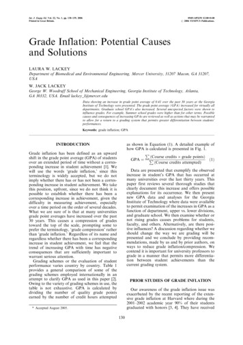
International Journal of New Technology and Research (IJNTR)ISSN:2454-4116, Volume-1, Issue-8, December 2015 Pages 53-57Etiological Causes of Intrahepatic and ExtrahepaticBile Duct DilatationYunfu Lv, Wan Yee Lau, Haiying Wu, Shunwu Chang, NingLiu, Yejuan Li, Jie Deng Abstract— This study aims to review the causes ofintrahepatic and extrahepatic bile duct dilatation. Based onreview of medical literature, a scientific classification of thecauses is proposed. Intrahepatic and extrahepatic bile ductdilatation can be divided into two categories: congenital andacquired. The former includes the five classical Todani’scongenital types. The latter can be sub-classified according tothe underlying pathogenesis, the relationship with bile ductlumen/wall/extralumen, or intrabiliary pressure.Index Terms— Bile duct dilatation; etiologic classification;diagnosis.I. INTRODUCTIONIn modern medicine, the bile duct is considered as animportant organ, rather than simply a duct [1]. The conditionof the bile duct, directly or indirectly, affects the function ofthe liver and other vital organs. The normal common bile ductmeasures 8 mm in diameter in adults, 1 mm in newborns, 3mm in infants, and 5 mm in children. A common bile ductdiameter 8 mm at its widest point in adult is considered ascommon bile duct dilatation. A diameter of 8-12 mm is calledmild, 12-16 mm moderate, 16-20 mm severe, and 20 mmextremely severe dilatation.The common hepatic duct with the left and right hepaticducts, the sectional bile ducts, and segmental bile ducts arecalled level-1, -2, and -3 bile ducts, respectively. The biliarytree exhibits a dendritic distribution [2]. Normally, thediameter of the right hepatic duct is 3.5 mm and the lefthepatic duct 3.3 mm. When the diameter of the left or righthepatic duct exceeds the normal range, and/or with wideningof the level-2 and -3 bile ducts, the condition is calledintrahepatic bile duct dilatation. An intrahepatic bile ductdiameter of 5 mm is called mild, 5-9 mm moderate, and 9mm severe dilatation.Intrahepatic and extrahepatic bile duct dilatation iscommon in clinical practice [3]. In addition to congenital andacquired factors like bile duct stones, parasites, and bile ductwall tumor, there are other uncommon causes of intrahepaticand extrahepatic bile duct dilatation. A comprehensiveunderstanding of intrahepatic and extrahepatic bile ductYunfu Lv1, Wan Yee Lau1,2, Haiying Wu1, Shunwu Chang1,NingLiu1, Department of Surgery, the People’s Hospital of HainanProvince, Haikou 570311, China.Wan Yee Lau2, Faculty of Medicine, The Chinese University of HongKong, Shatin, Hong Kong SAR, China.Yejuan Li3, Jie Deng3, Hainan University, No. 58, Renmin Avenue,Haikou, Hainan Province, 570228, Chinadilatation is necessary. In general, extrahepatic bile ductdilatation occurs before intrahepatic bile duct dilatation.However, hilar or intrahepatic lesions can cause intrahepaticbile duct dilatation without any extrahepatic bile ductdilatation. In this review, the causes of intrahepatic andextrahepatic bile duct dilatation are classified based on theiretiological causes.Ⅱ. CLASSIFICATION AND ETIOLOGICAL DIAGNOSISIntrahepatic and extrahepatic bile duct dilatation can bedivided into two categories: congenital and acquired.A. Congenital intrahepatic and extrahepatic bile ductdilatation (cyst) is a rare congenital malformation [4, 5]. Itwas first described by a German Abrahan Vater in 1713,then a British Todd in 1817, and an American Douglasin 1852. Subsequently, reports came from many othercountries. In 1958, Alonso-Lej and his associates fromAmerica classified congenital common bile ductdilatation into three types:- cystic dilatation of commonbile duct, diverticulum of common bile duct, and cyst ofthe terminal common bile duct [6]. A French Carolidescribed in 1959 multiple cystic dilatation in theintrahepatic bile duct (called Caroli’s disease) [7, 8],Flanigan in 1965 added two more types:- the type withboth intrahepatic and extrahepatic bile duct dilatation,and the type with intrahepatic but no extrahepatic bileduct dilatation, to the original three types to propose atotal of five types [9]. In 1977, Todani and his associates[10]modified the classification by Alonso-Lej to includethe types described by Caroli and Flanigan (Figure 1).This classification is still recognized and used byinternational counterparts today.B. Acquired intrahepatic and extrahepatic bile duct dilatationis also known as postnatal or secondary intrahepatic andextrahepatic bile duct dilatation. It is caused by diseasesor acquired conditions.There is no universally accepted classification ofacquired intrahepatic and extrahepatic bile duct dilatation. Aclassification can provide a better understanding of thiscategory of bile duct dilatation.i. According to the underlying pathogenesis which include(a) calculus-related dilatation (36%, including bileduct stones, Mirizzi syndrome, and chronic53www.ijntr.org
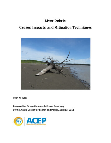
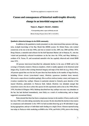
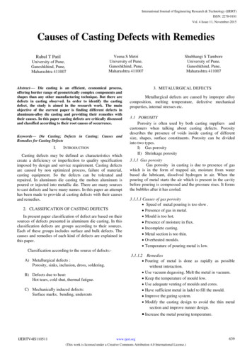
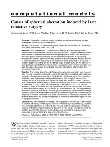
Please Enter Title Name of Your Paperechinococcosis); (d) inflammatory dilatation (5%, includingcholangitis, constrictive papillitis); (e) inoma,periampullary carcinoma, duodenal papillary carcinoma, andgallbladder carcinoma); (f) extrinsic compressionpressure-related dilatation (21%, including pancreatic headcarcinoma, tumor metastasis around the bile duct, chronicpancreatitis or pancreatic head cyst, peribiliary aneurysm,duodenal bulb ulcers, and parapapillary diverticulitis); (g)injury-related dilatation (2%, including bile duct stricture,and bilioenteric anastomotic stricture); and (h) others (2%,including bile duct bleeding, Oddi’s sphincter dysfunction,duodenal obstruction, and ectopic pancreas).ii. It can be divided into four types according to therelationship with the bile duct lumen/wall/extralumen:(1) In the lumen: Dilatation is caused by obstructionresulting from stones [12], parasites and other foreign bodieswithin the bile duct and bile duct parasites [13].(2) In the bile duct wall: Dilatation is caused by bile ductwall lesions, bile duct injury, or destruction of normal tissuestructure of bile duct wall due to inflammation, resulting instenosis at the lesion site. It includes bile duct polyps, bileduct adenoma [14], bile duct cancer, bile duct papilloma,ductal dilatation caused by ectopic pancreas, chroniccholangitis, periampullary carcinoma, duodenal papillarycarcinoma [15], gallbladder carcinoma invasion of bile ducts,bile duct wall hemangioma, bile duct injury, and bilioentericanastomotic stricture.(3) Extrinsic compression: Dilatation is caused bycompression or traction of the bile duct wall by externallesions. It is commonly found in patients with acute andchronic pancreatitis, pancreatic pseudocyst, pancreatic headcarcinoma, parapapillary diverticulum, bile duct compressionby metastatic carcinoma, polycystic liver disease [20, 21],duodenal postbulbar ulcer, or peribiliary aneurysm [22].(4) Other factors: It includes post-cholecystectomy bileduct dilatation [23], and bile duct dilatation caused by otherrare causes.iii. It can be divided into six types according to intrabiliarypressure:(1) Increase in intrabiliary pressure-related dilatation:Dilatation is caused by increased pressure in the bile duct(Figure 2) due to distal bile duct obstruction from any cause[24, 25]resulting in proximal bile duct dilatation andobstructive jaundice [26]. This type of dilatation is commonlyfound in patients with bile duct cystadenoma andcystadenocarcinoma [27, 28], sand-like stones, conical stones,impacted stones, tumors, bile duct diaphragm, or ectopicpancreas.(2) Expansion-related dilatation: It is the gradualdilatation of the bile duct during stone formation andexpansion without obstructive jaundice (Figures 3-5). In thiscase, there are many formed stones or parasites within the bileduct lumen. The bile can flow through the gaps between thecholecystitis with impacted stone at Hartmann’spouch); (b) compensatoryI Cystic dilatationII diverticulum-like dilatationIII Cyst of terminal common bile ductⅣ a. Dilatation of both intrahepatic and extrahepatic bileductsⅣ b. Multiple dilatation of extrahepatic bile ductsⅤ Multiple cysts in intrahepatic bile ducts (Caroli's disease)Figure 1. Todani’s modification of Alonso-Lej’sclassification of bile duct cysts (selected from the booktranslated by Li Yang and Wang Yi [11])Figure 2. Proximal bile duct dilatation and bilereflux caused by obstruction of the lower end of bile ductand increased pressure in the bile ductdilatation (6%, including post-cholecystectomy dilatation,chronic atrophic cholecystitis); (c) parasites-related dilatation(4%, including bile duct ascariasis, bile duct Fasciolopsis,bile duct schistosomiasis, clonorchis sinensis, and hepatic54www.ijntr.org
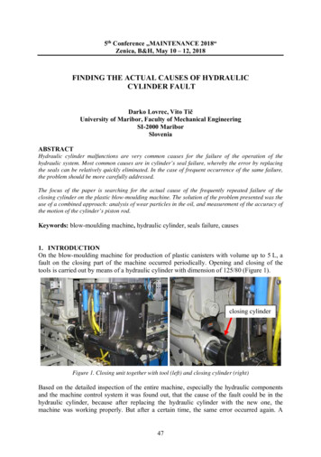
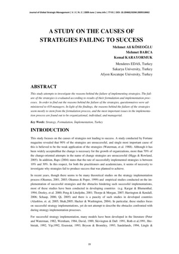
International Journal of New Technology and Research (IJNTR)ISSN:2454-4116, Volume-1, Issue-8, December 2015 Pages 53-57stones or parasites into the duodenum. Bile duct pressure isnot high or just slightly increased, but below the obstructivejaundice threshold of 20 mmHg 2.94 kpa [29].Common bile duct dilatation reported in the literature mostlyfall into this type.(4) Infectious dilatation: Dilatation is caused by increasedbile duct pressure (normal pressure in remission) or bile ductFigure 5. A common bile duct filled with stones had adiameter of 25 mm, but no jaundice was presentFigure 3. Bile duct dilatation caused by stones in thecommon bile duct, with bile flowing through the gaps, nohigh bile duct pressure, and no jaundiceFigure 6. Intrapancreatic bile duct was compressed into aline by cyst of the head of pancreasFigure 4. Intranperative cholangiography revealedthat a common bile duct filled with stones had a diameterof 20 mm, but no jaundice was present(3) Compressive dilatation: It is the dilatation of theproximal bile duct caused by compression of the distal bileduct with bile duct pressure varying at different levels, andfluctuating jaundice is present clinically. This type ofdilatation is commonly found in patients with a stone actingas a ball valve in the lower end of the common bile duct,sloughing tumors in the bile duct, pancreaticoduodenalaneurysm, acute and chronic pancreatitis [30], pancreaticcysts (Figure 6).wall thickening as a result of poor drainage of bile due torepeated infections of bile duct. This type of dilatation iscommonly found in patients with sphincter of Oddidysfunction, bile duct infection, or pyogenic cholangitis.(5) Compensatory dilatation: Patients may experiencecompensatory dilatation of common bile duct aftercholecystectomy because the gallbladder bile pool disappearsand the buffer storage function is lost. However, thesepatients seldom undergo investigations without symptoms.Therefore, it is difficult to know the exact number of cases.Chronic atrophic cholecystitis can also cause compensatorybile duct dilatation as a result of lost buffer storage functiondue to inelastic gallbladder wall.(6) Adhesive dilatation: After bile duct surgery,adhesion of tissues can cause traction of the bile duct wall,which can lead to bile tract compression (Figure 7) orangulation (Figure 8) in serious cases, thus resulting inproximal bile duct dilatation. This type of dilatation is55www.ijntr.org
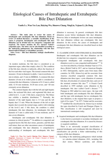
Please Enter Title Name of Your Papercommonly found in patients who have had several bile ductsurgeries and T-tube drainageREFERENCES[1] Wong ZQ. Development direction of hepatobiliary surgery. Journalof Surgery Concepts & Practice, 2011, 16 (4): 329-31.[2] Huang JF. Hepatobiliary and Pancreatic Surgery. 4th edition.Beijing: People's Health Publishing House, 2010: 81.[3] Chaudhary A, Dhar P, Sachdev A, Kumar N, Vij JC, Sarin SK,Broor SL, Sharma SS. Choledochal cysts - differences in 689160[PubMed - indexed for MEDLINE][4] Dowdy GS, Waldron GW, Brown WG. Surgical anatomy of thepancreato-biliary ductal system observations. Arch Surg, 1962,84:229. PMID:13887616[PubMed - indexed for MEDLINE][5] Okada A, Hasegawa T, Oguchi Y, Nakamura T. Recent advancesin pathophysiology and surgical treatment of congenital dilatationof the bile duct. J Hepatobiliary Pancreat Surg. 2002;9(3):342-51.PMID:12353145[PubMed - indexed for MEDLINE] .[6] Alonso Lej F, Revor WB, DJ. Congenital choledochal cyst, with areport of 2, and an analysis of 94 cases. Surg Gynecol Obstet,1959; 108:1-30[7] Caroli J, Soupault R, Kossakowski J, et al. La dilatation congenitaledes voies biliaires intrahepatiques. Sem hop Paris, 1958;34:488-95[8] Han Xiaoyu, Lv yunfu. Research progress of Caroli’s disease.International Journal of Surgery, 2010, 37(3):193-6.[9] Söreide K, Körner H, Havnen J, Söreide JA. Bile duct cysts inadults. Br J Surg. 2004 Dec;91(12):1538-48. PMID: 15549778[PubMed - indexed for MEDLINE][10] Todani T, Watanabe Y, Narusue M, Tabuchi K, Okajima K.Congenital bile duct cysts: Classification, operative procedures,and review of thirty-seven cases including cancer arising fromcholedochal cyst. Am J Surg, 889044[PubMed- indexed forMEDLINE][11] Li Y, Wang Y, chief translator. Diagnosis and treatment of bileduct disease. Beijing People's Health Publishing House, 2009,243.[12] Kim JH, Kim YS, Kim DK, Ha MS, Lee YJ, Lee JJ, Lee SJ, WonIS, Ku YS, Kim YS, Kim JH. Short-term Clinical OutcomesBased on Risk Factors of Recurrence after Removing CommonBile Duct Stones with Endoscopic Papillary Large MID:22741123 [PubMed][13] Wei JN, Zhu JF. Clinical analysis of 148 cases of pediatricascariasis. Journal of Clinical Pediatric Surgery, 2003, 2 (6):416-8.[14] Koga F, Tanaka H, Takamatsu S, Baba S, Takihara H, HasegawaA, Yanagihara E, Inoue T, Nakano T, Ueda C, Ono W. A case ofvery large intrahepatic bile duct adenoma followed for 7 years.World J Clin Oncol. 2012 Apr 10;3(4): 63-6. PMID:22574289[PubMed] PMCID:PMC3348183[15] Terada T. Non-invasive intraductal papillary neoplasms of thecommon bile duct: a clinicopathologic study of six cases. Int JClin Exp Pathol; 2012;5(7):690-7.[16] Lu FY, Zhang XX, Dong YH. Chronic pancreatitis-inducedcompressed relative stenosis of the distal common bile duct.HBPD Int, 2006, 5(1):119-22.[17] Repiso A, Gómez-Rodríguez R, García-Vela A, Pérez-GruesoMJ, Martín R, Romero M, Carrobles JM. Endosonographicexamination of the common bile duct in patients with acutebiliary pancreatitis. Rev Esp Enferm Dig. 2008Jun;100(6):337-42. PMID:18752362 [PubMed - indexed forMEDLINE][18] Sakai Y, Tsuyuguchi T, Ishihara T, Yukisawa S, Sugiyama H,Miyakawa K, Kuroda Y, Yamaguchi T, Ozawa S, Yokosuka O.Long-term prognosis of patients with endoscopically treatedpostoperative bile duct stricture and bile duct stricture due tochronic pancreatitis. J Gastroenterol Hepatol. 2009Jul;24(7):1191-7. PMID:19682193 [PubMed - indexed forMEDLINE][19] Lv Yunfu, Zhu Qinghua, Zhao Ge. The anatomicopathologicalclassification of Terminal stenosis of common bile duct caused bychronic pancreatitis and its treatment. Chinese Journal of GeneralSurgery, 2000, 15(7):417-9.[20] Everson G, Taylor M, Doctor R. Polycystic disease of the liver.Hepatology, 2004;40: 774-82.[21] Vauthey JN, Maddern GJ, Kolbinger P, Baer HU, Blumgart LH.Clinical experience with adult polycystic liver disease. Br JSurg,1992;79: 562-5[22] Yu YH, Sohn JH, Kim TY, Jeong JY, Han DS, Jeon YC, KimMY. Hepatic artery pseudoaneurysm caused by acute idiopathicFigure 7. Proximal bile duct dilatation caused by bileduct adhesion and compression after repeated surgeryA. Insertion of T-tube into bile duct B. Adhesion andangulation of bile duct after removal of T-tubeFigure 8. Adhesion and deformation of bile duct afterinsertion of T-tubeⅢ. CONCLUSIONFor more than 60 causes of intrahepatic and extrahepatic bileduct dilatation listed in this paper, they can be divided into thecongenital and acquired categories. The latter category canfurther be sub-classified according to the pathogenesis,relationship with bile duct wall or intrabiliary ductal pressure.ACKNOWLEDGMENTThis project is funded by the key clinical specialtyconstruction projects of China.56www.ijntr.org
International Journal of New Technology and Research (IJNTR)ISSN:2454-4116, Volume-1, Issue-8, December 2015 Pages tis. World J Gastroenterol. 2012 May 14;18(18):2291-4.PMID:22611325 [PubMed - indexed for MEDLINE]Park SM, Kim WS, Bae IH, Kim JH, Ryu DH, Jang LC, Choi JW.Common bile duct dilatation after cholecystectomy: a one-yearprospective study. J Korean Surg Soc. 2012 Aug;83(2):97-101.PMID:22880184 [PubMed]Mallet-Guy P.Value of preoperative manometric androentgenographic examination in the diagnosis of pathologicalchanges and functional disturbances of the biliary tract. SurgGynecol Obstet, 1952, 94:385-93.Dan JS, Wang BH. Preclinical and Clinical Biliary Surgery.Beijing: People's Health Publishing House, 2003, 113-5.Wang Bao-chun,LU Yun-fu,Han Xiao-yu.Etiology andclassification of cholangiectasisa:an analysis of 1098cases.Zhonghua Gandan Waike Zazhi, 2011, 17(9):752 754Ishak KG,Willis GW,Cummins SD,Bullock AA.Biliarycystadenoma and cystadenocarcinoma: report of 14 cases andreview of the literature.Cancer,1977,39: 322-38.Subramony C,Herrera GA,Turbat Herrera EA.Hepatobiliarycystadenoma.Astudy of five case with reference to histogenesis.Arch Pathol LabMed,1993,117: 1036-42.LU Yun-fu , Wang Bao-chun, Gon Xiao-Guang, HanXiao-Yu.correlation among pressure, diameter, and jaundice inacute obstruction of the common bile duct. Zhonghua shiyanwaike zazhi, 2011,28(11):1851 1852Aimoto T, Uchida E, Nakamura Y, Yamahatsu K, Matsushita A,Katsuno A, Cho K, Kawamoto M. Current surgical treatment forchronic pancreatitis. J Nippon Med Sch. 2011;78(6):352-9.PMID:22197867 [PubMed - indexed for MEDLINE]57www.ijntr.org
[10] modified the classification by Alonso-Lej to include the types described by Caroli and Flanigan (Figure 1). This classification is still recognized and used by international counterparts today. B. Acquired intrahepatic and extrahep