Transcription
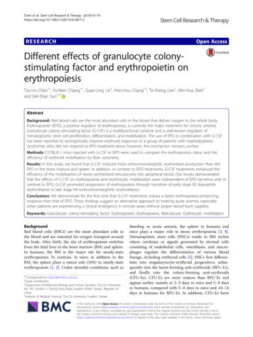
Chen et al. Stem Cell Research & Therapy (2018) ARCHOpen AccessDifferent effects of granulocyte colonystimulating factor and erythropoietin onerythropoiesisTzu-Lin Chen1†, Ya-Wen Chiang1†, Guan-Ling Lin2, Hsin-Hou Chang1,2, Te-Sheng Lien1, Min-Hua Sheh1and Der-Shan Sun1,2*AbstractBackground: Red blood cells are the most abundant cells in the blood that deliver oxygen to the whole body.Erythropoietin (EPO), a positive regulator of erythropoiesis, is currently the major treatment for chronic anemia.Granulocyte colony-stimulating factor (G-CSF) is a multifunctional cytokine and a well-known regulator ofhematopoietic stem cell proliferation, differentiation, and mobilization. The use of EPO in combination with G-CSFhas been reported to synergistically improve erythroid responses in a group of patients with myelodysplasticsyndromes who did not respond to EPO treatment alone; however, the mechanism remains unclear.Methods: C57BL/6 J mice injected with G-CSF or EPO were used to compare the erythropoiesis status and theefficiency of erythroid mobilization by flow cytometry.Results: In this study, we found that G-CSF induced more orthochromatophilic erythroblast production than didEPO in the bone marrow and spleen. In addition, in contrast to EPO treatments, G-CSF treatments enhanced theefficiency of the mobilization of newly synthesized reticulocytes into peripheral blood. Our results demonstratedthat the effects of G-CSF on erythropoiesis and erythrocytic mobilization were independent of EPO secretion and, incontrast to EPO, G-CSF promoted progression of erythropoiesis through transition of early stage R2 (basophilicerythroblasts) to late stage R4 (orthochromatophilic erythroblasts).Conclusions: We demonstrate for the first time that G-CSF treatments induce a faster erythropoiesis-enhancingresponse than that of EPO. These findings suggest an alternative approach to treating acute anemia, especiallywhen patients are experiencing a clinical emergency in remote areas without proper blood bank supplies.Keywords: Granulocyte colony-stimulating factor, Erythropoietin, Erythropoiesis, Reticulocyte, Erythrocytic mobilizationBackgroundRed blood cells (RBCs) are the most abundant cells inthe blood and are essential for oxygen transport aroundthe body. After birth, the site of erythropoiesis switchesfrom the fetal liver to the bone marrow (BM) and spleen.In humans, the BM is the major site for steady-stateerythropoiesis. In contrast, in mice, in addition to theBM, the spleen plays a minor role (10%) in steady-stateerythropoiesis [1, 2]. Under stressful conditions, such as* Correspondence: dssun@mail.tcu.edu.tw†Equal contributors1Department of Molecular Biology and Human Genetics, Tzu-Chi University,No. 701, Section 3, Zhong-Yang Road, Hualien 97004, Taiwan, Republic ofChina2Institute of Medical Sciences, Tzu-Chi University, Hualien, Taiwanbleeding or acute anemia, the spleen in humans andmice plays a major role in stress erythropoiesis [3, 4].Hematopoietic stem cells (HSCs) reside in BM nicheswhere cytokines or signals generated by stromal cellsconsisting of endothelial cells, osteoblasts, and macrophages regulate the differentiation of various bloodlineage, including erythroid cells [5]. HSCs first differentiate into megakaryocyte-erythroid progenitors, subsequently into the burst-forming unit-erythroids (BFU-Es),and finally into the colony-forming unit-erythroids(CFU-Es). CFU-Es are more mature than BFU-Es andappear earlier, namely at 2–3 days in mice and 5–8 daysin humans, compared with 5–8 days in mice and 10–14days in humans for BFU-Es. In addition, CFU-Es form The Author(s). 2018 Open Access This article is distributed under the terms of the Creative Commons Attribution 4.0International License (http://creativecommons.org/licenses/by/4.0/), which permits unrestricted use, distribution, andreproduction in any medium, provided you give appropriate credit to the original author(s) and the source, provide a link tothe Creative Commons license, and indicate if changes were made. The Creative Commons Public Domain Dedication o/1.0/) applies to the data made available in this article, unless otherwise stated.
Chen et al. Stem Cell Research & Therapy (2018) 9:119smaller colonies than do BFU-Es when cultured inmethylcellulose [2, 3]. Erythropoietin (EPO), a 30.4-kDaglycoprotein mainly synthesized by the kidneys, is themain regulator of erythroid cell proliferation, differentiation, and survival [6]. EPO production is upregulatedunder hypoxic conditions through the activity of thehypoxia-inducible transcription factor (HIF) [7, 8]. TheEPO receptor (EPOR) is expressed dominantly on CFUEs and gradually downregulated during erythroid differentiation [9, 10]. Upon stimulation initiated by EPObinding to the EPORs, CFU-Es develops into proerythroblasts, subsequently into basophilic erythroblasts, thenpolychromatic erythroblasts, and finally orthrochromaticerythroblasts. The final stage of erythroid differentiationinvolves the enucleation and maturation of reticulocytesinto circulating erythrocytes [11].Anemia can develop from loss of RBCs, a reduction inRBC production, increased destruction of RBCs, or ashorter RBC lifespan. The World Health Organization defines anemia as a hemoglobin level lower than 12 g/dl inwomen and lower than 13 g/dl in men [12]. Except for patients with inherited hematopoietic disease, the highestrates of anemia are observed in patients with chronic diseases such as those of the kidney and heart, cancer, inflammatory bowel disease, rheumatoid arthritis, and humanimmunodeficiency virus (HIV) [13]. Recombinant humanEPO (rhEPO) has been used medically for more than20 years and has generated a multibillion dollar market annually. However, since 1998, some severe adverse effectssuch as EPO-associated pure red cell aplasia by long-termEPO injection induced neutralizing antibodies have beenreported [14, 15]. Other side effects such as hypertension,increased risk of venous thromboembolism, stroke, anddeath have also been reported [16]. Currently availableerythropoiesis-stimulating agents (ESAs) are variations ofEPO generated by human cells or Chinese hamster ovarycells. New ESAs (e.g., peptide-based erythropoietic agents),activation of endogenous EPO production through HIFstabilization and GATA1 inhibition, and EPO gene therapyhave been developed; however, none of these new methodshave shown efficacy superior to that of existing ESAs [17].Taken together, the development of new anemia therapieswith satisfactory levels of efficacy and safety and faster action is required.Our previous study found that granulocyte colonystimulating factor (G-CSF) could mobilize newly synthesized erythrocytes to the peripheral blood (PB) and promoteerythrocytic differentiation and proliferation in vitro and exvivo [18]. The present study further investigated the differences between G-CSF and EPO on erythropoiesis. Aftermice had been treated with G-CSF and EPO, the erythropoiesis status in the BM and spleen were compared usingflow cytometry. The stimulation of early erythroid progenitor subsets and temporal regulation of erythroid progenitorPage 2 of 9mobilization were characterized. The mechanism of G-CSFpromotion of erythropoiesis was also discussed.MethodsToxins and miceB. anthracis lethal toxin (LT) was provided by the Instituteof Preventive Medicine, National Defense Medical Center(Taipei, Taiwan), and purified as previously described [19].EGFP mice [C57BL/6 J-Tg (Pgk1-EGFP) 03Narl] providedby Professor Chou CK [20] and C57BL/6 J mice were purchased from the National Laboratory Animal Center(Taipei, Taiwan). Animals were maintained in a specificpathogen-free environment in the experimental animalcenter of Tzu Chi University (Hualien, Taiwan).Analysis of erythropoiesis status in the BM and spleenC57BL/6 J mice (male, 10–12 weeks old) were retroorbitally injected with 2 IU/g rhEPO (Neorecormon ,Roche, Mannheim, Germany) or 55 μg/kg recombinant human G-CSF (Filgrastim, Kirin, Tokyo) once daily on 3 consecutive days. Animals treated with an identical volume ofsaline served as negative controls. BM cells were isolated aspreviously described [21]. Spleens were minced using theplunger of a 50-ml syringe and resuspended using a P1000Pipetman (Gilson, Middleton, WI, USA) to create a singlecell suspension. Cells were blocked with RPMI-1640medium (Gibco Laboratories) containing 5% bovine serumalbumin at 37 C for 1 h and subsequently incubated withrat anti-mouse fluorescein isothiocyanate-conjugated CD71antibody (BioLegend) and rat antimouse allophycocyanin(APC)-conjugated TER-119 antibody (BD Immunocytometry System) at 37 C for 1 h. After washing with phosphatebuffered saline (PBS), the cells were resuspended in 1 ml ofPBS and analyzed using a Beckman Coulter Gallios flowcytometer (Beckman Coulter, CA, USA).Flow cytometry analysis of mobilized erythrocytes inEGFP miceEnhanced green fluorescent protein (EGFP) mice (male,8 months old) were injected with 55 μg/kg G-CSF or2 IU/g EPO retro-orbitally once daily on 3 consecutivedays. PB was collected retro-orbitally at 20, 40, and 60 hafter initial injection. Cells were incubated with rat antimouse APC-conjugated TER-119 antibody at 37 C for1 h and subsequently with 5 μM of RNA-selective dyeF22 [22] at 37 C for 30 min. After washing with PBS,the cells were analyzed using a Beckman Coulter Gallios flow cytometer.EPO and soluble P-selectin immunoassayC57BL/6 J mice (male, 10–12 weeks old) were treated with55 μg/kg G-CSF administered by retro-orbital injectiononce daily on 2 consecutive days for the EPO immunoassayor 5 consecutive days for the soluble P-selectin
Chen et al. Stem Cell Research & Therapy (2018) 9:119immunoassay. Animals treated with an identical volume ofsaline served as negative controls. C57BL/6 J mice that wereretro-orbitally injected with 1.5 mg/kg LT and untreatedserved as positive controls for the EPO immunoassay andthe soluble P-selectin immunoassay, respectively. PB (serumfor the EPO immunoassay and plasma for the soluble Pselectin immunoassay) was collected retro-orbitally at 22,44, and 66 h after initial injection for the EPO immunoassay, and day 0 before initial injection and days 1–5 afterinitial injection for the soluble P-selectin immunoassay. TheEPO and soluble P-selectin immunoassays were performedusing an enzyme-linked immunosorbent assay (ELISA) inaccordance with the manufacturers’ instructions (Quantikine Mouse/Rat EPO immunoassay, R&D systems; MouseP-selectin ELISA kit, RayBiotech, Inc., USA).Hematopoietic parameter detectionC57BL/6 J mice (male, 10–12 weeks old) were treatedwith 55 μg/kg G-CSF or 1 mg/kg recombinant mouse Pselectin (purified mouse P-selectin–IgG fusion protein;BD Pharmingen) once daily on 2 consecutive daysthrough retro-orbital injection. The hematopoietic parameters were measured on day 2 after initial injectionusing an automated hematology analyzer (XP-300, Sysmex Corporation).StatisticsAll quantifiable data are presented as mean standarddeviation (SD). Statistical analysis was conducted byone-way analysis of variance (ANOVA) followed by thepost-hoc Bonferroni-corrected t test using the SPSS software, version 17.0 (SPSS Inc., Chicago, IL, USA). Pvalues lower than 0.05 indicated significant differences.ResultsG-CSF enhanced R4 erythroblast cell production to agreater extent than EPO did in the BM and spleenTo compare the erythropoiesis status after G-CSF andEPO treatments, C57BL/6 J mice were retro-orbitallyinjected with G-CSF or EPO once daily for 3 consecutivedays. Erythropoiesis progression was analyzed in the BM(Fig. 1a) and spleen (Fig. 2a) through flow cytometry.Using antibodies against the erythroid markers CD71 andTER119, erythroblasts were divided into the followingfour populations from early to late sequential differentiation stages: proerythroblasts (R1, CD71high/TER-119med),basophilic erythroblasts (R2, CD71high/TER-119high), latebasophilic and polychromatophilic erythroblasts throblasts (R4, CD71low or /TER-119high) (Figs. 1b and2b) [18, 21, 23]. In agreement with the findings of otherstudies, in mouse BM, G-CSF enhanced more nonerythroid cell populations (NE, TER-119 ) [24] than EPO did(Fig. 1c). In contrast, EPO substantially increased the totalPage 3 of 9number of erythroid cells [6] (Fig. 1d). Although totalerythroid cell counts in the BM of mice from the G-CSFtreated groups were lower than those of mice from theEPO-treated groups, G-CSF elicited a markedly larger R4population than EPO did (Fig. 1e).The spleen is a secondary organ for erythropoiesis butnot for granulopoiesis [2–4, 25]. In characterizing the differentiation progression in the mouse spleen, we foundthat G-CSF treatments did not increase the nonerythroidcell population or total erythroid cell number (Fig. 2c, d).Compared with the responses in the BM, EPO upregulated the percentage of early erythroblasts (R1 and R2)and downregulated the late stages of erythroblasts (R3 andR4) (Fig. 2e), whereas G-CSF reduced the percentage ofearly R1, R2, and R3 cells and considerably increased thenumber of late-stage R4 erythroblasts (Fig. 2e). These results collectively suggest that G-CSF enhances late-stageR4 erythroblast cell production to a greater extent thanEPO does in the BM and spleens of mice.G-CSF mobilized more newly synthesized reticulocytesthan EPO didUsing an EGFP transgenic mouse model, we found thatG-CSF caused newly synthesized erythrocytes tomobilize to the PB and induced erythrocytes to mobilizeinto the PB faster than EPO [18]. After using the sameexperimental strategy (Fig. 3a), our data revealed thatthe mobilization efficiencies of newly synthesized erythrocytes (R1, EGFPhigh/TER-119high) were higher at 20 hunder G-CSF treatment than under EPO, and were similar between G-CSF and EPO treatments at 40 and 60 hafter initial injection (Fig. 3b, c). Because G-CSF promoted more R4 erythroblast synthesis than EPO did inthe BM and spleen (Figs. 1 and 2), we hypothesized thatG-CSF may promote reticulocyte mobilization to agreater extent than EPO. Because only reticulocytes exhibit residue ribonucleic acid (RNA) expression amongnewly synthesized erythrocyte populations [26], F22, aRNA-selective dye [22], combined with TER-119 wasused to discriminate reticulocytes from matured erythrocytes in newly synthesized erythrocytes. Over two timecourses, 20 and 40 h, G-CSF preferred to mobilize newlysynthesized reticulocytes (F22high/EGFPhigh/TER-119high)to a greater extent than EPO did (Fig. 3b, d).G-CSF-dependent enhancement of erythropoiesis andRBC mobilization were not mediated by eliciting EPO andthe P-selectin pathwayThe transcription factor HIF-1α can be upregulated by GCSF and then binds to an EPO promoter to increase circulating EPO levels after five consecutive G-CSF injections[27]. Accordingly, we hypothesized that G-CSF may stimulate erythropoiesis and mobilize erythrocytes by increasingthe synthesis of EPO. Our experimental design is shown in
Chen et al. Stem Cell Research & Therapy (2018) 9:119Page 4 of 9Fig. 1 Granulocyte colony-stimulating factor (G-CSF) treatment induced more R4 erythroblast cells than erythropoietin (EPO) treatment did inbone marrow (BM). a The experimental outline used to measure erythropoiesis status after G-CSF (n 5) and EPO (n 5) treatments in the BM isdepicted. Saline-treated mice (n 5) were used as negative controls. Flow cytometry analysis was performed 72 h after initial injection. Theerythroblast cells were gated as R1 (CD71high, TER-119med), R2 (CD71high, TER-119high), R3 (CD71med, TER-119high), and R4 (CD71low or –, TER-119high)in all groups. Nonerythroid cells were gated as NE (TER-119 negative) (b). Cell numbers of nonerythroid (c) and total erythroid cells (sum of R1,R2, R3, and R4) (d), and the percentage of individual erythroblasts (R1, R2, R3, and R4) in the total number of erythroid cells in each group(e) were quantified. Data are reported as mean SD. *P 0.05, **P 0.01Fig. 4a. Treatment with Bacillus anthracis, a LT that can induce hypoxia-elicited EPO secretion in mice [28], was usedas a positive control. In agreement with a previous study[28], our results revealed that circulating EPO levels gradually increased following LT administration in mice [28]. Incontrast, circulating EPO levels were not upregulated at allexamined time points after G-CSF treatment in mice (Fig.4b–d). These results suggest that the G-CSF-dependent enhancement of erythropoiesis and RBC mobilization are notmediated by eliciting EPO.P-selectin is widely recognized as a cell adhesion receptor that mediates leukocyte rolling through bindingto glycosylated moieties of the main ligand P-selectinglycoprotein ligand 1 (PSGL-1) [29]. The same propertyof P-selectin enables HSCs to adhere to stromal cells, especially vesicular endothelial cells, through P-selectin(HSC)–PSGL-1 (stroma) interactions [30]. G-CSF induced greater and faster mobilization of myeloid cellsfrom BM in PSGL-1 knockout mice than in wild-typemice [31]. Studies have also suggested that G-CSF
Chen et al. Stem Cell Research & Therapy (2018) 9:119Page 5 of 9Fig. 2 Granulocyte colony-stimulating factor (G-CSF) treatment induced more R4 erythroblast cells than erythropoietin (EPO) treatment did in thespleen. a The experimental outline used to measure the erythropoiesis status after G-CSF (n 5) and EPO (n 4) treatments in the spleen isdepicted. Saline-treated mice (n 5) were used as negative controls. Flow cytometry analysis was performed 72 h after initial injection. Theerythroblast cells were gated as R1, R2, R3, and R4 in all groups (see Fig. 1 for details). Nonerythroid cells were gated as NE (b). Cell numbers ofnonerythroid (c) and total erythroid cells (sum of R1, R2, R3, and R4) (d), and the percentage of individual erythroblasts (R1, R2, R3, and R4) in thetotal number of erythroid cells in each group (e) were quantified. Data are reported as mean SD. *P 0.05, **P 0.01administration increases levels of circulating soluble Pselectin [32, 33]. This evidence collectively suggests thatP-selectin/PSGL-1-mediated binding between erythroidprecursors and stromal cells may partially control themobilization and release of progenitor cells from BMinto the blood stream, thereby indicating the possibilitythat G-CSF may enhance erythrocytic mobilization byeliciting circulating soluble P-selectin to compete ainteraction. To reproduce G-CSF-mediated elicitation ofcirculating soluble P-selectin, G-CSF was injected intoC57BL/6 J mice on 5 consecutive days (Fig. 5a). Thelevels of circulating soluble P-selectin increased significantly after G-CSF injection (Fig. 5b). Next, we designedan experiment to examine whether soluble P-selectincan increase the RBC counts in the PB (Fig. 5c). Although two doses of G-CSF increased the white bloodcell (WBC) and RBC counts in the PB, the same effectwas not observed when two equivalent doses of solubleP-selectin were applied (Fig. 5d, e). This finding suggests
Chen et al. Stem Cell Research & Therapy (2018) 9:119Page 6 of 9Fig. 3 Granulocyte colony-stimulating factor (G-CSF) treatment mobilized more newly synthesized reticulocytes than erythropoietin (EPO) treatment did. a The experimental outline for investigating erythrocyte mobilization in enhanced green fluorescent protein (EGFP) mice after G-CSF(n 4) and EPO (n 4) treatments is shown. Peripheral blood (PB) was collected 20, 40, and 60 h after initial injection. Flow cytometry analysiswas performed to analyze the relative fold of EGFP /TER-119 cells (gated R1 region) compared with the before experiment (Before exp.) groupsand the percentage of F22 cells in EGFP /TER-119 cells at 20 h (b). Quantitative results of the relative fold of EGFP /TER-119 cells (c) and thepercentage of F
PBS and analyzed using a Beckman Coulter Gallios flow cytometer (Beckman Coulter, CA, USA). Flow cytometry analysis of mobilized erythrocytes in EGFP mice Enhanced green fluorescent protein (EGFP) mice (male, 8 months old) were injected with 55 μg/kg G-CSF or 2 I