Transcription
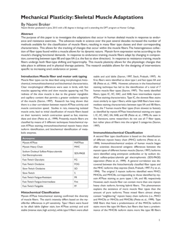
Mechanical Plasticity: Skeletal Muscle AdaptationsBy Naomi BrokerNaomi Broker graduated in June, 2015 with a BS degree in biology and is attending the DPT program at Hunter College.AbstractThe purpose of this paper is to investigate the adaptations that occur in human skeletal muscle in response to endurance and resistance exercises. The advances made in science over the past several decades increased the number ofmethods available for the classification of muscle fibers, resulting in many fiber type classes and their correspondingcharacteristics. This allows for the tracking of changes that occur within the muscle fibers. The heterogeneous collection of fiber types found within a muscle allows for its dynamic nature. Myosin form expression varies according to themuscle’s changing functional demands. In response to endurance training, muscle fibers adapt by changing in composition, converting between type IIB and type IIA (i.e. the fast to slow direction). In response to resistance training, musclefibers undergo both fiber-type shifting and hypertrophy. This muscle plasticity allows for the physiologic changes thattake place in athletes and in physical therapy patients. The research available allows for the designing of interventionsspecific to increasing one’s endurance or power.Introduction: Muscle fiber and motor unit typingMuscle fiber types can be described using morphological, histochemical, immunohistochemical and biochemical characteristics.Clear morphological differences were seen in birds, with fastmuscles appearing white and slow muscles appearing red. Theredness of the slow muscle is due to the greater myoglobinand capillary content that permits a greater oxidative capacityof the muscle (Staron, 1997). Research has long shown thatthere is a clear correlation between myosin ATPase activity andmuscle contraction speed. Studies in rats, measuring isolatedmuscle units, lead to initial classification of muscle fibers basedon their isometric twitch contraction speed as fast, intermediate and slow (Pette et. al., 1999). Presently, muscle fibers areclassified by means of 3 different techniques: histochemical myosin ATPase staining, immunohistochemical myosin heavy chainisoform identification, and biochemical identification of metabolic enzymes.Acronyms:Myosin ATPaseMATPaseMyosin Heavy ChainMHCSodium Dodecyl Sulfate-PolyacrylamideGel ElectrophoreticSDS-PAGEFast-Twitch GlycolyticFGFast-Twitch OxidativeFOGSlow-Twitch OxidativeSOSlow-TwitchSFast-Twitch Fatigue-ResistantFRFast-Twitch Fatigue-IntermediateFintFast-Twitch FatigableFFHistochemical ClassificationMyosin ATPase histochemical staining confirmed the diversityof muscle fibers. The stain’s intensity differs based on the myofibrillar differences in pH sensitivity. Type I fibers were foundto be alkali labile (lighter stain, low ATPase activity) and acidstable (intense stain, high activity), while type II fibers were alkalistable and acid labile (Staron, 1997, Sieck, Prakash, 1997). Atfirst, fibers were identified as slow type I, and fast types IIA andIIB (Pette et. al., 1999). However, advances in the histochemicalstaining technique has led to the identification of a total of 7human muscle fiber types (Staron, 1997). The newly identifiedfibers, types IC, IIC, IIAC, and IIAB, have intermediate myosinATPase staining characteristics. Type IC, the slowest fiber, stainsmost similarly to type I fibers, while type IIAB fibers have intermediate staining characteristics between type IIA and IIB fibers.Thus, the 7 human muscle fiber types (from slowest to fastest),as identified by myosin ATPase histochemical staining are: typesI, IC, IIC, IIAC, IIA, IIAB, and IIB (Pette et. al., 1999). As seen inthe literature, some researchers do not use all 7 fiber types,but rather place all fibers into the original 3 categories (Staron,1997).Immunohistochemical ClassificationA second fiber type classification is based on the identificationof different myosin heavy chain (MHC) isoforms (Pette et. al.,1999). Immunohistochemical analysis of human muscle beganafter scientists discovered antigenic differences between themyosin types of different human muscle (Staron, 1997). Isoformswere identified using antimyosin antibodies or by sodium dodecyl sulfate-polyacrylamide gel electrophoretic (SDS-PAGE)separation (Pette et. al., 1999). A general correlation was discovered between the histochemically classified fiber types andthe MHC isoforms they express (Sieck, Prakash, 1997, Fry et. al.,1994). The original 3 myosin isoforms identified were MHCI,MHCIIa, and MHCIIb, corresponding to those identified by myosin ATPase staining as pure types I, IIA, and IIB, respectively.However, each muscle fiber can contain more than one myosinheavy chain isoform, forming hybrid fibers. This phenomenonexplains the existence of more muscle fiber types than theamount of pure isoforms. These mixed fibers almost alwayscontain “neighboring” myosin heavy chain isoforms (i.e., MHCIand MHCIIa or MHCIIa and MHCIIb) (Pette et. al., 1999). TypeIIAB fibers that have a predominance of the MHCIIa isoformstains more like type IIA fibers, but fibers that have a predominance of the MHCIIb isoform stains more like type IIB fibers1
Naomi Broker(Staron, 1997). Consequently, the histochemical myosin ATPasetypes express their isoform genes to varying degrees, accordingto the variable ratio of isoforms present in the fiber. Due toits quantitative nature, the SDS-PAGE technique is perhaps thebest method for muscle fiber typing, as electrophoretic separation permits the recognition of the relative concentrations ofdifferent myosin heavy chain isoforms in mixed muscle fibers(Pette et. al., 1999, Fry et. al., 1994).A fourth myosin heavy chain isoform, MHCIIx or MHCIId, andits corresponding fiber type IIX, is present in small mammals.Evidence shows that MHCIIb in humans in homologous toMHCIIx/d of small mammals (Pette et. al., 1999, Hilber et. al.,1999). In actuality, MHCIIb in humans is really MHCIIx/d, as humans do not express MHCIIb, the fastest myosin heavy chainisoform (Pette et. al., 1999). In fact, recent data shows thatfibers that were histochemically identified as type IIB in humanscontain low amounts of MHCIIa, and therefore, in reality, arehybrid type IIAB fibers (Staron, 1997). Therefore, the 3 myosinheavy chain isoforms present in human limb muscles are (fromslowest to fastest): MHCI, MHCIIa, and MHCIIx/d (previouslyincorrectly known as MHCIIb).Biochemical ClassificationA third classification technique reflects the energy metabolismof the muscle fibers. Histochemical myosin ATPase fiber typing of type I or type II corresponds to slow and fast musclefibers, respectively, and the enzymes involved reflect the metabolic pathways that are either aerobic/oxidative or anaerobic/glycolytic (Pette et. al., 1999). This classification scheme leads to3 fiber types: fast-twitch glycolytic (FG), fast-twitch oxidative(FOG), and slow-twitch oxidative (SO) (Pette, Staron, 1997).Type I and IIA fibers have a greater mitochondrial density andoxidative capacity than type IIB and IIX fibers (Sieck, Prakash,1997). Accordingly, there is a good correlation between type Iand SO fibers, but correlations between type IIA and FOG andtype IIB and IIX and FG fibers are more diverse (Hamalainen,Pette, 1995). This can be explained by the fact that changesin oxidative capacity can take place without changes in MHCisoforms (Staron, 1997). Generally, type I fibers rely primarilyon aerobic/oxidative energy metabolism and type II fibers relyprimarily on anaerobic/glycolytic metabolism, but as it cannotbe assumed, the terms type IIB and FG or type IIA and FOGcannot be used interchangeably (Pette et. al., 1999).Myosin Light ChainsThe myosin light chains also exist in different isoforms, slowand fast, and affect the contractile speed of the fiber (Talmadge,Pette, 1995). A homogeneous/pure myosin heavy chain isoformcan be heterogeneous in regard to its myosin light chain isoform. Usually, however, fast heavy chain isoforms fuse with fastlight chain isoforms, and slow heavy chain isoforms fuse withslow light chain isoforms (Pette et. al., 1999, Jostarndt-Fogen et.al., 1998).Motor Unit ClassificationThe proper functional unit of the neuromuscular system, themotor unit, reflects the characteristics of its individual fibers.Motor units are categorized based on their contractile speedsand fatigue characteristics. Contractile speed classifies motorunits into slow-twitch (S), usually comprised of type I fibers, orfast-twitch (F), usually comprised of type II (Burke, 1999). TheTable 1: Summary of fiber types and their corresponding characteristicsType I fibers(I, IC)Type IIA fibers(IIC, IIAC, IIA, IIAB)Type IIX fibersType IIB fibers(not present in humans)MHC isoformsMHCIMHCIIaMHCIIx/dMHCIIbContraction timeSlowModerately FastFastVery FastOxidative CapacityHighHighIntermediateLowGlycolytic CapacityLowHighHighHighBiochemical FiberType (generally)SO(slow oxidative)FOG(fast-twitch oxidative)FG(fast-twitch glycolytic)FG(fast-twitch glycolytic)Resistance to Fatigue HighFairly HighIntermediateLowMotor Unit Class(generally)S(slow witchfatigue-intermediate)FF(fast-twitch fatigable)Activity used forAerobicLong-term anaerobicShort-term anaerobicShort-term owCapillary DensityHighIntermediateLowLow2
Mechanical Plasticity: Skeletal Muscle AdaptationsF motor units are additionally classified by their type II fibersubdivisions. Fast-twitch fatigue-resistant (FR) are comprised oftype IIA fibers, fast-twitch fatigue-intermediate (Fint) are comprised of type IIX, and fast-twitch fatigable (FF) are comprisedof type IIB (Burke, 1993). The fatigue resistance of the muscleunit is explained by the mitochondrial density and oxidative capacity of the muscle fibers. The motor unit typically reflectsthe function of the muscle. The diaphragm muscle employs itsfatigue-resistant S and FR units to produce normal breathing,but must recruit its Fint and FF units for actions that requiregreater force, such as gagging or sneezing. Contrariwise, theextensor digitorum longus muscle of the limb, which is used formotor activities demanding short periods of greater force production, is composed mostly of FF type IIB fibers. Consequently,type I and IIA fibers are intended to sustain longer periods ofactivation, while type IIX and IIB fibers are intended to produceshorter bursts of activation (Sieck, Prakash, 1997).regimes is evident in the vastus lateralis. In untrained, physicallyactive subjects, the vastus lateralis is made up of roughly 50%of slow type I fibers and 50% type II fibers (40% type IIA and10% IIX). Power athletes, such as sprinters, power weight liftersand throwers, are constituted chiefly of IIA and IIX fibers, witha percentage of type I as little as 20%. Ultra-endurance athletes,such as long-distance runners, have a 95% constitution of typeI fibers. The variability of the composition of the vastus lateralis muscle is due to fiber type shifting. Long-term endurancetraining induces a substantial transformation from fast musclefibers to slow muscle fibers. Slow muscle fibers rely mainly onaerobic metabolism and, for that reason, are vital for enduranceactivities such as swimming, cycling and marathon running. Theshift in MHC isoform type depends on changes at the molecularlevel. New slow MHC polypeptides, which are still categorizedas type IIA fibers, are produced, marking the start of their transition towards the slow type I fibers (Zawadowska et. al., 2004).Therefore, the overall properties of a muscle reflects the properties of its heterogeneous collection of fiber types and theiroverall proportions. It is this range of fiber types that allows forthe wide range of muscle function a single muscle can display.Muscle fibers adapt to their motor unit’s changing stimuli notonly by changing their size, but by converting their fiber typecomposition to suit the functional demands. This muscle plasticity serves as the physiologic basis for the adaptions of skeletalmuscle to resistance and endurance exercises.In response to training, the most common fiber conversions arebetween type IIB and type IIA. Slow to fast/ type I to type IIconversions are possible in response to the loss of function related to deconditioning, as shown in studies on humans with spinal cord injury and microgravity exposure during their time inspace. Detraining in humans (i.e., decreased use of skeletal muscle following a formerly high activity level) leads to shifts MHCIIato MHCIIx/d and possibly MHCI to MHCIIa. Additionally, thereis a related decrease in aerobic-oxidative metabolic enzymes(Pette, Staron, 1997). In short, immobilization of skeletal musclemay cause conversions in the slow to fast direction. Scientistsassumed that power training may also cause the transformationin slow to fast direction.This was a logical assumption, since fastmuscle fibers depend mainly on anaerobic metabolism and areconsequently essential for power training, such as sprinting andweight lifting. Additionally, IIX fibers produce a greater maximalpower output than IIA fibers and are therefore essential forsuch activities. However, studies have proven that all methodsof training (both endurance and power/resistance) cause a conversion toward the slow direction (Zawadowska et. al., 2004).MethodsEBSCO multi-search, ProQuest Medical Library, Wiley Library,Journal of Applied Physiology, MEDLINE, and PubMed databaseswere used to find material for this paper. Access was gained tothese databases through the Touro College Online Library andEinstein Online Library websites. Key words, such as “musclefiber types” and “effects of exercise on muscle” were used tosearch for scientific articles.After reading through these articles,other key words were identified, such as “fiber-type shifting”,and “resistance vs endurance”. Additionally, sources listed asreferences for review articles on the APTA website (AmericanPhysical Therapy Association) were searched for to find theoriginal papers.DiscussionThe plasticity of the heterogeneous composition of musclefibers allows for adaptations in the contractile and metabolicproperties of skeletal muscle. Various phenotypes are therefore possible in response to different workloads. This plasticityhas practical implications, both for athletes and rehabilitationpatients alike.This range of myosin form expression in response to exerciseEnduranceMany studies have proven that endurance exercises (low-resistance, repeated contractions that require the muscle to producea high aerobic metabolic rate) lead to several adaptations withinthe muscle to augment aerobic metabolism and resistance to fatigue (Staron et. al., 1990). Firstly, the oxidative capacity of all fibertypes will increase by increasing the amount of mitochondria, oxidative/aerobic enzymes and capillaries in the muscles being trained(Holloszy, Booth, 1976, Fitts, Widrick 1996). Based on metabolicclassification scheme, there is a transition from the FG fiber typeto the FOG fiber type without, necessarily causing a change in themyosin heavy chain composition (Pette, Staron, 1997).3
Naomi BrokerSecondly, there are alterations in myosin heavy chain isoform.It has been suggested that there is an increase in the percentage of type I fibers following aerobic training, and a subsequentdecrease following detraining in elite endurance athletes.Additionally, within type II fibers, MHCIIx/d (IIB) are convertedto MHCIIa (IIA). In other words, there is a decrease in thepopulation of pure type IIB fibers and an associated increase inthe population of pure type IIA fibers. It also seems that thereis an increase in the hybrid population of type I and type II fibers, known as type IIC (Staron et. al., 1990). Evidence as tothe conversion of type II to type I fibers, as stated previously,is lacking (Ricoy et. al., 1998). The transformation of type IIBfibers to type IIA in response to endurance training is fairlylogical. Although there are variations on the oxidative capacitieswithin a muscle type, as a class, type IIA fibers generally have agreater oxidative capacity than type IIB fibers. Therefore, anincrease in the population of type IIA fibers makes the musclemore oxidative. In fact, there is a negative correlation betweenthe percentage of type IIB fibers and maximum oxygen intake.Therefore, as stated previously, detraining and immobilizationcause a conversion in the slow to fast direction, from type IIAto type IIB (Staron et. al., 1990).Thirdly, there can be a transformation in myosin light chain isoforms. Type I fibers show an increase in contractile speed inresponse to endurance exercises, but show a decrease followingdeconditioning in humans. Logically, this change cannot be explained by a conversion of fiber type, but rather, by a conversionin the myosin light chain isoforms from slow to fast and from fastto slow, respectively (Larsson et. al., 1996, Widrick et. al., 1996).The conversion from slow to fast allows the fibers to maintaintheir properties of efficient energy usage while increasing theircontractile speed to keep up with the demands of the exercise(Fitts,Widrick, 1996). Such a conversion would not be detectedby the histochemical technique, as there is no change in the myosin ATPase (Pette, Staron, 1997). To summarize, muscle fiberadaptations to endurance exercise varies based on fiber type.In all types, the oxidative capacity of the fibers increases. TypeII fibers shift in the type I direction, leading to slower, oxidativetypes. Type I fibers convert their myosin light chains to increasetheir contractile speeds.by neural factors, rather than hypertrophy of the actual muscle fibers. However, adaptations may also be occurring in thecontractile proteins of skeletal muscle within a short durationof training, even two weeks, with sufficient intensity (McArdleet. al., 1994). Visible hypertrophy is not evident until later inthe training period ( 8 weeks) (Kraemer et. al., 1996), aroundthe same time researchers found a shift in muscle fiber typecomposition from MHCIIx/d to MHCIIa (Staron et. al., 1994,Kraemer et. al., 1995, Staron et. al., 1990).To explore this plasticity, twenty four male subjects were categorized into three groups according to their sports/physicalactivity. Group A was made up of untrained students, groupB of national and sub-national level endurance athletes (7.8 /2.9 years of specialized training) and group C of sprint-powerathletes (12.8 /-8.7 years of specialized training). Biopsies ofthe vastus lateralis muscle were obtained and immunohistochemically analyzed for fast/slow MHC composition. This muscle is easily accessible and easily trained. Most importantly, itexpresses all three myosin isoform types at specific amounts,and so its phenotype visibly mirrors any adaptive modificationsthat occur after different forms of exercises. Unpredictably,group C sprint-power athletes (such as ice hockey, volleyball,karate, soccer players and modern dancers), who were expected to display the highest percentage of MHCIIX, were nodifferent in this aspect from group B endurance athletes (suchas marathon runners, cyclists and cross country skiers). Themuscle phenotypes of both groups were similar, containing asmall proportion of the MHCIIX isoform and a predominanceof slow MHCI isoform. Clearly, the muscle phenotype wasadapted for long lasting, sustainable activities (similar to whathappens in endurance athletes), rather than activities that require a maximum power output in minimal time. Moreover,the fastest isoform, MHCIIX, was relatively lower in group Cathletes than in group A students. This myosin profile in groupC athletes is unfavorable to their sport. Muscles that containa higher percentage of type IIX fibers have a greater maximum shortening velocity, which is the most important factorin maximum power output and therefore vital for their sportsregime. This is a
Naomi Broker graduated in June, 2015 with a BS degree in biology and is attending the DPT program at Hunter College. Acronyms: Myosin ATPase MATPase Myosin Heavy Chain MHC Sodium Dodecyl Sulfate-Polyacrylamide Gel Electrophoretic SDS-PAGE Fast-Twitch Glycolytic FG Fast-Tw
