Transcription
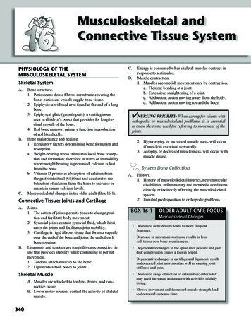
340CHAPTER 16Musculoskeletal and Connnective Tissue SystemMusculoskeletal andConnective Tissue SystemPHYSIOLOGY OF THEMUSCULOSKELETAL SYSTEMSkeletal SystemA.B.C.Bone structure.1. Periosteum: dense fibrous membrane covering thebone; periosteal vessels supply bone tissue.2. Epiphysis: a widened area found at the end of a longbone.3. Epiphyseal plate (growth plate): a cartilaginousarea in children’s bones that provides for longitudinal growth of the bone.4. Red bone marrow: primary function is productionof red blood cells.Bone maintenance and healing.1. Regulatory factors determining bone formation andresorption.a. Weight-bearing stress stimulates local bone resorption and formation; therefore in states of immobilitywhere weight bearing is prevented, calcium is lostfrom the bone.b. Vitamin D promotes absorption of calcium fromthe gastrointestinal (GI) tract and accelerates mobilization of calcium from the bone to increase ormaintain serum calcium levels.Musculoskeletal changes in the older adult (Box 16-1).Connective Tissue: Joints and CartilageA.B.Joints.1. The action of joints permits bones to change position and facilitate body movement.2. Synovial joints contain synovial fluid, which lubricates the joints and facilitates joint mobility.3. Cartilage is rigid fibrous tissue that forms a capsuleover the end of the bone and joins the end of eachbone together.Ligaments and tendons are tough fibrous connective tissue that provides stability while continuing to permitmovement.1. Tendons attach muscles to the bone.2. Ligaments attach bones to joints.Skeletal MuscleA. Muscles are attached to tendons, bones, and connective tissue.B. Lower motor neurons control the activity of skeletalmuscle.340C.D.Energy is consumed when skeletal muscles contract inresponse to a stimulus.Muscle contraction.1. Muscles accomplish movement only by contraction.a. Flexion: bending at a joint.b. Extension: straightening of a joint.c. Abduction: action moving away from the body.d. Adduction: action moving toward the body. NURSING PRIORITY: When caring for clients withorthopedic or musculoskeletal problems, it is essentialto know the terms used for referring to movement of thejoints.2. Hypertrophy, or increased muscle mass, will occurif muscle is exercised repeatedly.3. Atrophy, or decreased muscle mass, will occur withmuscle disuse.System Data CollectionA.History.1. History of musculoskeletal injuries, neuromusculardisabilities, inflammatory and metabolic conditionsdirectly or indirectly affecting the musculoskeletalsystem.2. Familial predisposition to orthopedic problems.BOX 16-1OLDER ADULT CARE FOCUSMusculoskeletal Changes Decreased bone density leads to more frequentfractures. Decrease in subcutaneous tissue results in lesssoft tissue over bony prominences. Degenerative changes in the spine alter posture and gait;disk compression causes a loss in height. Degenerative changes in cartilage and ligaments resultin decreased joint movement as well as causing jointstiffness and pain. Decreased range of motion of extremities; older adultmay need increased assistance with activities of dailyliving. Slowed movement and decreased muscle strength leadto decreased response time.
CHAPTER 16Musculoskeletal and Connnective Tissue Systemof normalactivity.BOX 16-13. LevelOLDERADULTCARE FOCUSa. Occupation, exercise, recreation.Musculoskeletal Changesb. Level of normal activity, ability to maintain ownADLs. Decreased bone density leads to more frequent4.Existenceof other chronic health problems.fractures.B. Physical assessment. Decrease 1.in subcutaneoustissue forresultsin lessInitial inspectiongrossdeformities, asymmetry,soft tissue over bony prominences.swelling, and edema. Degenerativechanges in thespineappropriatenessalter posture and ofgait;2. Nutritionalstatus:client’s weightdisk compressiona loss in height.and causesbody frame.3. Joints Degenerativechanges in cartilage and ligaments resultin decreased jointmovement aswell ascausingjointa. Movement:activeandpassive;examine activestiffness and pain.movement first; compare movement and range oftoofoppositeside.older adult Decreased range motionof motionextremities;b. with or withoutmay need increasedof dailymovement.living.c. Presenceof jointdeformitiesdislocations. Slowed movementand presenceofcrepitus.to decreased response time.4. Evaluate client’s spinal alignment, posture, and gait.5. Evaluate skeletal muscle.a. Muscle strength bilaterally.b. Coordination of movement.c. Presence of atrophy or hypertrophy.d. Presence of involuntary muscle movement.6. Assess peripheral pulses and peripheral circulation;capillary refill should normally take about 2 to 3BOX 16-3 seconds.OLDER ADULT CARE FOCUSMusculoskeletalNursingImplications of pain.7. Assessfor presenceof and characteristicsa. Most musculoskeletal pain is relieved by rest. It is more difficultto maintainimmobilityactivitiesin these and/or precipitatb. Identifyprecipitatingclients after fractures;thereforesurgicalinterventioning factors.(e.g., ORIF) is frequently used for treatment.c. Type of pain and location. The older8.clientheals formoreslowly,so useofofnormalthe affectedAssessanyalterationsensation in exextremity andtremities.weight bearing are frequently delayed.9. Assessfor use occurof propermechanics Complicationsof immobilitymorebodyfrequently;(Box 16-2).mobilize hospitalizedclients as early as possible.10. onAssessmusculoskeletalsystem re Do not relyfever ox16-1).infection; decreasing mental status is more common.11. Principles of body mechanics for health care Contractures are more common. Encourage use ofpersonnel (Box 16-2, Appendix 3-1).assistive devices (canes and walkers).TEST ALERT: Orthopedic questions may bebased on concepts of immobility, nursing assessment of an extremity, compromised circulation,and/or general perioperative care. Pay close attentionto the direction of the question.DISORDERS OF THEMUSCULOSKELETAL SYSTEMBOX 16-2341BODY MECHANICS The wider the base of support, the greater thestability. Position your feet wide apart. The lower the center of gravity, the greater the stability.Flex the knees; let the strong muscles of the legs dothe work. Position yourself close to object and/or client. Face the client; keep back, pelvis, and knees aligned;avoid twisting. Balance activity between arms and legs. Avoid bending to lift; this decreases strain on the back. Encourage client to assist. Pivoting, turning, rolling, and leverage requireless work. Person with heaviest load should coordinate teamefforts. Obtain assistance with heavy or difficult transfersor lifts. Teach clients proper body mechanics.TEST ALERT: Use good body mechanicswhen providing care – use assistive deviseswhen possible.Data Collection (Newborn)A.Ortolani sign: With the infant supine, knees flexed, andhips fully abducted, a click is heard or felt as the hip isabducted.B.BOXAsymmetricalgluteal andthigh folds.16-4 OLDERADULTCARE FOCUSC. Shortening of the leg on the affected side.Protecting JointsD. Limited hip abduction on affected side. If pain lasts longer than 1 hour after exercise, need toTreatmentchange exercises that involve that joint.A. PlanTreatmentbe initiatedasconservesoon as conditionactivityshouldand workschedule toenergy: doisidentified.tasks first.importantB. AlternateAbductiondevices.activities; do not complete all heavy tasks at1.Pavlikharnessis a fabric harness that maintains theone time.legs in the flexed, abducted position at the hip, it Minimizestressjoints: sithipratherthanstand, avoidwill holdtheonaffectedin anabductionposiprolonged repetitive movements, move aroundtion, and prevent extension and adduction. It may befrequently, avoid stairs or prolonged grasping.removed for bathing but the infant will wear it full Use largermusclessmaller ones: usetime untilthe ratherhip is thanstable.shouldersorarmsratherthanhandsto pushopen2. Hip spica cast may be used whenan adductioncondoors; pick up items without stooping or bending, usetracture is presentleg muscles; women can carry purses on their shouldersC. ratherSurgerybe doneif the correction is not feasiblethanmayin theirhands.with abduction devices. Painful, acutely swollen inflamed joints should not beexercised beyond basic ROM.Nursing InterventionsDevelopmental Dysplasia of the Hip Malformations of the hip that occur as a result ofimperfect development of the femoral head, the acetabulum,or both. Regularly exercise even when joints are slightly painfulv Goal: To identify problem in the newborn before disand stiff: swimming and bike riding maintain mobilitycharge.without weight bearing.v Goal: To assist parents to understand mechanism tomaintain reduction.A. Pavlik harness.
342B.CHAPTER 16Musculoskeletal and Connnective Tissue System1. Put an undershirt on the infant and place the braceon the outside of the shirt; always place the bracestraps on the outside of the diaper.2. Check the skin under the harness for irritation orpressure areas.3. Do not apply oils or lotions under the harness.4. Evaluate peripheral circulation and maintain cleanliness.Teach parents cast care if hip spica cast is applied.TEST ALERT: Apply or remove immobilizingequipment.v Goal: To facilitate developmental progress and adaptnurturing activities to meet needs of infant and parents.A. Provide appropriate stimuli and activity for developmental level.B. Encourage parents to hold and cuddle child.C. Maintain normal home routine.Herniated Intervertebral Disk The intervertebral disk forms a cushion betweenthe vertebral bodies of the spinal column. As stress on aninjured or degenerated disk occurs, the cartilage materialof the disk (nucleus pulposa) herniates inward towardthe spinal column, causing compression or tension on thespinal nerve root.Data CollectionA.B.C.D.The problem most commonly occurs in the lumbosacralarea.May be caused by an injury or stress to the lower back.Clinical manifestations.1. Low back pain radiating down one buttock and theposterior thigh (sciatica pain).2. Coughing, straining, sneezing, bending, twisting,and lifting aggravate the pain.3. Lying supine and raising the leg in an extended position will precipitate the pain.Diagnostics: Appendix 16-1TreatmentA.Medical.1. Analgesics, muscle relaxants, antiinflammatorymedications.2. Weight reduction if appropriate.3. Cool therapy (ice) may be used for the first 24 to 48hours after an injury, and then moist heat is applied.4. Physical therapy.5. Bed rest with good body alignment when pain isacute, then activity modification using good bodymechanics.B.Surgical.1. Laminectomy: removal of the herniated portion ofthe disk.2. Microlaminectomy (diskectomy): removal of theherniated disk with the use of a microscope tominimize the incision. There is less trauma in thedisk area, improved hemostasis, minimal nerve rootinvolvement, and quicker recovery using thisprocedure.Nursing Interventionsv Goal: To relieve pain via conservative measures andprevent recurrence of problem.A. Decrease muscle spasms with bed rest and medications.B. Begin ambulation slowly and avoid having client bend,stoop, twist, sit, or lift.C. Instruct the client regarding the principles of properbody mechanics (Box 16-2) and any prescribed mobilitylimitations.D. The client will need a firm mattress; client should notsleep or lie in the prone position.E. Encourage correct posture; avoid prolonged standing.F. Sit in straight-backed chairs.v Goal: To prepare client for laminectomy.A. Follow general preoperative nursing interventions.B. Have client practice logrolling preoperatively.C. Have a male client practice voiding from supineposition.D. Explain to client that postoperative pain is very similarto preoperative pain, due to temporary inflammationand edema of the area around the spinal cord.E. Evaluate bowel and bladder function.F. Record specific characteristics of pain to include in adatabase so that preoperative pain can later be compared with postoperative pain.G. Establish a baseline neurologic assessment for postoperative reference.v Goal: To maintain spinal alignment postoperativelaminectomy.A. Keep the bed in a flat position.B. Logroll client when turning.C. Keep pillows between the legs when positioned on theside; do not place pillow under the knees.D. Elastic stockings or pneumatic compression devicesmay be used to increase venous return.E. Encourage good pulmonary hygiene (e.g., increase fluidintake; perform coughing, deep breathing, and spirometry exercises).F. The client with microdisk surgery will have fewer limitations on mobility. Generally, the client may assume aposition of comfort.v Goal: To maintain homeostasis and assess for complications postoperative laminectomy.
CHAPTER 16Musculoskeletal and Connnective Tissue System NURSING PRIORITY: A hematoma at the incisionalarea may cause swelling and pressure resulting in neurological deficits in the lower extremities.A. Evaluate incision area for possible leakage of spinalfluid and bleeding. NURSING PRIORITY: Notify RN or surgeon if clearfluid is leaking from incision.B.C.D.E.F.G.Evaluate characteristics of pain, administer analgesics.Perform neurovascular checks on extremities.1. Evaluate sensation of extremities.2. Evaluate ability to move feet and toes.3. Evaluate vascular status of legs and feet.Assess for urinary retention and loss of sphincter control. Need to notify RN or physician immediately;it may be an indication of cord compression. Normalbladder function usually returns in 24-48 hours.Ambulate as soon as indicated (frequently on first postoperative day if no fusion was done). Client who has amicrodisk laminectony will have fewer limitations onmovement.If fusion was performed, often need to apply a backbrace or body brace before ambulation.The client with the microlaminectomy generally experiences less pain, is frequently out of bed the day of
orthopedic or musculoskeletal problems, it is essential to know the terms used for referring to movement of the joints. 2. Hypertrophy, or increased muscle mass, will occur if muscle is exercised repeatedly. 3. Atrophy, or decreased muscle mass, will occur with muscle disuse. System Data Collection A. History. 1. History of musculoskeletal injuries, neuromuscular
