Transcription
Int J Ophthalmol, Vol. 10, No. 2, Feb.18, 2017www.ijo.cnTel:8629-82245172 8629-82210956Email:ijopress@163.com·Clinical Research·Comparative analysis of the efficacy of astigmatic correctionafter wavefront-guided and wavefront-optimized LASIK inlow and moderate myopic eyesMounir A. Khalifa1,2,3, Mahmoud F. Alsahn3, Mohamed Shafik Shaheen4, David P. Pinero51Ophthalmology Department, Tanta University, Tanta 31527,Egypt2Horus Vision Correction Center, Alexandria 21311, Egypt3Alex LASIK Center, Alexandria 21311, Egypt4Department of Ophthalmology, University of Alexandria,Alexandria 21543, Egypt5Department of Optics, Pharmacology and Anatomy, Universityof Alicante, Alicante 03690, SpainCorrespondence to: Mounir A. Khalifa. Ophthalmology Department, Tanta University, Tanta 31527, Egypt. Mounir.khalifa100@gmail.comReceived: 2016-03-16Accepted: 2016-05-11less induction of high order aberrations were found withWFG treatments in both low and moderate myopia groups(P 0.006). CONCLUSION: A more efficacious correction of myopicastigmatism providing a better visual outcome is achievedwith WFG LASIK compared to WFO LASIK. KEYWORDS: myopia; wavefront-guided LASIK; wavefront-optimized LASIK; astigmatismDOI:10.18240/ijo.2017.02.17Khalifa MA, Alsahn MF, Shaheen MS, Pinero DP. Comparative analysis of the efficacy of astigmatic correction after wavefront-guided andwavefront-optimized LASIK in low and moderate myopic eyes. Int JAbstractOphthalmol 2017;10(2):285-292correction achieved with laser in situ keratomileusis (LASIK)INTRODUCTIONaser in situ keratomileusis (LASIK) has demonstratedto be a safe and efficacious technique for the correction of different levels of ocular astigmatism[1-12]. Two typesof ablation profiles can be used in LASIK for such purpose:wavefront-optimized (WFO) and wavefront-guided (WFG)ablation[13]. The aim of WFO algorithms is to minimize theinduction of spherical aberration (SA) through an approachbased on refraction and keratometry (aspheric profile)[13]. WFGprofiles are designed according to the preoperative magnitudeof low and high order aberrations (HOAs), providing a customized correction of the optical errors of the eye and maximizingthe postoperative level of visual quality[13]. WFO algorithmsare incorporated as a default option in most of currently available excimer laser platforms. However, WFG ablation profilesare calculated based on the ocular aberrometric and cornealtopographic data obtained by the clinician in the preoperativeexamination. These data must be exported and uploaded to theexcimer laser computer for their management, being a moretime-consuming procedure[13].Some studies have evaluated the potential benefit of WFGLASIK treatments over WFO for the correction of astigmatism[2,10,12]. Toy et al[2] compared the outcomes of WFO andWFG LASIK using the Alcon WaveLight Allegretto Eye-Q400-Hz excimer laser platform and found that the WFG profileprovided more predictable astigmatic corrections in eyes with AIM: To evaluate and compare the efficacy of the astigmaticin eyes with myopic astigmatism using wavefront-guided(WFG) and wavefront-optimized (WFO) ablation profiles. METHODS: Prospective study included 221 eyes undergoingLASIK: 99 and 122 eyes with low and moderate myopicastigmatism (low and moderate myopia groups). Twosubgroups were differentiated in each group according tothe ablation profile: WFG subgroup, 109 eyes (45/64, low/moderate myopia groups) treated using the AdvancedCustomVue platform (Abbott Medical Optics Inc.), andWFO subgroup, 112 eyes (54/58, low/moderate myopiagroups) treated using the EX-500 platform (Alcon). Clinicaloutcomes were evaluated during a 6-month follow-up,including a vector analysis of astigmatic changes. RESULTS: Significantly better postoperative uncorrectedvisual acuity and efficacy index was found in the WFGsubgroups of each group (P 0.041). Postoperative sphericalequivalent and cylinder were significantly higher in WFOsubgroups (P 0.003). In moderate myopia group, a higherpercentage of eyes with a postoperative cylinder 0.25 Dwas found in the WFG subgroup (90.6% vs 65.5%, P 0.002).In low and moderate myopia groups, the difference vectorwas significantly higher in the WFO subgroup comparedto WFG (P 0.001). In moderate myopia group, the magnitude (P 0.008) and angle of error (P 0.001) were alsosignificantly higher in the WFO subgroup. SignificantlyL285
Efficacy of wavefront-guided and optimized LASIKmyopia and astigmatism. Specifically, these authors found thatthe angle of error (AE) of the astigmatic treatment was 80%less with the WFG ablation profile compared to the WFO option[2].In contrast, He et al[14] did not find significant differences inthe level of residual astigmatism among a group of eyes withmyopic astigmatism treated with WFG LASIK using the Abbott Medical Optics (AMO) Visx CustomVue S4IR excimerlaser and another group of eyes treated with WFO LASIKusing the Alcon WaveLight Allegretto Eye-Q 400-Hz excimerlaser. In another study, Taneri et al[10] only obtained significantdifferences in terms of HOA correction between myopic eyesundergoing WFG and WFO LASIK using the Technolas Perfect Vision Zyoptix platform. Frings et al[6] found that the mostlimited correction of astigmatism with LASIK was present forcylinders of 0.50 D or less as they are commonly overcorrected.The purpose of the current study was to evaluate and comparethe clinical outcomes in eyes with low to moderate myopicastigmatism as well as the efficacy of the astigmatic correction evaluated by vector analysis after LASIK using WFG(Advanced CustomVue; AMO Inc., Santa Ana, CA, USA) andWFO (EX-500 platform, Alcon, Fortworth, Texas, USA) ablation profiles.SUBJECTS AND METHODSSubjects A prospective, consecutive, comparative, andmasked clinical trial was performed at Horus Vision CorrectionCentre (HVCC) to evaluate the visual and aberrometric outcomes of LASIK in 221 eyes with myopic astigmatism. Twogroups were differentiated according to the magnitude of thespherical equivalent (SE) treated: low (from -0.50 to -3.00 D)(99 eyes) and moderate (from -3.01 to -6.00 D) myopia (122eyes) groups. Likewise, each group was subdivided intotwo subgroups according to the ablation profile used for thetreatment: WFG subgroup including 109 eyes (45 low myopiavs 64 moderate myopia) treated using a WFG ablation profile(Advanced CustomVue platform, AMO Inc.), and WFOsubgroup including 112 eyes (54 low myopia vs 58 moderatemyopia) treated using a WFO profile (EX-500 platform,Allegretto Q-500 excimer laser, Alcon).Inclusion criteria for the study were patients with myopiaand/or myopic astigmatism, SE of -6.00 D or below, patientsrequiring spectacle independence and seeking for a laserrefractive surgery solution, and no previous ocular surgery oractive ocular disease. Exclusion criteria were myopic SE ofmore than -6.00 D, hyperopic astigmatism, unstable refractionfor the last 12mo, residual corneal bed thickness 300 mm,dry eye, any type of corneal or media opacity, active ocularpathology, previous intraocular or corneal surgery, history ofherpetic keratitis, immunodeficiency, systemic connectivetissue diseases, insulin-dependent diabetes mellitus, clinicalor subclinical corneal ectatic disease, history or suspect ofglaucoma, iris irregularities, and pregnancy or breastfeeding.286The study received the approval of the HVCC Ethics Committee. Patients were informed about the surgery and theclinical study, and provided informed consent to participate init in accordance with the tenets of the Declaration of Helsinki.Examination Protocol A preoperative ophthalmologicalexamination was performed in all patients that included ocularand medical history, measurement of uncorrected distancevisual acuity (UDVA) and corrected distance visual acuity(CDVA), manifest and cycloplegic refraction, slit-lamp examination of the anterior segment, Scheimpflug imagingbased corneal topography and pachymetry by means of thePentacam HR system (Oculus. Optikgeräte, GmbH Wetzlar,Germany) in the WFG subgroups and Placido ring-basedcorneal topography with the WaveLight Topolizer system(Alcon) in the WFO subgroups, applanation tonometry (AT900, Haag-Streit, Koeniz, Switzerland), contrast sensitivity(CS) testing (CVS-1000, Vistech), funduscopy and wavefrontaberration measurement using the iDesign system (AMO Inc.,Santa Ana, CA, USA) in both groups. With this aberrometer,the magnitude of the root mean square (RMS) for HOAs,primary coma and trefoil, and the magnitude of primary SA fora pupil aperture of 5 mm were calculated. Soft contact lensesand rigid gas permeable contact lenses were removed at leastone week and three weeks, respectively, prior to this completepreoperative examination.Postoperatively, clinical examinations were performed at 1d,1, 3 and 6mo after surgery. These postoperative examinationsincluded UDVA and CDVA measurement, manifest refraction,biomicroscopic examination, and topographic analysis. At thelast postoperative visit, CS and aberrometric outcomes werealso evaluated.Surgery All surgeries were performed by the same surgeon(Khalifa MA) under topical anesthesia. Before surgery, all eyeswere prepared by cleansing the periocular zone and two dropsof topical anesthesia were instilled. Corneal flaps were createdusing the M2 microkeratome (Moria, Antony, France), withan intended flap thickness of 110 µm. In the WFG subgroups,the STAR S4IR excimer laser was used, with a WFG ablationcalculated according to the iDesign aberrometric measurements. In the WFO subgroups, the Allegretto EX-500 excimerlaser platform was used. An iris-based torsional registrationwas previously obtained in all cases and used to controltorsional movements during surgery if necessary. Treatmentswere programmed with a 6-mm optical zone in both groupsand assuming a refractive target of emmetropia in all cases.Standard topical postoperative treatment was administered toall patients consisting of a combination of dexamethasone andtobramycin four times a day during one week. Also, patientswere instructed to use an artificial tear solution at least everytwo hours the day after the surgery and at least four times a dayduring one month. After this period, patients were instructed
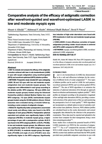
Int J Ophthalmol, Vol. 10, No. 2, Feb.18, 2017www.ijo.cnTel:8629-82245172 8629-82210956Email:ijopress@163.comTable 1 Preoperative clinical characteristics of the studied groupsParametersWFGWFOSphere (D)Mean (SD)Moderate myopiaLow myopiaPWFGWFO0.041-1.90 (0.58)-1.53 (0.74)-4.21 (0.83)-4.02 (0.89)-3.00 to -0.63-3.00 to -0.47-5.88 to -2.88-5.46 to -2.47Mean (SD)-1.20 (0.86)-1.12 (1.32)-1.31 (0.65)-1.47 (1.26)Range-3.25 to 0.00-4.50 to 0.00-2.75 to 0.00-4.75 to 0.00Range0.107Cylinder (D)Mean (SD)Range0.1070.023SE (D)0.098-1.99 (0.60)-2.15 (0.46)-4.36 (0.84)-4.67 (0.86)-3.00 to -0.63-3.00 to -1.38-6.00 to -3.13-6.00 to -3.380.041Decimal CDVAP0.0420.104Mean (SD)1.07 (0.11)0.99 (0.05)1.01 (0.11)1.00 (0.07)Range0.80 to 1.200.80 to 1.200.90 to 1.200.80 to 1.20CDVA: Corrected distance visual acuity; SD: Standard deviation.to use artificial tears when needed (foreign body sensation,dryness etc.).Data Analysis The Alpins vector analysis method wasused for the analysis of the astigmatic changes occurringafter surgery[15-16]. The following vectors were determinedand evaluated: targeted induced astigmatism (TIA) as thevector of intended change in cylinder for each treatment,surgically induced astigmatism (SIA) as the vector of the realchange achieved and difference vector (DV) as the additionalastigmatic change that would enable the initial surgeryto achieve its intended target. Additionally, the followingparameters derived from the relationship between these vectorswere calculated and analyzed at the last postoperative visit. AE:the angle described by the vectors of the achieved correction(SIA) and the intended correction (TIA). Negative: achievedcorrection is clockwise to its intended axis. Positive: achievedcorrection is counterclockwise to its intended axis. Correctionindex (CI): the ratio of the SIA to the TIA-what the surgeryactually induced versus what the surgery was meant to induce.The CI is preferably 1; it is greater than 1 if an overcorrectionoccurs and less than 1 if there is an undercorrection.Concerning statistical analyses, all were performed witha commercially available software package (SPSS forMac, Version 20.0; IBM Corporation, Armonk, NY, USA).Normality of data samples was evaluated by means of theKolmogorov-Smirnov test. When parametric analysis waspossible, the Student’s t-test for unpaired data was used forcomparisons between pairs of groups, whereas the MannWhitney test was applied to assess the significance of suchdifferences when parametric analysis was not possible. For theanalysis of differences between preoperative and postoperativevisits in each group, the Student’s t-test for paired data or theWilcoxon ranked sum test were used depending if the sampleswere normally distributed or not, respectively. For all statisticaltests, a P-value of less than 0.05 was considered as statisticallysignificant. Finally, the efficacy index was calculated as theratio of the postoperative UDVA to the preoperative CDVA, andthe safety index was calculated as the ratio of the postoperativeCDVA to the preoperative CDVA[17].RESULTSTable 1 summarizes the clinical characteristics of the studiedgroups.Low Myopia Group Outcomes Significantly better decimalpreoperative CDVA (WFG 1.07 0.11 vs WFO 0.99 0.05,P 0.041) and postoperative UDVA (WFG 1.07 0.13 vsWFO 0.95 0.14, P 0.011) was found in the WFG subgroupcompared to WFO. A significantly better efficacy indexwas found in the WFG subgroup (WFG 1.00 0.10 vs WFO0.96 0.11, P 0.041). A total of 41 (91%) and 47 eyes (87%)achieved a postoperative UDVA of 20/20 or better in the WFGand WFO subgroups, respectively (Figure 1). More patientsachieved postoperative UDVA of 20/16 in the WFG subgroupcompared to the WFO (44.4% vs 13.0%, P 0.001) (Figure 1).No differences in postoperative decimal CDVA (WFG1.12 0.11 vs WFO 1.03 0.10, P 0.107) and safety index(WFG 1.05 0.11 vs WFO 1.03 0.12, P 0.275) were foundamong subgroups. Gains of lines of CDVA were observedin 15 (33.3%) and 18 eyes (33.3%) in the WFG and WFOsubgroups, respectively (Figure 2). Losses of lines of CDVAwere only observed in the WFO subgroup (1 eye, 1.9%)(Figure 2).Manifest SE changed significantly from a mean preoperativevalue of -1.99 0.60 D to a mean postoperative value of-0.14 0.18 D (P 0.001) in the WFG subgroup and from-2.15 0.46 D to -0.49 0.39 D (P 0.001) in the WFO subgroup(Figure 3). No significant differences were observed betweenthe SE achieved at 1 and 6mo postoperatively in any subgroup(WFG, P 0.322; WFO, P 0.582). No significant differences287
Efficacy of wavefront-guided and optimized LASIKFigure 3 Change in the SE during the follow-up in the WFG andWFO subgroups of the low and moderate myopia groups.Figure 1 Distribution of preoperative CDVA and postoperativeUDVA at 6mo after surgery in the WFG and WFO subgroups A:Low myopia group; B: Moderate myopia group.Figure 4 Distribution of the 6-month postoperative SE in theWFG and WFO subgroups of the low and moderate myopia groups.Figure 2 Change in CDVA during the follow-up in the WFG andWFO subgroups of the low and moderate myopia groups.among groups were found in preoperative SE (P 0.098),but a higher postoperative value was present in the WFOsubgroup compared to WFG (P 0.002). More patients in theWFG subgroup achieved a postoperative SE within 0.25 D(WFG 84.4% vs WFO 66.8%, P 0.015) (Figure 4). Regardingmanifest cylinder, it was reduced significantly with surgery inboth subgroups (WFG: from -1.20 0.86 D preoperatively to-0.06 0.16 D at 6mo postoperatively, P 0.001; WFO: from-1.12 1.32 D preoperatively to -0.20 0.34 D at 6mo postoperatively, P 0.001). Significant differences between sub288groups were found in the magnitude of cylinder postoperatively(P 0.001) that were not present preoperatively (P 0.107).More patients had a 6-month postoperative cylinder of 0.25 D orbelow in the WFG subgroup compared to the WFO subgroup,but the difference was not statistically significant (WFG 88.9%vs WFO 74.1%, P 0.235) (Figure 5).No significant changes in CS for any spatial frequencyevaluated were found at 6mo postoperatively (P 0.107). Thechange induced with surgery in primary coma (P 0.006) andSA (P 0.005) was significantly lower in the WFG subgroupcompared to the WFO subgroup (Figure 6).Moderate Myopia Group Outcomes No statistically significant differences among WFG and WFO subgroups werefound in decimal preoperative CDVA (WFG 1.01 0.11 vsWFO 1.00 0.07, P 0.104). In contrast, significantly betterpostoperative UDVA (WFG 1.01 0.11 vs WFO 0.93 0.11,P 0.017) and better efficacy index (WFG 1.00 0.10 vsWFO 0.93 0.11, P 0.027) was found in the WFG subgroupcompared to WFO. A total of 59 (92.2%) and 52 eyes (89.7%)achieved a postoperative UDVA of 20/20 or better in the
Int J Ophthalmol, Vol. 10, No. 2, Feb.18, 2017www.ijo.cnTel:8629-82245172 8629-82210956Email:ijopress@163.comFigure 5 Distribution of the 6-month postoperative cylinder in theWFG and WFO subgroups of the low and moderate myopia groups.Figure 6 Distribution of the postoperative change in HOAs in theWFG and WFO subgroups of the low and moderate myopia groups.WFG and WFO subgroups, respectively (Figure 1). Morepatients achieved a postoperative UDVA of 20/16 in the WFGsubgroup compared to the WFO (17.2% vs 3.4%, P 0.001)(Figure 1). No significant differences among groups werefound in postoperative decimal CDVA (WFG 1.08 0.12 vsWFO 1.01 0.08, P 0.106) and safety index (WFG 1.07 0.12vs WFO 1.01 0.10, P 0.081). Gains of lines of CDVA wereobserved in 18 (27.9%) and 14 eyes (14.1%) in the WFG andWFO subgroups, respectively. Losses of lines of CDVA wereonly observed in the WFO subgroup (3 eyes, 5.1%).Manifest SE changed significantly from a mean preoperativevalue of -4.36 0.84 D to a mean postoperative value of -0.38 0.16 D (P 0.001) in the WFG subgroup and from -4.67 0.86 D to-0.56 0.31 D (P 0.001) in the WFO subgroup (Figure 3). Nosignificant differences were observed between the SE achievedat 1 and 6mo postoperatively in any subgroup (WFG, P 0.06;WFO, P 0.107). Lower SE was present preoperatively(P 0.023) and postoperatively (P 0.001) in the WFGsubgroup compared to the WFO subgroup. More patients in theWFG subgroup achieved a postoperative SE within 0.25 D(WFG 85.9% vs WFO 48.3%, P 0.001) (Figure 4). Regardingmanifest cylinder, it was reduced significantly with surgeryin both subgroups (WFG: from -1.31 0.65 D preoperativelyto -0.09 0.16 D at 6mo postoperatively, P 0.001; WFO:from -1.47 1.26 D preoperatively to -0.25 0.30 D at 6mopostoperatively, P 0.001). The magnitude of cylinder differedsignificantly among subgroups in the last postoperative visit(P 0.003), but not preoperatively (P 0.107). More patientshad a 6-month postoperative cylinder of 0.25 D or below in theWFG subgroup compared to the WFO subgroup (WFG 90.6%vs WFO 65.5%, P 0.002) (Figure 5).Regarding CS, only a significant improvement was foundin the WFG subgroup for the value obtained for the spatialfrequency of 18 cycles/degree (P 0.028). The change inducedwith surgery in trefoil (P 0.001) and SAs (P 0.001) wassignificantly lower in the WFG subgroup compared to theWFO subgroup (Figure 6).Vector Analysis of Astigmatic Changes Table 2 summarizesthe parameters derived from the vector analysis of ocularastigmatic changes at the end of the follow-up in the WFGand WFO subgroups of the low and moderate myopia groups.In the low myopia group, a significantly larger magnitudeof the DV was found in the WFO group compared to theWFG subgroup (P 0.001). In the moderate myopia group,a significantly larger magnitude of DV (P 0.001), ME(P 0.008), and AE (P 0.001) was found in the WFO subgroupcompared to the WFG subgroup. Likewise, in this same group,a significantly higher CI was found in the WFG subgroup(P 0.001).DISCUSSIONIn the
vs 64 moderate myopia) treated using a WFG ablation profile (Advanced CustomVue platform, AMO Inc), and WFO . subgroup including 112 eyes (54 low myopia vs 58 moderate myopia) treated using a WFO profile (EX-500 platform, Allegretto Q-500 excimer laser, Alcon). I