Transcription
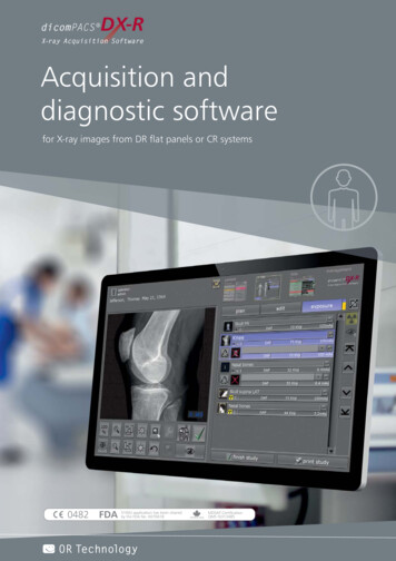
dicomPACSRDX-RX-ray Acquisition SoftwareAcquisition anddiagnostic softwarefor X-ray images from DR flat panels or CR systems0482FDA510(k) application has been clearedby the FDA No. K070618OR TechnologyHealth CanadaMDSAP CertificationQMS ISO13485
Professional
dicomPACSRDX-RX-ray Acquisition Softwareacquisition softwarefor static and dynamic X-ray imagesfor small medical practices & large hospital departments dicomPACS DX-R is a professional acquisition software for X-ray images fromflat panel systems (DR) and CR units (computed radiography with imaging plates)by any origin. In addition, the software controls X-ray generators and X-ray units ofvarious manufacturers, providing a smooth and systematic workflow. A simple and userfriendly GUI (graphical user interface) operated by touchscreen or mousecompletes the system. The professional dicomPACS DX-R image processing can be adapted to individual users‘needs and offers outstanding image quality. It has been specially developed to enable organspecific optimisation, guaranteeing the highest quality X-ray images.Many helpful integrated functions such as the radiographic positioning guide and intuitiveoperation greatly simplify daily routine tasks. In addition, dicomPACS DX-R allows integration with existing patient management systems. The integrated full dicomPACS viewer even enables the user to diagnose X-rayimages within the acquisition software. Therefore, the system can also be applied as fullyfledged diagnostic workstation with the option to upgrade to a PACS (Picture Archivingand Communication System).dicomPACS DX-R forms the core of a digital X-ray unit, whether it is a retrofit systemto upgrade existing X-ray units, a complete new unit including generator control,or a portable suitcase solution for mobile X-ray generators.Further information about acquisitionsoftware is available here:
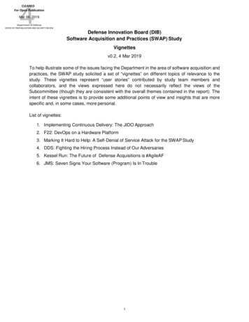
Easy switching betweenplanned examinations ifthe patient has to berepositioned frequentlyThe software can beoperated via touch screenand guarantees fast,efficient work and anoptimal workflow.Recording of recurringexamination procedures asa macro – simplifies workeven outside the routinehospital routine02
dicomPACSRDX-RX-ray Acquisition SoftwareGeneralOptimal workflowThe easy-to-use acquisition and diagnosticsoftware offers many advantages:Modern graphical user interface (GUI) adaptable to almost any languageTouchscreen operation – to ensure quick and efficient work anda smooth workflowCapture of patient data via DICOM Worklist, BDT/GDT, HL7or other protocols – data may also be captured manuallyUse of DICOM Procedure Codes for transferring of all relevant examinationdata directly from the connected patient management system (HIS/RIS)Freely configurable body parts with more than 400 projectionsand numerous possible adjustments in human and veterinary medicinealready includedSafe and fast registration of emergency patientsAllows the user to switch between examinations of a patient, forinstance to avoid having to re-position the patient frequentlyAllows the user to subsequently add images to an examination, evenafter that examination has already been completedEntry of recurring examination procedures as macros,e.g. thorax screeningsFully integrated radiographic positioning guide for each examinationincl. comprehensive notes, photos, videos and correct X-ray images03For more detailed informationplease see: www.or-technology.com
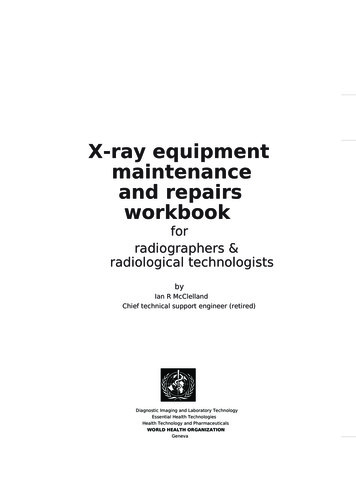
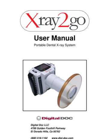
1Planning the X-rayexamination using thebody parts – switchto planning for childrenand infants with justone click2Fully integratedradiographicpositioning guide forcorrect adjustmentprocedure3Proposal of therecommended generatorvalues for the respectivestudy (kVp, mAs, focus,etc.) and worklist [right]04
dicomPACSRDX-RX-ray Acquisition SoftwareGeneralProcedure of an X-ray examination with dicomPACS DX-R4Preview image of theX-ray image (incl. variousdisplay options) [left] andwork list [right]5Findings in theprofessional PACS viewerincl. further processing,storage of images andmuch more6Extensive searchfunctions and displayof the result list05
06
dicomPACSRDX-RX-ray Acquisition SoftwareGeneralOptimal workflowModern graphical user interface with clearlyarranged functionalitiesJob creationThe cf o r a d u l o r r e c t s e t t in g sts andc h il d r e narea t a c l ic a v a il a b l ekChart forplanning anindividualX-ray jobS w itc h tobspl an ni ng X - ra y jofo r ch il dr enan d ba bi esRadiographic positioning guides te pS te p - b y - dsounv id e o w it htf o r p a ti e n n in gp o s it ioanShows mple of a ageexa -ray imXcorrectPresentatio n of helpfulhints for positioningthe patient, central beam,tips and tricks, frequenterrors etc.Opens examplesof inaccurateX-ray imageswith comments07For more detailed informationplease see: www.or-technology.com
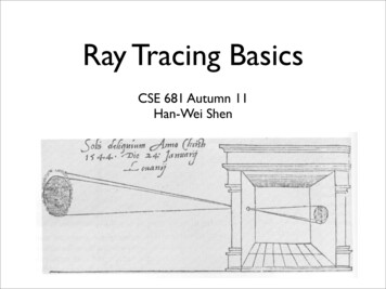
Function principlesThe acquisition software dicomPACS DX-Rguarantees an optimalworkflow in small practicesas well as in large hospitals.[dynamic X-ray]DynamicX-ray detectors(differentmanufacturers)[static X-ray]PanelcontrolFlat paneland CR systems(different manufacturers,also dental panel)RawimagesX-ray devicesheof t ,trol temConsed sys r etc.oriatomot collimRawimagesPanel controldicomPACSRDX-R(motorised)ingitionPos tocolproX-ray Acquisition SoftwareklistWopratientMfnOoatioDIC ery- operation softwarefor generator and panel- image processing- image managementinvDeli d exam uctionsan instrdataHIS/RIS etc.(Patient managementsystem)nations havefirmctionurotsC n inoutbodKVp, my pa AS,rt etc.Expprotosurecolwhe carriednbeeUnlessprovided ut of processedimages incl. all patientand exposure dataPACS(e.g. dicomPACS )08
dicomPACSRDX-RX-ray Acquisition SoftwareGeneralFlexible image acquisitionOptimum adaptation to all X-ray systemsIntegration of various flat panel and CR systems by different manufacturersfor static and dynamic X-ray [dynamic x-ray of image sequences see page 26/27]Option to connect up to 3 flat panels (bucky, wall stand and mobile)to one systemThe configurable generator interface enables the user to controlX-ray generators or X-ray systems by different manufacturers, deliveringthe generator settings directly from the software.Option for the parallel operation of a flat panel and a CR system included in thestandard package. The user has the choice to take the next image with either the flat panelor the integrated CR system. This flexibility also provides an excellent emergencyconcept in case of a defective flat panel.AEC (Automatic Exposure Control) and ARP (Anatomical Programmed Radiography)allow the user to automatically adjust all X-ray options for each projection with anoption to subsequently edit the image manually.Integration of dose area product meters (DAP) – the readings aresaved directly to the relevant imageElectronic X-ray logStatic DRX-ray detectorradiologyCR systemThe software allows simultaneous control of a CR systemand one or more X-ray detectors as standard.09For more detailed informationplease see: www.or-technology.comDynamic DRX-ray detectorNetwork
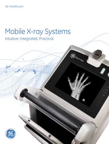
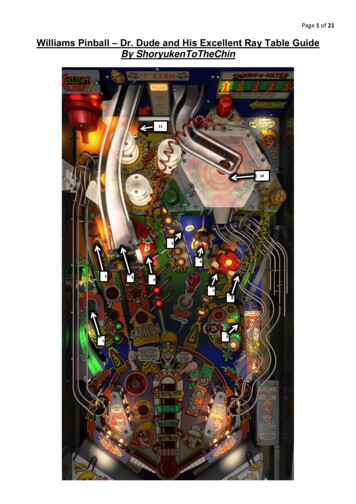
Image processingLeft:Conventional imageprocessingRight: dicomPACS DX-R ImageProcessingGLI image comparisonLeft:Exposure without gridRight:Same exposure with GLIscatter reduction10
dicomPACSRDX-RX-ray Acquisition SoftwareGeneralImage processingAutomatic image optimisation for perfect imagesPerfect images at all times – generally no adjustment requiredIntegrated software for automatic image optimisation with COP[see page 12/13]Professional, adaptable image processing for each individual examination toobtain best possible image settings for each customer‘s needsDue to specially developed processes, the image processing allows theuser to vary the X-ray settings on a large scale while the image qualityremains virtually the same (possibility of dosage reduction)Bones and soft tissue in one image – this enables the user tosignificantly improve the diagnosisDetails of bones and microstructures are very easy to recogniseNoise suppressionBlack border (automatic shutters)Automatic removal of grid lines when using fixed gridsGLI (gridless imaging) – reduction of scattered radiation:GLI: X-ray imaging without gridGrids are required for X-raying large body parts in order to focus the radiationand reduce scatter, thus improving the contrast and brilliance of X-ray images.The virtual scatter reduction GLI works like a grid and can be used instead of aphysical grid for all body regions, including thorax, abdomen, skull, spine,pelvis and upper and lower extremities.11For more detailed informationplease see: www.or-technology.com
Cognition Optimised Processing(COP)COP contains all calculation steps foran optimal image presentation, whichthe X-ray image passes through directlyafter its creation. Image contents areautomatically analyzed and thecalculation steps intelligently adapted.Thus, no post-processing of the imagesis necessary.12
dicomPACSRDX-RX-ray Acquisition SoftwareGeneralOptimisation of image data with the dicomPACS DX-R Cognition Optimised ProcessingThe automatic calculation steps of Cognition Optimised Processing (COP) include:ADPC – automatic dead pixel correctionAutomatically eliminates dead pixels – this reduces the need to calibrate the flat panelAIAA – automatic image area analysisAutomatically analyses each image for soft tissue and bone structures and applies the mostsuitable image processing algorithmsMFLA – multi frequency level analysisAnalyses each image on various frequency levels for ideal sharpness and high contrastANF – automatic noise filterAlgorithm for optimal noise reductionGLI – gridless imagingExposures without grid: enables the display of an image as if it had been taken with a grid –this is useful for supine chest exposures (bedside).AGLS – automatic grid line suppressionAutomatically removes gridlines from flat panel images – suitable for grids from 100 LPIto 200 LPIIBC – intelligent brightness controlAutomatically displays the image at the ideal level of brightnessACO – automatic contrast optimisationAutomatic contrast equalisation across the entire image – this enables the optimal displayof soft tissue and bones at the same timeABBS – automatic black border shutterAutomatically darkens all parts of an image outside the collimated area – varying degrees oftransparency are available and manual adjustments are easy to make.13For more detailed informationplease see: www.or-technology.com
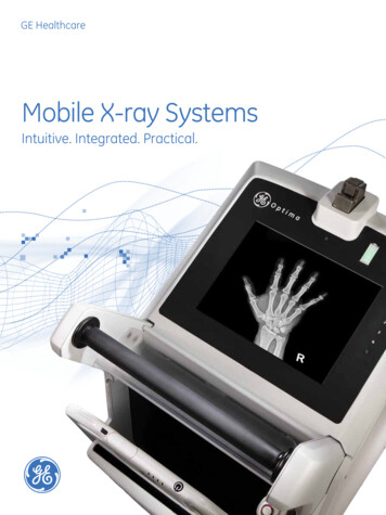
Professional diagnosiswith the completelyintegrated dicomPACS Viewer for imagediagnosisImage export:Send images by e-maildirectly from the softwareImage export:Print of X-ray imageson Windows printers(paper) and laserimagers (film)14
dicomPACSRDX-RX-ray Acquisition SoftwareGeneralImage diagnosisProfessional diagnosis and image processingwith the integrated viewer Completely integrated dicomPACS viewer for image diagnosis, further processingand storage of images in an SQL database incl. image manipulations, export options,layout adjustments, freely configurable user interface and much moreStepless zoom, PAN, magnifyer, ROI, crop, rotate, mirror etc.Insert image annotations, e.g. free texts, arrows, ellipses etc.Measure distances, angles, areas and densityExtended acquisition functionality for use in mammography*Adjust window/level options and gamma correction,sharpening filters, noise suppressionMany additional functions such as chiro tools, calculation of Cobb's angle, HDmeasurements, pelvic obliquity measurements, integrated capturing ofdiagnostic reports etc.Easily upgradable to the integrated image management system (PACS)Image exportExport images to JPEG, TIFF, BMP and DICOM formatsPrint images both on Windows printers and laser imagers via DICOM Basic PrintCreat DICOM patient CDs with free WEB viewerInbuilt email tool to image distribution - no external email application necessary*Pending CE and FDA conformity approval for the module.15For more detailed informationplease see: www.or-technology.com
Integrated ViewerIntegrated dicomPACS Viewer for image diagnosis, further processingand storage of imagesin an SQL database incl.image manipulations,export options etc.The integratedprosthesis moduleallows preoperativeplanning (optional)The system enables quickand easy adaptation ofthe user interface toindividual customerrequirements16
. with extensive functionsUseful tools, such as theconfigurable measuringmagnifier, make reportingsignificantly easierThe stitching modulecreates a single imagefrom separate digital X-rayimages (optional)Comprehensivesearch tools allow thecomparison of X-rayexaminations, even ofdifferent patients17
CR systemsIn combination with theprofessional image acquisition software dicomPACS DX-R, a CRsystem combines all necessaryimage processing functions.DR systemsDR X-ray systems can becontrolled by the professionalacquisition and diagnostic software dicomPACS DX-R.18
dicomPACSRDX-RX-ray Acquisition SoftwareGeneralModalitiesWhich flat panels and CR systems does dicomPACS DX-R support? dicomPACS DX-R is a generally open system. Its conception and developmentwas independent of hardware manufacturers.Components from the following manufacturers have already been integrated (We arecontinuously working on the integration of new models and manufacturers):X-ray detectors (DR)CareRayCR systemsDivario CR SystemsFireCRCETD19For more detailed informationplease see: www.or-technology.com
Individual adaptationof the graphical userinterface according tothe specifications of theOEM partnerComplete control ofX-ray generators and X-raysystems from variousmanufacturersOrderly and optimalworkflow&Simple and user-friendlyuser interface20
dicomPACSRDX-RX-ray Acquisition SoftwareGeneralOEM: Software wanted? Who is interested in dicomPACS DX-Rfrom OR Technology?OEM partnerships provide numerous benefits to manufacturers who are interested in combing their X-ray systems with our dicomPACS DX-Racquisition software under
dicomPACSR DX-R X-ray Acquisition Software OR Technology 510(k) application has been cleared 0482 FDA by the No. K070618FDA Health Canada MDSAP Certification QMS ISO13485. Professional . In addition, allowsdicomPACS DX-R integration with existing patient management systems. The integrated full viewer even enables the user to diagnose X-ray images within the acquisition software.