Transcription
DIAGNOSIS OF CANINE HYPOTHYROIDISM: CASE-BASED APPROACHEllen N. Behrend, VMD, PhD, Diplomate ACVIM (Small Animal Internal Medicine)CASE 1Signalment: 8 yr old, CM, English bulldog; History: Presented for annual exam. Low activity and obesitydespite limited feedings only problems noted.; Physical examination: Obese; Laboratory data:3Complete CBC, profile, urinalysis done. Abnormalities were: WBC 17.5 x 10 /µl (6.0-17.0); Neutrophils:33314.6 x 10 /µl (3.0-11.5); Lymphocytes 0.7 x 10 /µl (1.0-4.8); Monocytes 1.7 x 10 /µl (0.2-1.4)The approach to a dog with no known non-thyroidal illness (e.g. renal disease, neurological disease,neoplasia, etc.) vs. a dog with non-thyroidal illness is a bit different. In dogs with no known non-thyroidalillness the diagnosis is more straightforward. Starting with a measurement of total T4 alone is reasonable1-4and economical. If total T4 is normal, it is highly unlikely that the dog is hypothyroid.Since nonthyroidal factors such as drugs and illness affect T4, if the T4 is below normal, the dog may or may not be1-3hypothyroid , and further testing is required.CASE SUMMARY: Serum T4 concentration 28 nmol/L (reference range 20-50 nmol/L). Based on minimalclinical signs and normal serum T4 concentration, diagnosis of hypothyroidism ruled out.CASE 2Signalment: 8 yr old, CM, English bulldog; History: Presented for decreased activity, obesity despitebeing on a weight-loss program, thinning haircoat, heat-seeking behavior.; Physical examination:Obese; partial, bilaterally symmetrical alopecia; Laboratory data: Complete CBC, profile, urinalysis done.6Abnormalities were: RBC x 10 /µl 5.0 (5.5-8.5); Hemoglobin 11.2 g/dL (12-18)); PCV 33 % (37-55);3Lymphocytes 0.5 x 10 /µl (1.0-4.8); Cholesterol 470 mg/dL (130-370)The clinical findings suggestive of hypothyroidism are much stronger in this dog than in Case 1.Starting with measurement of serum T4 is still a good first choice, but be prepared to do further testing ifthe serum T4 concentration is below normal.Free T4 (fT4) is the portion of total T4 not bound to protein and represents about 0.1% of total T4.Since the pituitary-thyroid axis functions to maintain free, not total, T4 within a certain range, fT4 isaffected less by non-thyroidal factors and measurement of fT4 is a better test of thyroid function.1Accordingly, fT4 is a more sensitive and more specific test for diagnosis of hypothyroidism , but it is alsonot as good a stand-alone test as once believed (see below). It can be the initial test for the diagnosis ofhypothyroidism or can be used in dogs that have been found to have low total T4 concentrations.Free T4 should always be measured by the equilibrium dialysis method. The other technique formeasuring fT4, analogue RIA, is not reliable and provides no additional diagnostic value over5measurement of total T4. Equilibrium dialysis is also the only RIA for measuring fT4 that is unaffected by6the presence of autoantibodies.7Primary thyroidal failure is believed to be the cause of canine hypothyroidism in 99% of cases.Accordingly, negative feedback of thyroid hormones on the pituitary would be lost and TSH should1,4,8-10increase. However, an elevated serum TSH occurs in only 63-85% of hypothyroid dogs,and ifmeasurement of canine TSH were used alone for diagnosis of hypothyroidism, up to 37% of cases wouldbe missed. Serum TSH can also be elevated in approximately10% of euthyroid dogs with non-thyroidal1,9,11,12illness.Therefore, measurement of TSH is best used not as a sole test but in conjunction with T4or, ideally, fT4. Use of the combination will aid in identifying false-positive and false-negative results seenwith assessment of TSH alone.Measurement of baseline serum T3 is of little value in differentiating hypothyroid from normal dogs.1,2,5There is no apparent difference in serum T3 concentrations between these groups.CASE SUMMARY: Serum T4 concentration was 13 nmol/L (borderline range: 12-19; reference range 20-50nmol/L). Measurement of serum fT4 concentration (by equilibrium dialysis) and serum TSH concentrationwere requested. The serum fT4 concentration was 8 pmol/L (reference range 15-45 pmol/L, 10-14 pmol/Lborderline) and serum TSH concentration was 0.25 ng/ml (normal 0.5 ng/ml).
The interpretation of the case now becomes a clinical dilemma. The question is whether this is ahypothyroid dog with a normal TSH or whether this is a euthyroid sick dog whose TSH has remainednormal while the fT4 is falsely lowered. The danger of falsely diagnosing a dog with hypothyroidism arethreefold: 1. If clinical signs are incorrectly attributed to hypothyroidism, then the true diagnosis will bedelayed or never sought. 2. Thyroxine is a catabolic hormone. Administering a catabolic hormone to anill patient may be detrimental. 3. The patient will needlessly be treated with thyroid hormone for the restof its life. On the other hand, the danger of not treating hypothyroidism is that the clinical signs willprogress. However, in a case such as this one, the clinical signs are relatively mild and benign andprogression is typically insidious, i.e. not treating for a month will most likely not be detrimental in thelong-term.At this point there are 2 choices: 1. Retest in 4-8 weeks. 2. Start trial therapy forhypothyroidism. If choosing option 2, make sure that you have OBJECTIVE measures of the endpointdetermined beforehand, i.e. normalization of serum cholesterol concentration and return to normal weight.Regrowth of hair is not a good endpoint to choose; the haircoat of euthyroid dogs will improve in responseto thyroid supplementation. Be prepared to stop giving the thyroxine if the clinical signs do not improvegiven adequate post-pill levels and time. (You must measure post-pill levels to determine if the trial issuccessful or not.)CASE 3Signalment: 8 yr old, CM, English bulldog; History: Presented for lethargy, weight gain and obesitydespite a poor appetite, bilaterally symmetrical alopecia (non-pruritic) that has been progressive over thepast year, heat-seeking behavior; Physical examination: Obese; partial, bilaterally symmetrical alopecia;6Laboratory data: Complete CBC, profile, urinalysis done. Abnormalities were: RBC x 10 /µl 5.0 (5.538.5); Hemoglobin 11.2 g/dL (12-18)); PCV 33 % (37-55); Lymphocytes 0.5 x 10 /µl (1.0-4.8); Cholesterol470 mg/dL (130-370)In a case that seems to be textbook for hypothyroidism, starting with measurement of serum T4concentration is reasonable. If no other abnormalities are found other than those that can be explainedby hypothyroidism and the serum T4 concentration is very low, a presumptive diagnosis of hypothyroidismcan be made. It would be ideal to measure a fT4 concentration by dialysis for confirmation, but it may beunnecessary. Measurement of serum TSH concentration is not worth the money in this situation. Giventhat the sensitivity of measuring serum fT4 concentration is much higher than that of serum TSHconcentration measurement, in a case such as this if serum fT4 concentration were low but serum TSHconcentration was normal, I would believe the serum fT4 concentration and start treatment forhypothyroidism.CASE SUMMARY: Serum T4 concentration was measured and was non-detectable. Due to financialconsiderations, fT4 concentration was not measured. Therapy with L-thyroxine was instituted. Post-pilltesting was done to ensure adequate serum T4 concentration was achieved. Within 3-4 months clinicalsigns had resolved.CASE 4Signalment: 8 yr old, CM, English bulldog; History: Originally presented to his veterinarian for a geriatricscreen and then was referred to the Auburn University Small Animal Clinic for evaluation of an incidentalfinding of proteinuria. On a urinalysis, a 2 proteinuria was noted with a specific gravity of 1.014. A urineprotein/creatinine ratio (UP/C) was determined to quantify the protein loss and was found to be 5.8(normal 0.5); The dog had received regular veterinary care at a private veterinary clinic. He lived inAlabama with no history of travel out of state. His vaccinations were up-to-date, and he was receivingInterceptor for heartworm prevention. He spent his days outdoors, but was a house dog at night.The owners reported no problems, and had seen no evidence of coughing, sneezing, vomiting, diarrhea,polydipsia, polyuria or weight loss. The dog’s activity had decreased slowly over the past year and wasattributed to aging. His appetite was normal.; Physical examination: On PE, the dog was noted to beobese, and he had moderate to severe dental tartar and gingivitis. Chest auscultation and abdominalpalpation were within normal limits.; Laboratory data: Complete CBC, profile, urinalysis done.33Abnormalities were: WBC 17.5 x 10 /µl (6.0-17.0); Neutrophils: 14.6 x 10 /µl (3.0-11.5); Lymphocytes 0.72
33x 10 /µl (1.0-4.8); Monocytes 1.7 x 10 /µl (0.2-1.4); Albumin 2.2 g/dL (2.7-4.5); 4 proteinuria in urine with1.021 specific gravity; urine protein/creatinine ratio 7.5 (normal 0.5); Blood pressure: normalDue to the magnitude of the UP/C, a possible diagnosis of immune-complex glomerulonephritis(ICGN) was made. ICGN can be idiopathic or secondary to chronic immune stimulation. As the dentaldisease could be a source of antigens, a dental procedure was recommended and was performed. Onemonth post-dental, the UP/C was essentially unchanged at 7.2 and blood pressure remained normal.Further diagnostics were initiated to find possible underlying disease processes that could initiateimmune stimulation. Three-view chest radiographs were obtained to rule out neoplasia (primary ormetastatic) as well as other pulmonic diseases, and they were within normal limits. Abdominal ultrasoundwas normal. An occult heartworm test was negative. Serology for Ehrlichia canis, Bartonella and Lyme’sdisease was negative. PCR for Bartonella spp. and Ehrlichia spp. was negative. Urine culture yielded nogrowth.At re-evaluation approximately 4 weeks later, after the results of all the tests had been obtained, theUP/C was 8.6, systolic blood pressure was moderately elevated at 190 mm Hg and the cholesterol wasmoderately elevated (412 mg/dl). In order to determine the pathology underlying the proteinuria (e.g.glomerulonephritis vs. amyloidosis vs. structural glomerulopathy) and whether the disease process wasreversible, an ultrasound-guided renal biopsy was performed. The histopathological diagnosis wasglomerulonephritis. Enalapril was prescribed (0.5 mg/kg daily) to decrease proteinuria and bloodpressure.On subsequent rechecks the dog was doing well. Systolic blood pressure was 140-150 mm Hg andthe UP/C was approximately 4.3. However, persistent hypercholesterolemia (persistent), obesity andpoor hair regrowth after abdominal ultrasound were noted, and a diagnosis of hypothyroidism wasconsidered.Given the complexity of the case, I would start with measurement of serum total T4, fT4, and TSHconcentrations.The effect of non-thyroidal illness on testing for hypothyroidism is quitesignificant. Two hundred twenty-three dogs with normal thyroidal function but with non-thyroidal illnesswere divided into those with mild, moderate and severe disease. Mildly ill dogs were considered to haveclinical signs of disease but could be treated as outpatients, moderately ill dogs were sick enough togenerally require hospitalization and more aggressive treatment and severely ill dogs required intensive12care and advanced treatment. Interesting results were obtained.Disease severityAll dogsMild diseaseModerate diseaseSevere diseaseTotal T43182860T31631827% abnormalFree T42281744TSH81168Of the 69 dogs with low T4, 45% also had a low fT4 whereas only 8.7% also had a high TSH. Only 1.8%12of sick dogs had a low T4 and fT4 in combination with a high TSH.Similar results have been obtained from other studies as well. In 49 dogs with a variety of illnesses,9T4 was low in 22% but TSH was elevated in only 12%. In 127 dogs with “severe disease” that causedthe owners to euthanatize their dog, 65% had at least one abnormal value. Overall, 59% had decreased11T4, 32% had decreased fT4 and 8% had increased TSH. Forty percent had only a decreased T4, 5%had only a decreased fT4, 5% had increased TSH, 43% had decreased T4 and fT4, and 7% had11decreased T4 and increased TSH. No dog had decreased fT4 and increased TSH.Possibly, in order affect thyroid hormones, a non-thyroidal illness must cause metabolic or systemic13problems. For example, moderate to severe arthritis had no effect on thyroid testing. However, eventransient systemic illness can have effects on thyroid testing and the alterations may be prolonged. Twostudies have been done in which endotoxin was administered to dogs to cause transient illness. After 114dose, 25% of dogs had decreased T4. fT4 was normal, but TSH was not measured. In dogs givenendotoxin every 12 hours for 8 doses , T4 decreased during treatment. Interestingly, T4 then returned to3
normal but decreased again into the hypothyroid range at days 2-16 after administration ceased. TSH15and fT4 were not affected.Therefore, in sick dogs, the first choice for diagnosis of hypothyroidism would be a combination ofndTSH, fT4 and T4, 2 choice is a combination of TSH and fT4 and third choice is a combination of TSH andT4. If the results are conflicting (some parameters are suggestive of hypothyroidism while others are not),the ideal would be to resolve the non-thyroidal illness, if possible, and retest the dog at that time.Alternatively, a TSH stimulation test could be done with recombinant human TSH (rhTSH), if available(Thyrogen , Genzyme Corp.). The exact recommendations are unknown. Previous information statedthat a dose of 50 mcg rhTSH should be used IV for dogs 29 kg and 100 mcg IV for dogs 29 kg, andserum T4 concentration measured before injection and 4 hr post. In previous information, in normal dogs,serum T4 concentration should increase by 20 nmol/L or to at least 40 nmol/L (to convert to mcg/dl, dividevalue in nmol/L by 12.87). If either one or both of the criteria is met, the dog is not hypothyroid. If neithercriteria is met, the dog is hypothyroid (S. Daminet, personal communication). Studies are ongoing todetermine if this protocol is optimal.A vial of Thyrogen contains 1100 mcg of rhTSH. Once reconstituted, the vial should be divided into50 mcg aliquots and frozen in syringes at -20 C. At this temperature, the rhTSH is stable for at least 8weeks.If resolution of the other disease is not possible or testing with rhTSH is not available, the diagnosis ofhypothyroidism poses a clinical dilemma as in Case 2. It is up to the clinician to decide how high theirindex of suspicion is for hypothyroidism, e.g. what clinical signs are present that could be attributed tohypothyroidism alone and not to the other disease process. The same drawbacks to treating or nottreating exist as before but not treating could have more devastating consequences if some of the severeclinical signs are caused by the hypothyroidism, e.g. neuropathy. It may be best to treat the dog forhypothyroidism while still looking for other possible etiologies of the clinical signs.CASE SUMMARY: A total serum T4, fT4 and TSH concentrations were measured. Serum T4 concentrationwas 13 nmol/L (normal 20-55 nmol/L; borderline 12-19 nmol/L), serum fT4 was 11 pmol/L (normal 15-45pmol/L, borderline 10-14 pmol/L) and the TSH was 0.04 ng/ml (normal 0.5 ng/ml). Due to the effect thatnon-thyroidal illness can have on thyroid function testing, the dog was judged to be most likely euthyroidbased on a normal TSH and minimal clinical signs. A recheck was recommended in 4-6 weeks.The dog improved on treatment. Blood pressure remained normal on enalapril, the UP/C stabilized atapproximately 3.2 and cholesterol remained very mildly elevated (380-400 mg/dl) as well. Two monthsafter stabilization, the thyroid panel was repeated. Total T4 was still below normal (16 nmol/L), but the fT4(18 pmol/L) and TSH (0.02 ng/ml) were within normal. Hypothyroidism was ruled out.CASE 5Signalment: 8 yr old, CM, English bulldogHistory: Presented for geriatric examination. Doing well at home.Physical examination: Normal (for a bulldog )Laboratory data: Complete geriatric profile done. All within normal limits except ALP 254 IU/L (normal10-95) and T4 12 nmol/L (normal 20-50).What to do now with this case? I believe in geriatric screening in some scenarios - thyroid testing indogs is not one of them. One thing to consider with random testing (i.e. not testing based on thepresence of clinical signs), what is the predictive value of a test? Sensitivity and specificity look at a testfrom the viewpoint of the patient. Sensitivity is the chance that an animal with the disease will testpositive and specificity is the chance that an animal that doesn't have the disease will test negative.Sensitivity and specificity are NOT affected by the prevalence of the disease in a population tested. PPVand NPV look more at a test from the viewpoint of the test. Positive predictive value (PPV) tells you howlikely it is that an individual with a positive test result actually has the disease. Negative predictive value(NPV) tells you the likelihood that an animal with a negative test result doesn't have the disease.Let’s look at the following chart regarding using fT4ed for testing for hypothyroidism. The chartassumes that there is a 50% prevalence in the population, i.e. 50% of tested dogs are truly hypothyroid.Since the prevalence is 50%, in a population of 200 dogs, 100 are hypothyroid (left hand column) and 100are normal (right hand column). In the chart, it is the left column that is used to calculate sensitivity (that's4
why it says sensitivity below that column) and the right hand column that is used to calculate specificity.Calculations:Prevalence: 100/200 50%Sensitivity True positive / (True positive false negative) 98/100 98%Spec True negative / (True negative false positive) 93/100 93%PPV True positive / (True positive false positive) 98/105 93%NPV True negative /( True negative false negative) 93/95 98%So in this population of 200 dogs, an animal with a low ft4ed is truly hypothyroid about 93% of the timeand an animal with a normal ft4ed is normal about 98% of the time. That's a pretty darn good test.However, these values for PPV and NPV are dependent upon the prevalence of the disease in thepopulation in question. The higher the prevalence of a disease in a population being tested, the higherthe PPV and NPV. In dogs that have actual clinical signs of a disease being tested for, it is likely that theprevalence is relatively high - if there are clinical signs of a disease, then there is a higher likelihood ofthat disease being present than in a random population of dogs. Although hypothyroidism is one of the 2most common endocrine diseases, in the dog population as a whole it is not THAT common. Let's justsay for sake of argument that it is 5% (which is WAY too high). Now we test the next 200 dogs forhypothyroidism that walk through our door. If the prevalence of the disease in general is 5%, thenchances are 5% of the next 200 dogs are hypothyroid. Based on those percentages, we could constructa 2x2 chart (below). Now the calculations are:Prevalence: 10/200 5%Sensitivity True positive / (True positive false negative) 10/10 100%Spec True negative / (True negative false positive) 177/190 93%PPV True positive / (True positive false positive) 10/23 43%NPV True negative /( True negative false negative) 177/177 100%5
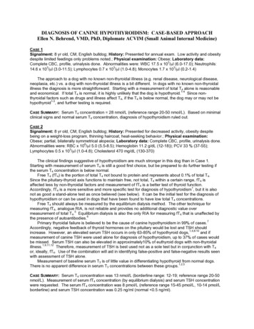
So when you randomly test dogs or cats for a disease without carefully selecting the population, i.e.those with clinical signs of the disease, you HUGELY decrease the PPV of the test. In our example, theprevalence was 5% and that is high for many of the disease we deal with, so PPV would be even lower.In addition, T4 is not that easy to interpret and depends on so many things - age being one of them!In a recent paper, Purina kept Labs from birth until death and looked at the effect of lifetime food16restriction on variables. They also looked at effect of age . The figure below shows the mean total T4from the paper in one group (but the other group was statistically similar):T4 over time30T4 (nmol/L)2520151050567891011Age (yrs)So there appears to be an effect of age that no laboratory takes into consideration in their referenceintervals. In addition, MANY things besides age can make T4 go down.CASE SUMMARY: I would not recommend further treatment of this dog without clinical signs, regardless ofthe T4 measurement.MEASUREMENT OF THYROID AUTO-ANTIBODIESExcept in the evaluation of breeding dogs, measurement of thyroid auto-antibodies does not addmuch to evaluation of dogs for possible hypothyroidism. If a hypothyroid dog has auto-antibodies then itcan be determined that the underlying etiology is lymphocytic thyroiditis as compared to idiopathichypothyroidism. However, the management of the hypothyroidism does not differ.17,18In general, the clinical and prognostic significance of autoantibodies is unknown.If autoantibodies6are suspected, measure fT4 for the best assessment of function. If the fT4 concentration is normal,6
thyroid function is normal at that time but the patient should be re-evaluated periodically (e.g. q. 3 mths)for development of hypothyroidism. If fT4 is low, the dog is likely hypothyroid. One study followed 234dogs with normal T4 and TSH levels and elevated anti-thyroglobulin antibodies (TGAA) for 1 year. Only19% developed clinical signs of hypothyroidism or consistent laboratory values. Another 57% remainedTGAA positive without signs or laboratory evidence of hypothyroidism, 8% went from positive to19borderline results and 15% became TGAA negative. The final outcome of all the dogs is unknown (i.e.how many would become hypothyroid if followed for more than one year), but it can be said that not alldogs with autoantibodies will become hypothyroid, as at least 15% do not.References available from author upon request.7
The danger of falsely diagnosing a dog with hypothyroidism are threefold: 1. If clinical signs are incorrectly attributed to hypothyroidism, then the true diagnosis will be delayed or never sought. 2. Thyroxine is a catabolic hormone. Administering a catabolic hormone to an ill patient may be detrimental. 3.