Transcription
Adult HypothyroidismLast Updated: December 12, 2013Wilmar M. Wiersinga, M.D.Department of Endocrinology F5-171 Academic Medical Center Meibergdreef 9 NL-1105 AZAmsterdam, The Netherlands Tel: 011-31-20-566-6071 Fax: 011-31-20-566-44409.1 HISTORICALThe full-blown expression of hypothyroidism is known as myxedema. Adult myxedemaescaped serious attention until Gull described it in 1874 1. That it was a state resembling thefamiliar endemic cretinism, but coming on in adult life, was what chiefly impressed Gull. Ord 2invented the term myxedema in 1873. The disorder arising from surgical removal of thethyroid gland (cachexia strumipriva) was described in 1882 by Reverdin 3 of Geneva and in1883 by Kocher of Berne 4. After Gull's description, myxedma aroused enormous interest,and in 1883 the Clinical Society of London appointed a committee to study the disease andreport its findings 5. The committee's report, published in 1888, contains a significant portionof what is known today about the clinical and pathologic aspects of myxedema. It is referredto in the following discussion as the Report on Myxedema. The final conclusions of the 200page volume are penetrating. They are as follows:1. That myxedema is a well-defined disease.2. That the disease affects women much more frequently than men, and that the subjects arefor the most part of middle age.3. That clinical and pathological observations, respectively, indicate in a decisive way that theone condition common to all cases is destructive change of the thyroid gland.4. That the most common form of destructive change of the thyroid gland consists in thesubstitution of a delicate fibrous tissue for the proper glandular structure.5. That the interstitial development of fibrous tissue is also observed very frequently in theskin, and, with much less frequency, in the viscera, the appearances presented by this tissuebeing suggestive of an irritative or inflammatory process.6. That pathological observation, while showing cause for the changes in the skin observedduring life, for the falling off the hair, and the loss of the teeth, for the increased bulk of body,as due to the excess of subcutaneous fat, affords no explanation of the affections of speech,movement, sensation, consciousness, and intellect, which form a large part of the symptomsof the disease.7. That chemical examination of the comparatively few available cases fails to show thegeneral existence of an excess of mucin in the tissues adequately corresponding to theamount recorded in the first observation, but that this discrepancy may be, in part, attributedto the fact that tumefaction of the integuments, although generally characteristic ofmyxedema, varies considerably throughout the course of the disease, and often disappearsshortly before death.8. That in experiments made upon animals, particularly on monkeys, symptoms resemblingin a very close and remarkable way those of myxedema have followed complete removal of
the thyroid gland, performed under antiseptic precautions, and with, as far as could beascertained, no injury to the adjacent nerves or to the trachea.9. That in such experimental cases a large excess of mucin has been found to be present inthe skin, fibrous tissues, blood, and salivary glands; in particular the parotid gland, normallycontaining no mucin, has presented that substance in quantities corresponding to what wouldbe ordinarily found in the submaxillary gland.10. That following removal of the thyroid gland in man in an important proportion of thecases, symptoms exactly corresponding with those of myxedema subsequently develop.11. That in a considerable number of cases the operation has not been known to have beenfollowed by such symptoms, the apparent immunity being in many cases probably due to thepresence and subsequent development of accessory thyroid glands, or to accidentallyincomplete removal, or to insufficiently long observation of the patients after operation.12. That, whereas injury to the trachea, atrophy of the trachea, injury of the recurrentlaryngeal nerves, injury of the cervical sympathetic, and endemic influences, have been byvarious observers supposed to be the true cases of experimental or of operative myxedema(cachexia strumipriva), there is, in the first place, no evidence to show that, of the numerousand various surgical operations performed on the neck and throat, involving various organsand tissues, any, save those in which the thyroid gland has been removed, have beenfollowed by the symptoms under consideration; that in many of the operations on man, and inmost, if not all, of the experimental operations made by Professor Horsley on monkeys andother animals, this procedure avoided all injury of surrounding parts, and was perfectlyantiseptic; that myxedema has followed removal of the thyroid gland in persons neither livingin nor having lived in localities the seat of endemic cretinism; that, therefore, the positiveevidence on this point vastly outweighs the negative; and that it appears strongly proved thatmyxedema is frequently produced by the removal, as well as by the pathological destruction,of the thyroid gland.13. That whereas, according to Clause 2, in myxedema women are much more numerouslyaffected than men, in the operative form of myxedema no important numerical difference isobserved.14. That a general review of symptoms and pathology leads to the belief that the diseasedescribed under the name of myxedema, as observed in adults, is practically the samedisease as that named sporadic cretinism when affecting children; that myxedema isprobably identical with cachexia strumipriva; and that a very close affinity exists betweenmyxedema and endemic cretinism.15. That while these several conditions appear, in the main, to depend on, or to beassociated with, destruction or loss of the function of the thyroid gland, the ultimate cause ofsuch destruction or loss is at present not evident.9.2 DEFINITION AND EPIDEMIOLOGY OF HYPOTHYROIDISMHypothyroidism is traditionally defined as deficient thyroidal production of thyroid hormone.The term primary hypothyroidism indicates decreased thyroidal secretion of thyroid hormoneby factors affecting the thyroid gland itself; the fall in serum concentrations of thyroidhormone causes an increased secretion of TSH resulting in elevated serum TSHconcentrations. Decreased thyroidal secretion of thyroid hormone can also be caused byinsufficient stimulation of the thyroid gland by TSH, due to factors directly interfering withpituitary TSH release (secondary hypothyroidism) or indirectly by diminishing hypothalamic
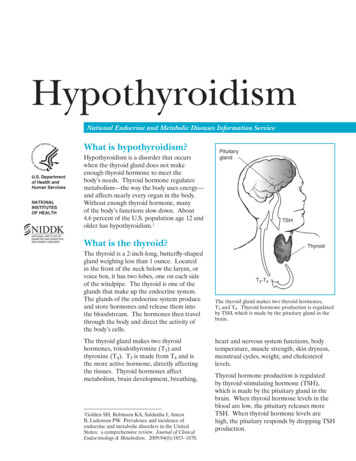
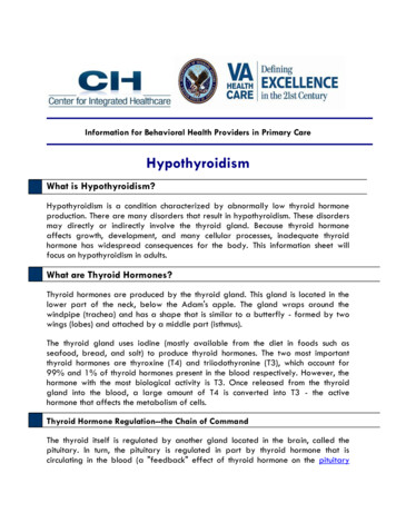
TRH release (tertiary hypothyroidism); in clinical practice it is not always possible todiscriminate between secondary and tertiary hypothyroidism, which are consequently oftenreferred to as central hypothyroidism. In rare cases, symptoms and signs of thyroid hormonedeficiency are caused by the inability of tissues to respond to thyroid hormone by mutationsin the nuclear thyroid hormone receptor TRß; this condition, known as thyroid hormoneresistance (see Ch. 16), is associated with an increased thyroidal secretion of thyroidhormones and increased thyroid hormone concentrations in serum in an attempt of the bodyto overcome the resistance to thyroid hormone. Mutations in other genes involved withextrathyroidal metabolism and action of thyroid hormones in target tissues may also cause ahypothyroid state. Such cases could be labelled as peripheral (extrathyroidal)hypothyroidism. It thus seems more appropriate to define hypothyroidism as thyroid hormonedeficiency in target tissues, irrespective of its cause.9.2.1. GRADES OF HYPOTHYROIDISMHypothyroidism is a graded phenomenon, ranging from very mild cases in which biochemicalabnormalities are present but the individual hardly notices symptoms and signs of thyroidhormone deficiency, to very severe cases in which the danger exists to slide down into a lifethreatening myxedema coma. In the development of primary hypothyroidism, the transitionfrom the euthyroid to the hypothyroid state is first detected by a slightly elevated serum TSH,caused by a minor decrease in thyroidal secretion of T4 which doesn't give rise to subnormalserum T4 concentrations. The reason for maintaining T4 values within the reference range isthe exquisite sensitivity of the pituitary thyrotroph for even very small decreases of serum T4,as exemplified by the log-linear relationship between serum TSH and serum FT4 1. A furtherdecline in T4 secretion results in serum T4 values below the lower normal limit and evenhigher TSH values, but serum T3 concentrations remain within the reference range. It is onlyin the last stage that subnormal serum T3 concentrations are found, when serum T4 hasfallen to really very low values associated with markedly elevated serum TSH concentrations(Figure 9-1). Hypothyroidism is thus a graded phenomenon, in which the first stage ofsubclinical hypothyroidism may progress via mild hypothyroidism towards overthypothyroidism (Table 9-1)3.
Figure 9-1. Individual and median values of thyroidfunction tests in patients with various grades ofhypothyroidism. Discontinuous horizontal linesrepresent upper limit (TSH) and lower limit (FT4,T3)of the normal reference ranges. (Reproduced withpermission) (2)Table 9-1. Grades of hypothyroidismGrade 1Subclinical hypothyroidismTSH FT4 NT3 N( )Grade 2Mild hypothyroidismTSH FT4 -T3 NGrade 3Overt hypothyroidismTSH FT4 -T3 - , above upper normal limit; N, within normal reference range; -, below lower normal limit.Maintenance of a normal serum T3 concentration until a relatively late stage in thedevelopment of hypothyroidism obviously serves as an appropriate mechanism of the bodyto counteract the impact of diminishing production of T4. It is accomplished by a preferentialthyroidal secretion of T3: the increased secretion of TSH enhances the synthesis of T3 morethan that of T4 and stimulates thyroidal 5'-monodeiodination of T4 into T3 4,5. It explains whysometimes a slightly elevated serum T3 is found in the early stage of development of
hypothyroidism. About 80% of the daily production rate of T3 is generated in extrathyroidaltissues via the conversion of T4 into T3. The peripheral tissues also have a defensemechanism against developing hypothyroidism by increasing the overall fractionalconversion rate of T4 into T3 6.9.2.2. EPIDEMIOLOGY OF HYPOTHYROIDISMThyroid hormone resistance syndromes are seldom the cause of hypothyroidism; the numberof registered patients approximates one thousand (see Ch. 16). Central hypothyroidism isalso rare; its precise prevalence is unknown, but has been estimated as 0.005% in thegeneral population 7. Primary hypothyroidism, in contrast, is a very prevalent diseaseworldwide. It can be endemic in iodine-deficient regions (see Ch. 20), but it is also a commondisease in iodine-replete areas as evident from prevalence and incidence figures reported ina number of population-based studies 8-14. The most extensive data has been obtained fromthe Whickham Survey, a study of 2779 adults randomly selected of the general population inGreat Britain who were evaluated between 1972 and 1974 and again twenty years later 8,9.Most striking are the high prevalence of thyroid microsomal (peroxidase) antibodies and of(subclinical) hypothyroidism, and the marked female preponderance (Table 9-2).Table 9-2. Prevalence and incidenceof thyroid antibodies andhypothyroidism in the Whickhamsurvey (8,9). thyroglobulin antibodiesPrevalence microsomal (TPO)antibodies subclinical hypothyroidism hypothyroidismIncidence hypothyroidismWomenMen30 per 10009 per 1000103 per 100027 per 100075 per 100028 per 100018 per 10001 per 10004.1 per 1000 per yr0.6 per 1000 per yrThe mean incidence of spontaneous hypothyroidism in women was 3.5/1000 survivors/year,that of hypothyroidism after destructive treatment for thyrotoxicosis 0.6/1000 survivors/year;similar figures were obtained in those who had deceased during follow-up. The hazard rate(the probability to develop hypothyroidism) increased with age; the mean age at diagnosis ofhypothyroidism in women was 60 years. Studies from other countries like the USA 10,11,Japan 12 and Sweden 13 report essentially similar data.Of particular interest are risk factors for development of hypothyroidism. In women survivorsof the Whickham Survey, the risk of developing overt hypothyroidism was 4.3% per year ifboth raised serum TSH and thyroid antibodies were present initially, 2.6% per year if raisedserum TSH was present alone, and 2.1% per year if thyroid antibodies were present alone 9.At the time of follow-up twenty years later, hypothyroidism had developed in these threegroups in 55%, 33% and 27% respectively, but only in 4% if initial serum TSH was normaland thyroid antibodies were absent. The probability of developing hypothyroidism already
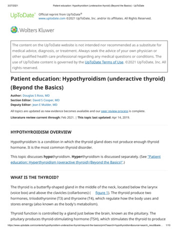
increases at a rise in serum TSH above 2 mU/L as shown in Figure 9-2, in thyroid antibodypositive as well as in thyroid antibody negative women; it also increases with higher titres ofthyroid microsomal antibodies 9,15. These data are confirmed by two other more recent largepopulation-based longitudinal surveys with a mean follow-up of 11-13 years. A figure almostidentical to figure 9.2 was obtained in an Austalian study, in which the odds ofhypothyroidism increased at TSH 2.5 mU/L, being always higher in the presence of TPOantibodies 16 . Increasing TSH values within the reference range of 0.2-4.5 mU/L graduallyincreased the risk of future hypothyroidism in the Norwegian HUNT study: odds ratio’s weresignificantly higher at baseline TSH 1.5 mU/L in women and at TSH 2.0 in men 17 .Figure 9-2. Logit probability (log odds) for the development ofhypothyroidism as a function of TSH values at first surveyduring a 20-year follow-up of 912 women in the WhickhamSurvey. (Reproduced with permission)(9).9.3 CAUSES OF HYPOTHYROIDISMA variety of functional or structural disorders may lead to hypothyroidism, the severity ofwhich depends on the degree and duration of thyroid hormone deprivation. A classificationaccording to etiology appears in Table 9-3. The two principal categories are primary (orthyroprivic) hypothyroidism caused by an inherent inability of the thyroid gland to supply asufficient amount of the hormone, and central (or trophoprivic) hypothyroidism due toinadequate stimulation of an intrinsically normal thyroid gland resulting from a defect at thelevel of the pituitary (secondary hypothyroidism) or the hypothalamus (tertiaryhypothyroidism). In a third (uncommon) form of hypothyroidism, regulation and function ofthyroid gland are intact. Instead, manifestations of hormone deprivation arise from a disorderin the target tissues that reduces their responsiveness to the hormone (peripheral tissueresistance to thyroid hormone) or that inactivates the hormone (in massive infantilehemangiomas).The most common cause of hypothyroidism is destruction of the thyroid gland by disease oras a consequence of vigorous ablative therapies to control thyrotoxicosis. Primary
hypothyroidism may also result from inefficient hormone synthesis caused by inheritedbiosynthetic defects (see Ch. 16), a deficient supply of iodine (see Ch. 20), or inhibition ofhormonogenesis by various drugs and chemicals (see Ch. 5). In such instances,hypothyroidism is typically associated with thyroid gland enlargement (goitroushypothyroidism).Table 9-3. Causes of hypothyroidism1. Central (hypothalamic/pituitary) hypothyroidism1. Loss of functional tissue1. tumors (pituitary adenoma, craniopharyngioma, meningioma,dysgerminoma, glioma, metastases)2. trauma (surgery, irradiation, head injury)3. vascular (ischemic necrosis, hemorrhage, stalk interrruption,aneurysm of internal carotid artery)4. infections (abcess, tuberculosis, syphilis, toxoplasmosis)5. infiltrative (sarcoidosis, histiocytosis, hemochromatosis)6. chronic lymphocytic hypophysitis7. congenital (pituitary hypoplasia, septooptic dysplasia, basalencephalocele)2. Functional defects in TSH biosynthesis and release1. mutations in genes encoding for TRH receptor, TSHß, pituitarytranscription factors (Pit-1, PROP1, LHX3, LHX4, HESX1), orLEPr, IGSF12. drugs: dopamine; glucocorticoids; bexarotene; L-T4 withdrawal2. Primary (thyroidal) hypothyroidism1. Loss of functional thyroid tissue1. chronic autoimmune thyroiditis2. reversible autoimmune hypothyroidism (silent and postpartumthyroiditis, cytokine-induced thyroiditis).3. surgery and irradiation (131I or external irradiation)4. infiltrative and infectious diseases, subacute thyroiditis5. thyroid dysgenesis2. Functional defects in thyroid hormone biosynthesis and release1. congenital defects in thyroid hormone biosynthesis2. iodine deficiency and iodine excess3. drugs: antithyroid agents, lithium, natural and syntheticgoitrogenic chemicals, tyrosine kinase inhibitors3. "Peripheral" (extrathyroidal) hypothyroidism1. Consumptive hypothyroidism (massive infantile hemangioma)2. Mutations in genes encoding for MCT8, SECISBP2, TRα or TRβ(thyroid hormone resistance)9.3.1. CENTRAL HYPOTHYROIDISMHypothalamic disorders cause reduced TSH secretion by impairing the production ortransport of TRH to the pituitary gland. Hypothyroidism may occur because the pituitarysecretes TSH in insufficient quantities, or secretes TSH with an abnormal glycosylationpattern which reduces the biologic activity of TSH 1,2,3. Treatment with oral TRH restores thebiologic activity of TSH, suggesting that deficient hypothalamic TRH release induces bothquantitative and qualitative abnormalities of TSH secretion. TSH molecules with reducedbiologic activity may retain their immunologic reactivity in TSH immunoassays, explaining the
sometimes observed slightly increased values of serum TSH (up to 10 mU/l) in centralhypothyroidism18, 23.The term central hypothyroidism is preferred because it is not always possible to distinguishbetween hypothalamic and pituitary causes. Central hypothyroidism is also associated with adecreased nocturnal TSH surge (due to loss of the nocturnal increase in TSH pulseamplitude under preservation of the nighttime increase in TSH pulse frequency), whichfurther hampers maintenance of a normal thyroid function 4,5.Central hypothyroidism is a relatively rare condition occurring about equally in both sexes.Congenital cases of central hypothyroidism are due to structural lesions like pituitaryhypoplasia, midline defects and Rathke's pouch cysts, or to functional defects in TSHbiosynthesis and release like loss-of-function' mutations in genes encoding for the TRHreceptor 6, the TSH-beta subunit 7,8, pituitary-specific transcription factors ( POU1F1 ,PROP1, LHX3, LHX4 or HESX1), and LEPR or IGSF1 9. Familial hypothyroidism due toTSHβ gene mutations follows an autosomal mode of inheritance. The β-subunit (118 aa)heterodimerizes noncovalently with the α-subunit through a segment called the seat-belt (aa88-105). The described mutations of the TSHβ gene hamper dimerization with the α-subunitand thereby the correct secretion of the mature TSH heterodimer: Q42X and Q29X introducea premature stop codon resulting in a truncated TSHβ subunit, G29R is a nonsense mutationpreventing dimer formation, and C105V, 114X is a frameshift mutation causing disruption ofone of the two disulfide bridges stabilizing the seat belt region7,8,19,20. Plasma TSH levels arevariable, the TSH response to TRH is impaired but PRL secretion is normal, and plasmaglycoprotein hormone α-subunits are high19. Mutations in pituitary transcription factors likePOU1F1 and PROP1 are associated with deficiencies of TSH, GH and PRL 9. Loss-offunction mutations in the membrane glycoprotein IGSF1 cause an X-linked syndromecharacterized by central hypothyroidism, hypoprolactinemia, delayed puberty,macroorchidism and increased body weight; it is hypothesized that central hypothyroidism inthese cases is secondary to an associated impairment in pituitary TRH signalling 32,33 . Casesof central hypothyroidism in childhood are mostly caused by craniopharyngioma (TSHdeficiency in 53%) or cranial irradiation for brain tumors like dysgerminoma (TSH deficiencyin 6%) or hematological malignancies24. Prophylactic cranial irradiation of the central nervoussystem in children with acute lymphoblastic leukaemia did not have an adverse effect onthyroid function within a median follow-up time of 8 years21.Central hypothyroidism in adults is most frequently due to pituitary macroadenomas andpituitary surgery or irradiation 22. The occurrence of TSH deficiency occurs usually after lossof GH and gonadotropin secretion. Return to euthyroidism is sometimes observed afterselective adenomectomy 10. Radiotherapy of brain tumors or pituitary adenomas is followedby hypothyroidism in up to 65%; the onset of hypothyroidism may be seen many years afterradiotherapy 11,12. Less common causes of adult central hypothyroidism are head injury 13, 25,ischemic necrosis due to postpartum hemorrhage (Sheehan's syndrome), pituitary apoplexy,infiltrative diseases, and lymphocytic hypophysitis 14. Lymphocytic hypophysitis seems to bean autoimmune disease; it occurs predominantly in women, especially during and afterpregnancy, and the clinical picture is characterized by a pituitary mass and hypopituitarism26.A systematic review of articles published between 2000 and 2007 reported frequencies ofanterior hypopituitarism in adults in the chronic phase after traumatic brain injury orsubarachnoid hemorrhage (27): TSH deficiency was observed in 5.9% (95% CI 1.3-10.5)after subarachnoid hemmorrhage, and in 4.1% (95% CI 2.9-5.7) after traumatic brain injury.In prospective studies after traumatic brain injury TSH deficiency was observed in 3.9%,6.8%, 2.1% and 4.2% at the acute phase and after 3, 6 and 12 months respectively (27).Dopamine infusion inhibits the release of TSH, which may decrease T4 production rate by56% 15. Supraphysiological amounts of endogenous or exogenous glucocorticoids also
dampen the release of TSH, but give seldom rise to decreased serum T4 values. The sameis true for treatment with long-acting somatostatin analogs. A transient decrease of TSHsecretion can be observed after withdrawal of TSH-suppressive doses of L-thyroxine, whichmay last up to 6 weeks 16.A new and novel cause of iatrogenic central hypothyroidism is from the administration of theRXR-selective ligand, bexarotene (Targretin). This medication is highly effective in cutaneousT cell lymphoma, but as reported by Sherman et al, up to 70% of patients treated with dailydoses 300 mg/m2 had symptoms and signs of hypothyroidism. This was associated withreduction of serum TSH to an average of 0.05 mU/l, and reduction of free T4 from 12.9pmol/l to 5.8 pmol/l17. A single dose of bexarotene rapidly and significantly suppresses serumTSH in healthy subjects, without an effect on serum prolactin or cortisol, suggesting aspecific effect on thyrotropes (28). In vitro studies have shown that activity of the TSHβsubunit gene promoter is suppressed by 9-cis-retinoic acid and bexarotene17, but otherstudies have not confirmed this29. Rexinoids may further increase thyroid hormonemetabolism through deiodination, sulfation and possibly glucuronidation (30,31).Thecondition can be appropriately treated by administration of thyroid hormone.(17)9.3.2 CHRONIC AUTOIMMUNE THYROIDITISChronic autoimmune thyroiditis may eventually cause hypothyroidism, mainly via destructionof thyrocytes (see also Ch. 7). In goitrous autoimmune hypothyroidism (the classical variantoriginally described by Hashimoto) the histology of the thyroid gland is characterized bymassive lymphocytic infiltration with formation of germinal centers and oxyphilic changes ofthyrocytes. In atrophic myxedema fibrosis is predominant, next to lymphocytic infiltration. Thediffuse Hashimoto goiter has a peculiar firm consistency like rubber; the goiter may regresswith time but can persist in many cases 1. In some instances the patient presents with aninitial transient hyperthyroid stage, called Hashitoxicosis'. The term Hashimoto's disease isgenerally used to indicate auto-immune destruction of thyrocytes which may eventually resultin hypothyroidism although many cases remain euthyroid (see also Ch. 8). The serologicalhallmark of Hashimoto's disease is the presence of high titers of thyroid peroxidase (TPO)autoantibodies, formerly known as thyroid microsomal antibodies. The opposite ofHashimoto's disease is Graves' disease characterized by the presence of TSH receptorstimulating antibodies resulting in hyperthyroidism. The two disease entities frequentlyoverlap, and can be viewed as the opposite ends of a continuous spectrum of autoimmunethyroid disease. Indeed, many patients with Graves' disease have TPO antibodies, and somecase reports mention classical features of Graves' disease like exophthalmos and pretibialmyxedema in the presence of hypothyroidism without any previous thyrotoxicosis 2. TSHreceptor blocking antibodies do occur in Hashimoto's disease, contributing to thyroid atrophyand hypothyroidism; they are more prevalent in Japanese than in Caucasian patients 3,4.TSH receptor antibodies in Hashimoto's disease are negatively correlated to serum FT4 andthyroid size 5.The clinical manifestation of Hashimoto's disease with respect to thyroid function and thyroidsize depends on the net effect of the various immunologic effector mechanisms involved inchronic autoimmune thyroiditis. Genetic and environmental factors may modulate theexpression of the disease (6). Autoimmune hypothyroidism is associated with a number ofsingle nucleotide polymorphisms in susceptibility genes (HLA-DR3, CTLA-4, PTPN22, Tg)(7). The prevalence of Hashimoto’s thyroiditis is higher in regions with a high ambient iodineintake than in iodine-deficient areas 8,9,10. Smoking to a certain extent protects against thedevelopment of TPO antibodies and overt autoimmune hypothyroidism (11,12,13).9.3.3 REVERSIBLE AUTOIMMUNE HYPOTHYROIDISM
Chronic autoimmune thyroiditis. Conventional wisdom has it that ‘once hypothyroid meansalways hypothyroid'. Indeed, the vast majority of patients with hypothyroidism due to chronicautoimmune thyroiditis require life-long thyroxine replacement therapy, but spontaneousrecovery does occur in about 5% 1. Return to the euthyroid state is apparently more frequentin countries like Japan, where - at a high ambient iodine intake - restriction of dietary iodinealone may induce a remission 2.Conditions that increase the likelihood of spontaneousrecovery are the presence of a goiter, a relatively high thyroidal radioiodine uptake, and apreserved increase of T3 after the administration of TRH during thyroxine treatment 3,4,5.Thespontaneous evolution from hypothyroidism back to euthyroidism has been related to thedisappearance of TSH receptor blocking antibodies 6. Changes in the titers of co-existingTSH receptor blocking and stimulating antibodies explain the sometimes observedalternating course of hypothyroidism and hyperthyroidism in the same subject 7.Silent thyroiditis and postpartum thyroiditis. Silent or painless thyroiditis and postpartumthyroiditis are variant forms of chronic autoimmune thyroiditis. The autoimmune reactioncauses a mainly T-cell mediated destructive thyroiditis, which however is self-limiting. Thecharacteristic course of the disease is thus first a thyrotoxic stage due to the release ofstored hormone from the disrupted follicles, followed by a hypothyroid stage during therecovery towards a normal thyroid architecture; usually euthyroidism is restored within a fewmonths (see also Ch. 8). In many cases the disease remains unnoticed, as clinical symptomsand signs are mostly limited. In the postpartum period it is also quite natural to attributeemerging complaints - especially if they are nonspecific in nature - to the aftermath ofpregnancy and the work load of having a baby. Postpartum thyroiditis is, however, a rathercommon event, with an incidence of 4-6% as evident from several population-based studies8,9. The incidence in type I diabetes mellitus is four times higher, up to 25% 10. Postpartumthyroiditis can be predicted to a certain extent from the presence of TPO antibodies in theserum of pregnant women in the first trimester: a titer of 100 kU/l at 2 weeks has a positivepredictive value of 0.50 and a negative predictive value of 0.98 in this respect 9. The titer ofTPO antibodies decreases in the second and third trimester, and increases again in thepostpartum period .Women who have experienced postpartum thyroiditis, have a 40% risk to develop againpostpartum thyroiditis after a following pregnancy. About 20-30% of women with postpartumthyroiditis will develop permanent hypothyroidism within 5 years; the risk is higher in womenwith high titers of TPO-antibodies 11. A subset of women with postpartum thyroiditisexperience only a thyrotoxic phase; they are less at risk for later development ofhypothyroidism 12. Maternal TPO antibodies are associated with depression in thepostpartum period 13 and with impaired child development 14. A low maternal FT4concentration during early pregnancy is also associated with impaired psychomotordevelopment in infancy 15,16.Cytokine-induced thyroiditis. Cytokines are heavily involved in immune reactions (see Ch. 7),and it is thus not surprising that treatment with pharmacological doses of cytokines mayinduce autoimmune diseases in susceptible subjects. Treatment with interleukin-2 orinterferon-α (IFNα) of patients with malignant tumors or hepatitis B or C is causally related tothe occurrence of TPO-antibodies and the development of abnormal thyroid function 17,18,19,20.The incidence is about 5-20%. Two types of IFNα-induced hypothyroidism have beenrecognized: autoimmune and non-autoimmune (21). Interferon-α induced autoimmunehypothyroidism is characterized by the presence of TPO antibodies. Elevated TPOantibodies before start of IFNα therapy increases the r
(subclinical) hypothyroidism, and the marked female preponderance (Table 9-2). The mean incidence of spontaneous hypothyroidism in women was 3.5/1000 survivors/year, that of hypothyroidism after destructive treatment for thyrotoxicosis 0.6/1000 survivors/year; similar figures were obtained in those who had deceased during follow-up. The hazard rate