Transcription
This article was downloaded by:[Joelsson, Ingemar]On: 10 January 2008Access Details: [subscription number 789458366]Publisher: Informa HealthcareInforma Ltd Registered in England and Wales Registered Number: 1072954Registered office: Mortimer House, 37-41 Mortimer Street, London W1T 3JH, UKJournal of Psychosomatic Obstetrics& GynecologyPublication details, including instructions for authors and subscription information:http://www.informaworld.com/smpp/title content t713634100Autonomic nervous system activity in the late lutealphase of eumenorrheic women with premenstrualsymptomatologyTamaki Matsumoto a; Takahisa Ushiroyama b; Mina Morimura c; Toshio Moritanid; Tatsuya Hayashi d; Takashi Suzuki a; Noriyuki Tatsumi aaDepartment of Health Science, International Buddhist University, Osaka, JapanbDepartment of Obstetrics and Gynecology, Osaka Medical College, Osaka, JapancDepartment of Graduate Medical Education and General Practice, Osaka CityUniversity Graduate School of Medicine, Osaka, JapandGraduate School of Human and Environmental Studies, Kyoto University, Kyoto,JapanOnline Publication Date: 01 September 2006To cite this Article: Matsumoto, Tamaki, Ushiroyama, Takahisa, Morimura, Mina, Moritani, Toshio, Hayashi, Tatsuya,Suzuki, Takashi and Tatsumi, Noriyuki (2006) 'Autonomic nervous system activity in the late luteal phase of eumenorrheicwomen with premenstrual symptomatology', Journal of Psychosomatic Obstetrics & Gynecology, 27:3, 131 - 139To link to this article: DOI: 10.1080/01674820500490218URL: http://dx.doi.org/10.1080/01674820500490218PLEASE SCROLL DOWN FOR ARTICLEFull terms and conditions of use: f-access.pdfThis article maybe used for research, teaching and private study purposes. Any substantial or systematic reproduction,re-distribution, re-selling, loan or sub-licensing, systematic supply or distribution in any form to anyone is expresslyforbidden.The publisher does not give any warranty express or implied or make any representation that the contents will becomplete or accurate or up to date. The accuracy of any instructions, formulae and drug doses should beindependently verified with primary sources. The publisher shall not be liable for any loss, actions, claims, proceedings,demand or costs or damages whatsoever or howsoever caused arising directly or indirectly in connection with orarising out of the use of this material.
Downloaded By: [Joelsson, Ingemar] At: 11:30 10 January 2008Journal of Psychosomatic Obstetrics & Gynecology, September 2006; 27(3): 131–139Autonomic nervous system activity in the late luteal phaseof eumenorrheic women with premenstrual symptomatologyTAMAKI MATSUMOTO1, TAKAHISA USHIROYAMA2, MINA MORIMURA3,TOSHIO MORITANI4, TATSUYA HAYASHI4, TAKASHI SUZUKI1, &NORIYUKI TATSUMI11Department of Health Science, International Buddhist University, Osaka, Japan, 2Department of Obstetrics and Gynecology,Osaka Medical College, Osaka, Japan, 3Department of Graduate Medical Education and General Practice, Osaka CityUniversity Graduate School of Medicine, Osaka, Japan, and 4Graduate School of Human and Environmental Studies,Kyoto University, Kyoto, Japan(Received 15 April 2005; accepted 22 November 2005)AbstractThe majority of women of reproductive age experience a regular recurrence of various symptoms in the premenstrual phase.The etiopathogenesis of premenstrual symptomatology, however, remains inconclusive. The present study was proposed toevaluate whether the activity of the autonomic nervous system (ANS), which largely contributes to the relative stability of ahuman’s internal environment, is altered during the menstrual cycle of women with premenstrual symptomatology. Thirtyeumenorrheic young women participated in this study. All subjects were investigated during the follicular and late lutealphases. The ANS activity was assessed by means of heart rate variability power spectral analysis during supine rest. Nointramenstrual cycle differences in the ANS activity were found in women experiencing no or small increases in premenstrualsymptoms. In contrast, the sympathetic nervous system (SNS) activity significantly increased and the parasympatheticnervous system (PNS) activity apparently decreased in the late luteal phase in subjects whose premenstrual symptomatologywas not unbearable, but substantially increased (420%) compared to the symptom-free follicular phase. The women withgreater degrees of premenstrual distress possessed higher SNS activity and lower PNS activity in the late luteal phase than thewomen with less symptomatology. The ANS activity in the follicular phase did not differ among the subjects regardless oftheir premenstrual symptoms. Although causes and consequences continue to elude, the present study provides additionalintriguing evidence that the altered functioning of ANS in the late luteal phase could be associated with diversepsychosomatic or behavioral symptoms appearing premenstrually.Keywords: Premenstrual symptomatology, menstrual cycle, autonomic nervous system, sympathetic nervous system,parasympathetic nervous system, heart rate variability, power spectral analysisIntroductionPremenstrual syndrome is defined as the cyclicrecurrence of a constellation of physical, psychological,and/or behavioral symptoms that occur in the lutealphase of the menstrual cycle and resolve within afew days after onset of menstruation [1]. Over 150complaints have been associated with premenstrualsyndrome, but the symptoms commonly noted include: abdominal bloating, weight gain, fluid retention,breast swelling, headaches, fatigue, irritability, moodswings, depression, tension, and food cravings. According to epidemiological reports, including those inJapan, up to 90% of women of reproductive age sufferfrom some degree of premenstrual symptomatology,with 2–10% having disabling, incapacitating symptoms[2,3]. Despite its high prevalence, no specific symptoms or signs appear, nor are any recognizableanatomical factors identified in women suffering frompremenstrual syndrome. Clinical research on themenstrual cycle has been conducted from variousperspectives, and several theories, such as estrogenexcess, progesterone deficiency, serotonergic abnormalities, and opioids withdrawal, have been proposed asan etiology of premenstrual syndrome [4]. However,the exact pathophysiological mechanisms of symptomatology appearing in the late luteal phase, which ismultidimensional and might affect diverse neurophysiologic systems, remain unknown.Correspondence: Tamaki Matsumoto, Department of Health Science, International Buddhist University, 3-2-1 Gakuenmae Habikino Osaka 583-8501, Japan.Tel: þ81 729 56 3181. Fax: þ81 729 56 6011. E-mail: tamaki@mail.shitennoji.ac.jpISSN 0167-482X print/ISSN 1743-8942 online Ó 2006 Informa UK Ltd.DOI: 10.1080/01674820500490218
Downloaded By: [Joelsson, Ingemar] At: 11:30 10 January 2008132T. Matsumoto et al.Although all body systems contribute, the relativestability of the human internal environment dependslargely on the orchestrations of the autonomic nervoussystem, the system of motor neurons that innervatesorgans and glands throughout the body. The autonomic nervous system has two divisions: thesympathetic nervous system (SNS) and the parasympathetic nervous system (PNS). The two systemsgenerally counterbalance each other’s activities tokeep nearly every important homeostatic processgoing within the body. Thus, even a slight disorderof the autonomic nervous system could inducebroadly ranged psychophysiological phenomena,such as premenstrual symptomatology, and ultimately, far-reaching adverse effects on health.Although endocrinological and other physiologicaleffects of a menstrual cycle on the autonomic functionhave been examined, the findings remain inconclusive[5–14]. This might be partly attributable to thedifferences in methodology including the assessmentof autonomic nervous system activity, the selection ofsubjects, and the definition of menstrual cycle phases.In addition, despite the findings that the autonomicfunction is altered in people with psychosomaticsymptoms, such as depressed mood, anxiety, orchronic fatigue [15–17], a paucity of information isavailable regarding a potential association of premenstrual symptomatology and the SNS and/or PNSactivity.With the progress of information technology, itis now possible to explore the functioning of theautonomic nervous system reliably and noninvasively using comprehensive and functional analysisof heart rate variability (HRV). In general, powerspectral analysis of HRV has shown at least twodistinct regions of periodicity in electrocardiogram(ECG) R–R intervals. The high-frequency component (40.15 Hz) is a major contributor toreflecting the PNS activity, and the low-frequencycomponent (50.15 Hz) is dually mediated by theSNS and PNS activities [18,19]. Because it isan easy-to-use and patient-friendly method, HRVspectral analysis has gained popularity in broadapplications as a functional indicator of the autonomic nervous system [20–22]. In addition, HRVpower spectral analysis lightens the burden imposedon subjects during an experiment, unlike invasivemeasurement, i.e., plasma catecholamine concentration and muscle sympathetic nerve activity. It alsooffers a practical and valuable approach to evaluatingthe sympatho-vagal activity in gynecological research[6,7,9,12].Accordingly, the present study was proposed toevaluate the resting autonomic nervous systemactivity by means of the HRV power spectral analysisof eumenorrheic women and to examine whether thesympatho-vagal activities change in the late lutealphase of women with premenstrual symptomatology.We further investigated sympatho-vagal activities inthe symptomatic late luteal phase and/or the nonsymptomatic follicular phase for variation amongwomen with different degrees of premenstrualsymptomatology.MethodsSubjectsThirty young women (mean age, 20.6 0.2 years)volunteered to participate in this research. The studyprotocol was approved in advance by the InstitutionalReview Board of International Buddhist Universityand was performed in accordance with the Declaration of Helsinki. All subjects received an explanationof the nature and purpose of the study, and all gavetheir written informed consent to participate in thestudy. Prior to obtaining any data from experimentsin the laboratory, the subjects completed a standardized health questionnaire regarding medical history,medications, current health condition, menstrualcycle (the length of cycle, length of menstrual flow,and regularity of cycle), premenstrual discomfort,diet, physical activity, and lifestyle. All subjectsself-reported regular menstrual cycles of between25 and 35 days for at least three cycles. None ofthe subjects was clinically diagnosed with diabetesmellitus, hypertension, cardiovascular disease, or anyother endocrine or systemic disorders that couldaffect the autonomic nervous system. The subjectswere nonobese (body mass index 525 kg/m2 ) nonsmokers who were not taking any medications,including oral contraceptives. A pregnancy test wasnot performed in our study; however, regularmenstrual cycle subsequently resumed in all subjectsafter completion of the study. Thus, none were foundto be pregnant during the study period. All subjectswere asked not to consume any food or beveragescontaining alcohol or caffeine after 9:00 p.m. ofthe day preceding the experiment. The subjectswere also instructed to abstain from alcohol useand excessive physical activity for 24–48 hours beforetesting.Experimental procedureAll subjects were examined on two separate occasionsacross two consecutive menstrual cycles: once duringthe follicular phase, within five days after menstruation, and once during the late luteal phase, withinseven days before the next menstruation. The orderof testing was counterbalanced so that equal numbersof subjects were studied first in each phase. Cyclephase was determined by the onset of menstruationand oral temperature and verified by concentrationsof ovarian hormones in a urine sample taken early inthe morning.
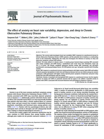
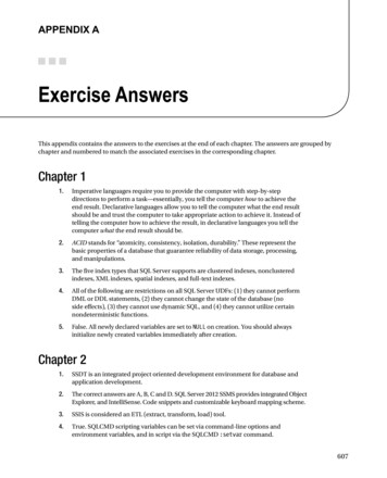
Downloaded By: [Joelsson, Ingemar] At: 11:30 10 January 2008ANS activity in premenstrual symptomsOn the days of testing, subjects came to thelaboratory between 9:00 and 11:00 a.m. after havingfasted overnight. All experiments were performed inthe morning. The room was temperature controlledat 258C, quiet and comfortable, with minimization ofarousal stimuli. After weighing each subject, thepercentage of body fat was determined by meansof a bioelectrical impedance analyzer (TBF-305,TANITA, Japan). Body mass index (BMI) wascalculated as body weight divided by height squared.After accurate skin preparation, the subjects werefitted with ECG electrodes and then rested for atleast 20 minutes before the start of the experiment.After the resting period, the CM5 lead ECGwas continuously recorded for 20 minutes duringsupine rest. All subjects breathed in synchrony to ametronome at 15 beats min71 (0.25 Hz) to ensurethat respiratory-linked variations in heart rate did notoverlap with lower-frequency heart rate fluctuations(below 0.15 Hz) from other sources.After the subjects completed the experiment forthe autonomic nervous system activity, each filled outthe Menstrual Distress Questionnaire (MDQ) [23]which evaluated their physical, emotional, andbehavioral symptoms with their menstrual cycle.The MDQ has been widely used in menstrual cycleresearch [9,24,25], and a recent study has reconfirmed the validity and applicability of thequestionnaire for evaluating menstrual symptoms[26]. The MDQ consists of 46 symptoms in eightcategories: pain, concentration, behavioral change,autonomic reactions, water retention, negative affect,arousal, and control. The subjects were asked to ratetheir experience of all 46 symptoms on the questionnaire on a six-point scale ranging from noexperience of the symptom to so extremely severeas to cause disruption of their daily activities. Thetotal score could, therefore, range from a minimumof 46 points to a maximum of 276 points.133Before the R-R spectral analysis was performed,the stored R-R interval data were displayed andaligned sequentially to obtain equally spaced sampleswith an effective sampling frequency of 2 Hz [35]and displayed on a computer screen for visualinspection. Then, the direct current componentand linear trend were eliminated by digital filteringfor the band-pass between 0.007 and 0.5 Hz. Theroot mean square value of the R-R interval wascalculated as representing the average amplitude.After passing through the Hamming-type datawindow, power spectral analysis by means of a fastFourier transform was performed on a consecutive1024-sec time series of R-R interval data obtainedduring the test.The spectral powers in frequency domain werequantified by integrating the areas under the curvesfor the following respective band widths: thevery low frequency (VLF) power (0.007–0.035 Hz)reflecting thermoregulatory sympathetic function;the low frequency (LF) power (0.035 and0.15 Hz), an indicator of both SNS and PNSactivity; the high frequency (HF) power (0.15 and0.5 Hz), which solely reflects the PNS activity; andthe total power (0.03 and 0.5 Hz, TOTAL) representing the overall autonomic nervous systemactivity (Figure 1) [21,27–34]. In addition, indicesof the global SNS and PNS activities were calculatedas the quotient of (VLF þ LF)/HF and HF/TOTALaccording to previous studies, respectively [29,31].The mean heart rate of each 1024-sec segment was also calculated together with standarddeviation.R-R spectral analysis procedureThe autonomic nervous system activity was noninvasively measured by HRV power spectral analysis. Thetechnique of the analysis for the present investigationhas been applied in basic physiological and clinicalresearch fields, and its validity and reliability hasbeen previously confirmed [21,27–34]. The procedure of R-R interval power spectral analysis has beenmentioned in great detail elsewhere [27–29,31].Briefly, the ECG signal was amplified (MEG-6108,Nihon Kohden, Tokyo, Japan) and digitized via a 16bit analog-to-digital converter (Model PS-2032GP,TEAC, Tokyo, Japan) at a sampling rate of 1000 Hz.The digitized ECG signal was differentiated, andthe resultant QRS spikes and the intervals of theimpulses (R-R intervals) were stored sequentially ona hard disk for later analyses.Figure 1. Examples of ECG R-R interval changes and thecorresponding power spectra for a woman in the resting condition.The technique of heart rate variability power spectral analysis usedin the present study identifies three separate frequency components, very low (0.007–0.035 Hz), low (0.035–0.15 Hz), and high(0.15–0.5 Hz), represented by the black, striped, and white areas,respectively.
Downloaded By: [Joelsson, Ingemar] At: 11:30 10 January 2008134T. Matsumoto et al.Urinary analysisEach subject collected urine at the first urine void inthe morning. Refrigerated 10-mL aliquots of urinewere immediately frozen and stored at 7208C untilassay. Urine samples were then analyzed for estroneconjugate (E1C) and pregnanediol-3-glucuronide(PdG) by radioimmunoassay as described by Munroet al. [36] and De Souza et al. [37] E1C and PdGwere both indexed to creatinine (Cr) excretion in thesame sample to control for variations in urinevolume. E1C and PdG are expressed as nanogramsand micrograms per mg Cr, respectively.Statistical analysesAll data are expressed as mean SE. Paired t-testwas performed to assess statistical differences of theparameters, including the autonomic nervous systemactivity, symptoms with menstrual cycles, and otherclinical features, between the follicular and the lateluteal phases. A group comparison among threegroups (Low, Middle, and High) was made withANOVA using post hoc Tukey’s test for subsequentpairwise comparisons. Correlation coefficients andsignificance values between two variables werecalculated by linear regression analysis. P values50.05 were considered statistically significant. Allstatistical analysis was performed using a commercialsoftware package (SPSS version 12.0 for Windows,SPSS inc., Illinois).ResultsThe length of the menstrual cycle and the duration ofmenstrual flow of all subjects during the study were28.9 0.5 days and 6.2 0.2 days, respectively.The days of the experiments were 10.4 0.3th dayin the follicular phase and 26.0 0.4th day in thelate luteal phase from the first day of menstruation.Statistical analysis of data of all subjects hasshown a significant increase in the following clinicalcharacteristics in the late luteal phase compared tothat in the follicular phase: basal body temperature(36.55 0.04 vs. 36.18 0.048C, p 5 0.01), concentration of ovarian hormones in urine (E1C22.6 2.3 vs. 11.9 0.7 ng/mg Cr, p 5 0.01; PdG3.3 0.3 vs. 0.5 0.04 mg/mg Cr, p 5 0.01),and total MDQ scores (68.3 3.2 vs. 59.4 1.7,p 5 0.01). No significant effects of menstrual cyclephase were detected on any parameters of HRVpower spectral analysis.Concerning menstrual cycle symptomatology, wenoticed that the rate of increase in scores on theMDQ from the follicular to late luteal phase variedamong the subjects from 0 to 87.7%. To scrutinizethe potential influence of premenstrual discomforton the autonomic nervous system activity, we thusdivided the subjects into three groups based on therate of increase in scores on the MDQ, i.e., LowGroup (less than 10%), Middle Group (greater than10 to less than 20%), and High Group (greater than20%). It should be mentioned that, according to theresults of a standardized health questionnaire andindividual interviews, none of the subjects in theHigh Group experienced distressing physical, psychological, and/or behavioral changes of sufficientseverity to result in deterioration of interpersonalrelationships and/or interference with normal activities. In addition, no subjects in the present studysought medical treatment.Table I shows clinical characteristics of the Low,Middle, and High Groups. Basal body temperatureand concentration of E1C and PdG in urine weresignificantly more elevated in the late luteal phasethan in the follicular phase in all three groups.Comparing the clinical characteristics among thethree groups, no statistical difference was foundeither in the follicular or in the late luteal phase.As Figure 2 represents, total scores on the MDQsignificantly increased from the follicular to late lutealphase in all three groups (Low Group 55.2 1.7vs. 56.9 1.9, p 5 0.01; Middle Group 57.0 2.8vs. 64.6 2.9, p 5 0.01; High Group 71.9 4.0 vs.99.1 7.8, p 5 0.01). Within the High Group, scoresof each factor increased from the follicular to the lateluteal phase and significant changes were detected inthe following factors: pain ( p 5 0.05), concentrationTable I. Clinical characteristics of subjects in the follicular and late luteal phase.Low Group (n ¼ 11)Basal body temperature (8C)Estrone conjugates (ng/ml Cr)Pregnanediol-3-glucuronide(mg/ml Cr)Weight (kg)Body Mass Index (kg/m2)Body fat (%)Middle Group (n ¼ 11)High Group (n ¼ 36.17 0.0511.2 1.00.6 0.136.59 0.04**26.2 3.1**3.7 0.4**36.09 0.0812.7 1.40.5 0.0436.42 0.06**19.9 3.9*3.2 0.5**36.31 0.0812.5 1.60.4 0.136.59 0.12*18.6 6.0*2.3 0.5**50.8 1.019.7 0.324.7 0.751.1 1.019.8 0.3*24.9 0.651.6 1.219.9 0.624.6 1.254.1 1.920.5 0.724.9 1.254.4 1.920.6 0.624.9 1.3Values are means SE. **p 5 0.01; *p 5 0.05 (follicular vs. late luteal).51.9 1.220.0 0.624.7 1.3
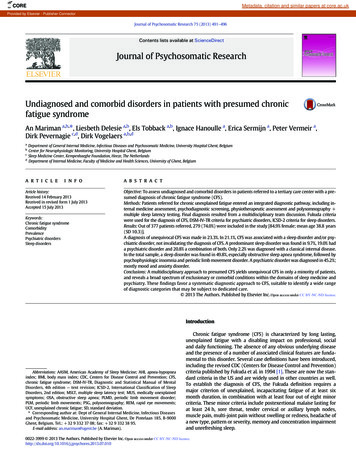
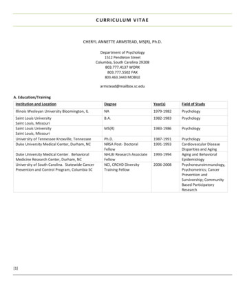
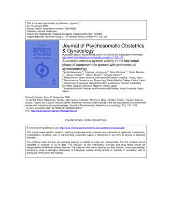
Downloaded By: [Joelsson, Ingemar] At: 11:30 10 January 2008ANS activity in premenstrual symptoms135Figure 2. Comparison of scores on the menstrual distress questionnaire (MDQ) between the follicular and late luteal phases among the Low,Middle, and High Groups. Low Group N ¼ 11, Middle Group N ¼ 11, and High Group N ¼ 8. Results are expressed as mean SE. for eachgroup. **p 5 0.01; *p 5 0.05.( p 5 0.01), behavioral change (p 5 0.01), negativeaffect (p 5 0.01), and control ( p 5 0.01). Groupcomparison revealed no significant differences eitherin subscores or in total scores on the MDQ betweenthe Low and Middle Groups in any menstrualphases. The total scores on the MDQ in the lateluteal phase were, however, significantly greater inthe High Group than in the Low or Middle Group.All subscores of the late luteal phase were higher inthe High Group compared to those in the other twogroups, and statistical analysis further demonstratedsignificant differences in pain (p 5 0.01), concentration ( p 5 0.01), behavioral change ( p 5 0.01), waterretention ( p 5 0.01), negative affect (p 5 0.01),arousal (p 5 0.05), and control ( p 5 0.01) factors.With respect to the autonomic nervous systemactivity, there were no statistical differences in anyparameters of HRV between the follicular and lateluteal phases both in the Low and the MiddleGroups. As Figure 3 shows, however, significantchanges were found between the menstrual phases inthe High Group; Heart rate (p 5 0.05) and the SNSIndex ( p 5 0.05) were higher in the late luteal phasethan those in the follicular phase. In contrast, the HFpower ( p 5 0.05) and PNS Index ( p 5 0.05) apparently decreased in the late luteal phase compared tothe follicular phase. Group comparison of theautonomic nervous system activity revealed nosignificant differences in any parameters of HRVbetween the Low and the Middle Groups in anymenstrual phases. In the late luteal phase, however,the SNS Index (p 5 0.05) markedly increased, andthe PNS Index ( p 5 0.05) significantly decreased inthe High Group compared to the other two groups.The HF power in the late luteal phase was also lowerin the High Group than in the Low or Middle Group.This difference, however, did not reach statisticalsignificance. Further data analysis revealed nosignificant correlation between urine ovarian hormone concentration and both indexes of sympathovagal activities regardless of menstrual phases in eachsubject group.DiscussionAutonomic nervous system activity has been extensively examined during the menstrual cycle. Severalstudies have found the variation of sympatho-vagalactivities during the menstrual cycle, but the resultsare not consistent. Goldstein et al. [5] reported thatplasma norepinephrine concentration is significantlyhigher in the luteal phase than in the follicular phase.Recent research conducted by Minson et al. [6] andHirshoren et al. [7] support that finding; however, nosignificant difference was reported during the menstrual cycle in another study [8]. Time and frequencydomain variables of the HRV measurement havebeen applied to investigate the autonomic nervoussystem activity during the menstrual cycle. As to thesympathetic branch of the system, several studies havedemonstrated that an index of SNS activity is predominant in the luteal phase as compared to the follicularphase [7,9–11]. In contrast, research on autonomicresponse reported that the SNS index in supineposition remains unchanged along the menstrualcycle [12,13]. Phase-dependent changes of PNS
T. Matsumoto et al.Downloaded By: [Joelsson, Ingemar] At: 11:30 10 January 2008136Figure 3. Comparison of heart rate, high power, sympathetic nervous system index (SNS Index), and parasympathetic nervous system index(PNS Index) between the follicular and late luteal phases among the Low, Middle, and High Groups. Low Group N ¼ 11, Middle GroupN ¼ 11, and High Group N ¼ 8. Results are expressed as mean SE for each group. *p 5 0.05.activity during the menstrual cycle have also beendemonstrated [9–11]; however, they failed to bedetected in studies measuring the time [14] orfrequency [13] domain variables of HRV during amenstrual cycle. Although study designs and objectives are not always consistent with the presentinvestigation, the discrepancy might partly be causedby the differences in assessing the activity of bothbranches of autonomic nervous system in humansubjects. It also could arise largely from the difficultyin controlling the array of variables, including age,number of subjects, determination of menstrual cyclephases, eating habits, physical activity level, medicalcomplications, emotional stress, and psychosocialand other environmental factors. In addition, despitethe fact that a majority of women of reproductive ageexperience premenstrual symptomatology regardlessof the degree and/or the duration it lasts, few studieshave evaluated in detail the symptoms accompanyingthe menstrual cycle and reflected them when interpreting the results of autonomic function.In the present study, all participants were nonobese eumenorrheic young women without anymedical history or complications. Subjects werecategorized into three groups depending on thedegree of premenstrual symptomatology, and nointramenstrual cycle differences in the parameters ofautonomic nerve activity were found among thewomen in the Low and Middle Groups. Since thedata was obtained at two time points during amenstrual cycle, the findings could not completelyrule out the menstrual cyclicity of autonomic nervoussystem activity. Although fluctuations in the ovarianand/or other hormones along the menstrual cyclehave been suggested to influence autonomic nervoussystem [6,7,9–11], the present investigation detectedno significant correlation between ovarian hormoneconcentration and the sympatho-vagal indexes ofHRV either in follicular or late luteal phases. Exactneurophysiological and/or hormonal effects of themenstrual cycle could not be explicated from thisstudy. Considering the present findings obtainedfrom the women of fertile age with homogeneousclinical characteristics, however, the resting cardiacsympatho-vagal activity does not differ between thefollicular and late luteal phases, at least in healthyyoung women who experience no or small increasesin premenstrual symptoms.In contrast to the Low and Middle Groups, amenstrual-phase effect on the autonomic nervoussystem activity was discernible in the High Group:higher SNS activity and lower PNS activity in the late
Downloaded By: [Joelsson, Ingemar] At: 11:30 10 January 2008ANS activity in premenstrual symptomsluteal phase. In addition, the SNS activity was higherand the PNS activity was lower during the late lutealphase in the High Group than the other two groups.It should be restated that scores of premenstrualsymptoms were markedly higher in the High Groupthan in the other two groups, but the symptoms werenot as severe as to cause disruption of the dailyactivities or to need medical consultations, accordingto the questionnaires and individual interviews. Asto pathogenesis of premenstrual symptomatology,whether the autonomic changes are primary orsecondary could not be discerned based on theevidence presently available. The present study,however, suggests that the autonomic nervous systemactivity is altered in the symptomatic late luteal phasewhen premenstrual distress is not unbearable, butsubjectively increases over a certain degree, i.e., 20%increases from the nonsymptomatic follicular phase.The physiological stability of a human’s internalenvironment depends largely on the orchestrations ofthe autonomic nervous system. Previous studiessuggest that a dysfunction of both sympathetic andparasympathetic systems contributes to psychosomatic disorders as well as to cardiovascular andmetabolic malfunctions. Patients with anxiety disorder or depression exhibit reduced heart ratevariability [16]. Yeragani et al. [38] evaluated autonomic responsiveness in patients with panic disorderand indicated that the patients had increasedadrenergic and decreased cholinergic responsiveness.Sloan et al. [39] showed men with hostility as apersonality trait have lowered HRV associated withdecreased vagal modulation and sympathetic predominance. Altered autonomic functions have alsobeen reported in healthy individuals under negativepsychological situations.
University Graduate School of Medicine, Osaka, Japan d Graduate School of Human and Environmental Studies, Kyoto University, Kyoto, . Journal of Psychosomatic Obstetrics & Gynecology, September 2006; 27(3): 131-139 . Review Board of International Buddhist University and was performed in accordance with the Declara-