Transcription
J Head Trauma RehabilVol. 22, No. 3, pp. 141–155c 2007 Wolters Kluwer Health Lippincott Williams & WilkinsCopyright Objective Documentation of TraumaticBrain Injury Subsequent to Mild HeadTrauma: Multimodal Brain ImagingWith MEG, SPECT, and MRIJeffrey David Lewine, PhD; John T. Davis, PhD; Erin D. Bigler, PhD; Robert Thoma, PhD;Dina Hill, PhD; Michael Funke, MD, PhD; John Henry Sloan, MD; Sandra Hall, PhD;William W. Orrison, MDObjective: To determine to what extent magnetic resonance imaging (MRI), single photon emission computed tomography (SPECT), and magnetoencephalography (MEG) can provide objective evidence of brain injury in adultpatients with persistent ( 1 year) postconcussive symptoms following mild blunt head trauma. Design: A retrospective and blind review of imaging data with respect to the presence of specific somatic, psychiatric, and cognitivecomplaints. Setting/Participants: Thirty complete data sets (with MRI, SPECT, MEG, and neuropsychological testing results) were collected between 1994 and 2000 from the MEG programs at the Albuquerque VAMC and theUniversity of Utah. Main Outcome Measures: MRI data were evaluated for focal and diffuse structural abnormalities, SPECT data for regions of hypoperfusion, and resting MEG data for abnormal dipolar slow wave activity(DSWA) and epileptiform transients. Results: Structural MRI was abnormal for 4 patients. SPECT showed regionsof hypoperfusion in 12 patients, while MEG showed abnormal activity in 19 patients. None of the imaging methodsproduced findings statistically associated with postconcussive psychiatric symptoms. A significant association wasfound between basal ganglia hypoperfusion and postconcussive headaches. For patients with cognitive complaints,abnormalities were more likely to be detected by MEG (86%) than either SPECT (40%) or MRI (18%) (P .01).MEG also revealed significant (P .01) associations between temporal lobe DSWA and memory problems, parietalDSWA and attention problems, and frontal DSWA and problems in executive function. Conclusions: Functionalbrain imaging data collected in a resting state can provide objective evidence of brain injury in mild blunt headtrauma patients with persistent postconcussive somatic and/or cognitive symptoms. MEG proved to be particularly informative for patients with cognitive symptoms. Keywords: head trauma, magnetoencephalography, MEG, MRI,postconcussive syndromes, SPECT, traumatic brain injuryIN the United States alone, it is estimated that emergency departments treat more than 2 million casesFrom the Department of Radiology, the University of Utah School ofMedicine, Salt Lake City (Drs Lewine, Davis, Bigler, Thoma, Hill, Funke,and Orrison); Hoglund Brain Imaging Center (Drs Lewine and Hall),Department of Neurology and the Department of Psychiatry and BehavioralSciences (Dr Lewine), and Department of Preventative Medicineand Public Health (Dr Hall), University of Kansas Medical Center,Kansas City; Department of Psychology, Brigham Young University,Provo, Utah (Dr Bigler); and Department of Psychiatry, Universityof New Mexico (Dr Thoma and Hill) and St. Joseph’s RehabilitationCenter, St. Joseph’s Hospital (Dr Sloan), Albuquerque, NM.This work was supported by research grants from NSF (CCR-0105504), TheUniversity of Kansas Research Institute, Picker International, and NeuromagLtd to J.D.L. The authors are grateful to K. Paulson, M. Johnson, K. Hatch,D. Detmers, J. Meyer, and R. Christensen for technical support and to MichaelHartshorne for the evaluation of SPECT data. The authors also thank thereviewers and JHTR editors for their valuable and insightful comments.Corresponding author: Jeffrey David Lewine, PhD, Illinois MEG Center,The Alexian Neurosciences Institute, Alexian Brothers Medical Center, 800Biesterfield Rd, Suite 610, Elk Grove Village, IL 60007 (e-mail: Jeffrey.Lewine@abbhh.net).of head trauma each year. When blunt trauma is severe, many patients will show a wide range of persistentposttraumatic problems that may include headache, fatigue, impaired memory, reduced concentration and attention, reduced information-processing capacity, poorimpulse control, personality changes, depression, irritability, sleep disturbances, and sexual dysfunction.1,2Similar symptoms may be reported initially after mildhead trauma (mild HT), but in most cases, all symptoms subside within a few weeks. However, 15% to 30%of patients who suffer minor trauma report postconcussive symptoms that persist for many years after thetraumatic incident.3–6 Given that more than 80% of allhead traumas are of the mild type, this translates into200,000 to 400,000 new patients in the United Stateseach year with a persistent postconcussive syndrome resulting from mild HT. Available data suggest that manyof these patients will experience vocational and interpersonal distress that place a significant economic and141
142JOURNAL OF HEAD TRAUMA REHABILITATION/MAY–JUNE 2007social burden on our society, with many cases giving riseto litigation.3–6In considering both treatment and legal ramifications of head trauma, objective documentation oftraumatic brain injury (TBI) subsequent to mild HTwould be of utility, especially in view of the common assertion that cognitive symptoms following mildHT reflect “compensation neurosis” rather than braindamage.7 In cases of moderate-severe head trauma,there is a very high likelihood of TBI, with structuralbrain imaging via computed tomography (CT) or magnetic resonance imaging (MRI) often demonstratingchronic abnormalities including focal encephalomalacia, diffuse axonal injury, ventricular dilation, and cortical atrophy.8,9 Consequently, many researchers and clinicians use the term TBI interchangeably with the termhead trauma. Unfortunately, this can lead to confusionwhen considering mild HT because clinical CT and MRIare typically negative for mild HT patients, especiallyseveral months after the traumatic event.9 Thus, objective evidence of actual TBI subsequent to mild HT isoften lacking. TBI is a potential consequence of headtrauma, but it is not a necessary one. Throughout thisarticle, special care is therefore taken to distinguish mildHT from mild-TBI, with a major goal of the study beingto provide objective documentation of actual mild-TBIin cases of observed mild HT.Because routine clinical MRI rarely reveals grossstructural abnormalities following mild HT (even incases with significant and persistent postconcussive complaints), many research teams are exploring the possibility that functional brain imaging methods will bemore sensitive to subtle TBI. Methods of particular interest include hemodynamic techniques such as positronemission tomography (PET) and single photon emissioncomputed tomography (SPECT), and electrophysiological methods such as electroencephalography (EEG) andmagnetoencephalography (MEG).In cases of moderate and severe head trauma withclear structural lesions on MRI, resting-baseline PET andSPECT data generally demonstrate concordant perfusion and metabolic deficits. These perfusion deficits mayextend into normal appearing tissue, and in some cases,very diffuse hemodynamic abnormalities consistent withthe concept of diaschisis have been reported.10,11 Someresting PET studies reveal a high sensitivity to mildHT,10,12,13 but this is not always the case.14 SPECTstudies in mild trauma often show focal regions of hypoperfusion consistent with expectations based on neuropsychological testing,15,16 but not all groups have reported a strong correlation between specific regionalSPECT findings and specific cognitive test results.2,17,18Overall, resting PET and SPECT demonstrate abnormalities in 25% to 80% of head trauma survivors—thelikelihood of a positive finding being dependent on theseverity of initial trauma, the presence of MRI findings, the severity of postconcussive compromise, and thetime lag between imaging and the traumatic event.2,10–18When scanning is performed several months after mildtrauma, most studies report resting hemodynamic abnormalities in fewer than 50% of subjects, even whenconsidering only patients who report persistent postconcussive problems. Another limitation of the restinghemodynamic approach to mild HT is a general lackof specificity for the methods. Although normal comparison participants rarely show hemodynamic anomalies, hypoperfusions can be seen in almost all neurologicand psychiatric diseases, including stoke, epilepsy, depression, schizophrenia, attention-deficit disorder, anddementia.Most hemodynamic imaging studies in head traumahave focused on resting hemodynamic profiles, but thereis a growing trend in functional brain imaging to examine activity profiles during performance of specificcognitive tasks. Recently, several investigators have usedPET, SPECT, and/or functional MRI (fMRI) to examine hemodynamic responses in patients with mild HTperforming working memory tasks.14,19,20 These studiesindicate that mild HT can lead to activation deficits, butmost deficits are subtle and apparent only in group data,with critical observations failing to be sufficiently robustfor diagnostic use on a case-by-case basis.Turning toward electrophysiological methods (EEGand MEG), there are also 2 general types of studies—those that use evoked response paradigms to assess specific cognitive processes and those that examine “resting”activity patterns. In general, simple sensory and motorevoked responses (potentials and fields) are within normal limits following mild HT,21 although some recentpilot data indicate that somatosensory evoked magneticresponses show depressed M30 responses following headtrauma (J.D. Lewine, J.T. Davis, E. Bigler, et al, unpublished data, 2006). Some data indicate that evoked responses related to mnemonic processing (eg, N2 and P3)are abnormal following mild HT,21 but these are verynonspecific findings common to many disease conditions. Also, there is a general lack of diagnostic utilityand validity on a case-by-case basis.Considering “resting-state” studies, routine clinicalEEG is very unlikely to reveal abnormalities (and therebysupport the existence of TBI) subsequent to mild HT.22In contrast, quantitative EEG (qEEG) often revealsabnormalities subsequent to mild HT. For example,Thatcher and colleagues23,24 have developed qEEG discriminant functions that allow for the identificationof individual patients with a history of mild HT withbetter than 90% accuracy. However, it has been difficult to place specific qEEG discriminant factors (eg,changes in coherence in a narrow frequency band) ina functional neuroscience framework that allows for an
Objective Documentation of Traumatic Brain Injury Subsequent to Mild Head Traumaunderstanding of the relations between regional braindysfunction and specific symptoms following headtrauma.Using MEG, we have previously reported that amoderately high percentage of patients with persistentcomplaints subsequent to mild HT demonstrate focaldipolar slow wave activity (DSWA).25 MEG assessesthe pattern of magnetic flux associated with intradendritic currents. Mathematical methods for inferring thelocation of discrete neuronal populations that generate abnormal MEG signals of interest provide dataon brain dysfunction within a framework that allowsfor explicit assessment of the relations between specific post–head trauma symptoms and specific clinicalfindings.Presented herein is a retrospective review of integratedMRI, SPECT, and MEG data from patients with persistent psychiatric, somatic, or cognitive complaints subsequent to mild HT. A primary goal of the study was todetermine to what extent imaging methods could provide, on a case-by-case basis, objective evidence of braininjury. A secondary goal of the study was to determinethe relations between specific patterns of imaging abnormalities and specific postconcussive symptoms. If it canbe demonstrated that functional brain imaging methods provide a sensitive and specific measure of braininjury following mild HT, these imaging methods arelikely to emerge as critical in the future developmentof rational and evidence-based therapeutic and medicalmanagement plans for patients with persistent postconcussive symptoms.METHODSParticipantsBetween 1994 and 2000 the first author and his colleagues performed whole-head MEG examinations andMRI examinations on 58 adult patients with persistent( 1 year) psychiatric, somatic, and/or cognitive complaints that had developed within the first few weeks following emergency department–documented mild blunthead trauma. This article presents a retrospective reviewof data from the 30 patients in this group who had detailed neuropsychological testing (completed within 2months of the neuroimaging procedures) and an additional, clinically ordered SPECT examination. All participants were 18 years or older at the time of MEGevaluation, and none were younger than 14 years at thetime of head trauma.Mild-HT was defined by a Glasgow Coma Scale scoreof 13 or higher and a loss of consciousness of not morethan 20 minutes. In all cases, there was evidence that thehead had actually struck (or was struck by) a blunt object. Patients with whiplash injury without evidence ofexternal head trauma were excluded. Most participants143(67%) had suffered their trauma in a motor vehicle accident, with 18% being the victims of violent crime and15% of home or industrial accidents. At the time of referral to the imaging centers, approximately half weretaking regular doses of some form of psychoactive medication. Participants were asked to refrain from takingthese medications on the days of imaging.Evaluation of post–head trauma symptomsInformation on post–head trauma symptoms was obtained from several sources, including review of all medical and psychological records and a patient interviewand self-report post-concussion symptom checklist completed on the day of MEG examination. (Prior to 1998,this was a 25-item questionnaire designed by our team.After 1998, we switched to the European Brain InjuryQuestionnaire, which provides for a clear separation ofmood, cognitive, and social factors.26 ) All participantsalso had detailed neuropsychological evaluations performed independent of the imaging studies and completed within 2 months of the imaging. In most cases(N 25), neuropsychological assessment had been completed before the brain imaging, and in fact, mild deficitson cognitive testing were one of the main reasons for referral for imaging. Because referrals were acquired fromseveral different clinical contacts, the exact neuropsychological test protocol that had been used varied somewhatfrom patient to patient. Nevertheless, all had data available from the following instruments: (1) the Vocabularyand Block Design Subtests of the WAIS-R, (2) the VisualReproduction and Verbal Paired Associates subtests ofthe WMS-R, (3) a word fluency test, (4) the Trail MakingTest (parts A & B), and (5) the Wisconsin Card Sort Test.All patients also had data from (1) the Beck or Hamilton Depression and Anxiety Inventories, (2) Conner’sContinuous Performance Task (CPT) and/or the Test ofVisual Attention (TOVA), and (3) the Annett or groovedpegboard.While blind to neuroimaging results, the investigativeteam used data from these tests to define for each participant a 3-domain bioclinical profile of symptoms participant: (1) psychiatric, (2) somatic, and (3) cognitive.Psychiatric symptoms were considered present if the patient reported problems in mood and responses on theBeck/Hamilton inventories indicated a mood disorder.Patients were considered to have somatic symptoms ifthey reported unusually frequent and severe headaches,pain, and/or dizziness. Cognitive impairments wereevaluated with respect to 4 subdomains: (1) attentionproblems (CPT or TOVA), (2) memory problems (WMSR), (3) executive function problems (WCS and the FASword generation task), and (4) a generalized reduction ininformation-processing speed (various time-dependenttasks including the Trails Test [parts A& B], the groovedwww.headtraumarehab.com
144JOURNAL OF HEAD TRAUMA REHABILITATION/MAY–JUNE 2007or Annett pegboard, and the CPT [when available,N 26]).Performance in a particular cognitive subdomain wasconsidered impaired if the average performance acrossrelevant tests was more than 1 SD below the mean andthe patient (while unaware of test results) had explicitlyindicated during the interview that performance in thatdomain was impaired since the traumatic event. A 1 SDthreshold is admittedly quite liberal for defining impairment, but deficits in mild HT are often subtle on formal testing, even when patients report that the traumaticevent caused a significant decline relative to their concept of pretrauma abilities.MEG examinationMEG data were collected using either a 122-channelor a 306-channel biomagnetometer system (both manufactured by Neuromag Ltd, Finland). Studies conductedbetween January 1994 and January 1997 (n 10) wereperformed with a 122-channel system located at theAlbuquerque Veteran’s Affairs Medical Center. Studiesconducted between January 1997 and November 1998(n 9) were performed with a 122-channel system located at the Center for Advanced Medical Technologiesof the University of Utah in Salt Lake City. Studies conducted between November 1998 and July 2000 (n 11)were performed with a newly installed 306-channel system at the Center for Advanced Medical Technologies.The 122-channel systems use 61 pairs of orthogonallyoriented planar gradiometers to characterize extracranialmagnetic field patterns. The 306-channel unit has 102gradiometer pairs, with an additional magnetometer ateach recording location. Because MEG is a somewhatless familiar technology than MRI and SPECT, we provide Figure 1 to illustrate the basic principles of MEGdata acquisition and analysis.For each participant, 10 minutes of data were collectedwhile the participant sat quietly with eyes closed. Datawere collected with a bandpass of 0.1 Hz to 100 Hz, at adigitization rate of 300 Hz. Following additional digitalfiltering (1–70 Hz) and removal of eye blink and cardiacartifacts via a signal space projection method,27 the 10minute block of MEG data was inspected visually forepileptiform transients and processed via an automatedcomputer routine for abnormal DSWA.When interictal spikes were identified, a multipledipole, spatiotemporal source modeling algorithm wasused to elucidate the spatial position of the spike initiation zone. Most slow waves (in the 1- to 6-Hz upper deltaand lower theta bands) show very complicated magneticfield patterns that cannot be explained by even 5 or 6dipole sources. While these may still represent significant pathophysiology, for the purposes of this study wefocused only on that subset of large amplitude ( 200 fT)slow waves whose generation could be reasonably wellexplained by a single focal dipole source. The basic dataanalysis strategy used by our group for examining slowwaves in patients with head trauma has been describedin detail.25Briefly, following artifact removal, data are filtered between 1 and 6 Hz, and large amplitude ( 200 fT) slowwaves are identified. A 50-millisecond window about thepeak of each slow wave is marked and a single equivalentcurrent dipole is fit every 10 milliseconds. The single bestfit during within each event window is then retained forfurther consideration. If the goodness of fit for the dipolemodel is greater than 0.8 (with all planar sensors includedduring the fitting procedure), the dipole is considereda viable model, and its spatial parameters are noted. Aspatial clustering algorithm is then used to determine foreach viable dipole how many neighbor dipoles are seenwithin a 2 2 2 cubic centimeter region. If this valueexceeds 1 dipole per minute of recording time, then allmembers of the dipole cluster are retained and plottedon spatially aligned MRI to generate magnetic sourcelocalization images. For regional analyses in the presentstudy, the brain was divided into 8 regions (right andleft frontal, temporal, parietal, and occipital lobes). Aregion was coded as demonstrating abnormal functionif it contained 10 or more dipolar slow wave sources.MEG is relatively insensitive to subcortical activity withnone of the participants in this study showing a substantive number ( 10) of slow wave dipole sources localizing to subcortical nuclei. Exploratory data analysesshowed no significant right-left differences, so findingswere collapsed with respect to right hemisphere versusleft hemisphere.Magnetic resonance imagingFollowing MEG, all participants also underwent astructural MRI evaluation on the same or following day.In each case, MRI data were collected using a 1.5-Twhole-body imaging system. Acquisition sequencesincluded a T1-weighted 3D volumetric sequence(1.5-mm contiguous sagittal slices, no interslice gap),T2-weighted and FLAIR sequences (5-mm axial slices,no interslice gap), and a gradient recalled echo sequence(5-mm axial slices, no interslice gap). Data were clinically evaluated by a board-certified neuroradiologist,and the report and data were available for review in thisstudy. While blind to all other imaging and clinical observations, data sets were coded as showing evidence ofatrophy, diffuse axonal injury, or focal encephalomalacia in right/left frontal, temporal, parietal, occipital,or subcortical regions. In the majority of cases, CT orMRI reports were also available from the time of thetraumatic event. These findings were also reviewed andtabulated. Exploratory analyses showed no significant
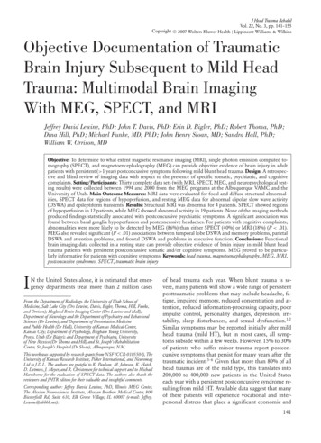
Objective Documentation of Traumatic Brain Injury Subsequent to Mild Head Trauma145Figure 1. Basic principles of magnetoencephalography: Currents flowing within the apical dendrites of pyramidal cells orientedparallel to the skull surface give rise to a magnetic field that can be measured outside of the head using supercooled sensorsconnected to SQUIDs (superconducting quantum interference devices). The sensors are arrayed within a head-shaped cryogenicvessel. During testing, the participant sits underneath the sensor unit. A 122-channel unit is pictured. The output of each sensor isa waveform that shows how the local magnetic flux changes in time. Representative data are shown for a normal comparison andmild head trauma patients. A 6.8-second long epoch of data is shown for 10 sensors over the left posterior temporal region. For thecontrol participant, the bulk of activity is of low amplitude and high frequency. In contrast, the patient with head trauma displaysprominent, large amplitude slow waves (1–6 Hz, each approximately 800 fT, peak-to-peak). Most slow waves have complicatedmagnetic field patterns as revealed by iso-field contour maps that show multiple regions of emerging (gray contour lines) andentering flux (black contour lines). In contrast, some slow waves have a dipolar field pattern characterized by single regions ofemerging and entering flux. The source location for the neuronal currents that contribute to dipolar slow waves can be inferredusing mathematical techniques that identify the position, orientation, and time course for the best-fitting dipole source. Thislocation can be plotted on a spatially aligned magnetic resonance image to generate a composite magnetic source localizationimage.right-left differences, so findings were collapsed acrossthe hemispheres.SPECT imagingIn all cases, SPECT imaging of baseline regional brainperfusion was performed on the same day or the dayimmediately before or after MEG and MRI. Following10 minutes of rest, a 30-mCI dose of Tc-99m-HMPAO(Ceretec, used for studies in Albuquerque) or a 25-mCIdose of Tc-99m-ECD (used for studies in Salt Lake City)was administered intravenously. Fifteen minutes later,SPECT imaging was performed using a triple-headedgamma camera equipped with high-resolution fan-beamcollimators (Albuquerque—Picker, Odyssey 3000, SLC—Picker, IREX). Data were acquired using a simultaneouswww.headtraumarehab.com
146JOURNAL OF HEAD TRAUMA REHABILITATION/MAY–JUNE 2007transmission and emission protocol. A total of 120 rotation stops were acquired using a 128 128 acquisitionmatrix. Images were reconstructed using a Butterworthor Weiner filter. Data were reformatted in axial, coronal,and sagittal planes, and evaluated for regions of hypoperfusion by a board-certified radiologist specializing innuclear medicine. The radiology report and all imageswere available for review by the investigative team. Raterswho were blind to other imaging and behavioral findingscoded SPECT data sets as showing evidence of hypoperfusion in right/left frontal, temporal, parietal, occipital,or subcortical basal ganglia nuclei, or in a diffuse patternconsistent with generalized brain atrophy. Exploratoryanalyses showed no significant right-left differences, sofindings were collapsed across the hemispheres.Image integrationAfter MEG, MRI, and SPECT data were initially evaluated in an independent manner, image fusion was performed to explore the relations between modalities. Image fusion was accomplished by first sending DICOMimages from the MR scanner to the MEG workstation.Using proprietary software (Neuromag Ltd), the fiducialpoints used during MEG to define the head-centered coordinate system were identified on the MR images. Thesoftware then aligned MEG and MRI coordinate systemsso that MEG source locations could be plotted on theMR images to form magnetic source localization images.These images, with MEG source points now an inherent part of the data set, were then DICOM transferredto the SPECT workstation, which treated the magneticsource localization images as though they were simplyMR images. A 9 df volume registration protocol with interactive processing was then used to align SPECT andMRI/MEG coordinate frames. The magnetic source localization images were reformatted in planes centered onthe SPECT slices and image blending was performed.RESULTSNeuroimagingTable 1 summarizes demographic, neuroimaging, andclinical observations for each patient. As previously mentioned, there were no significant laterality effects, so aregion was coded as being abnormal if an abnormalitywas observed for either hemisphere. A descriptive summary of findings is provided below.CT/MRIStructural imaging performed at the time of accidentrevealed abnormalities in only 5 of the 30 patients (allwith subdural hematomas). MR follow-up at the time ofMEG and SPECT (performed, on average, 33.2 monthsafter trauma) revealed chronic MR findings (mild atrophy) in only 2 of these 5 cases. There were also 2 caseswith initially normal MR scans that showed mild cortical atrophy and some ventricular dilation at follow-up.Hence, there were 4 cases with evidence of atrophy onMRI at the time of MEG and SPECT imaging. No patient showed focal lesions or clear MR evidence of diffuse axonal injury. Ten patients had history of more than1 traumatic event, including each of the 4 patients withchronic MR findings.SPECTPerfusion imaging using SPECT revealed abnormalities in 12 participants (40%), including each of the 4 withchronic MR findings and 8 others. SPECT was abnormal for 7 of 9 patients with histories of multiple mildtraumatic events but only 5 of 21 patients with singletraumas (P .05 by the Fisher exact test, odds ratio [OR] 11.2, 95% confidence interval [CI] 1.7–100.1). Inall cases with abnormal SPECT findings, focal perfusiondeficits were seen in regions not explicitly identified asabnormal by MRI.MEGSix patients showed intermittent epileptic spikes, eventhough none had reported clinical seizures. MEG alsorevealed the presence of abnormal DSWA in 19 patients(63%). All 4 patients with structural findings and 9 of the12 patients with SPECT findings showed MEG abnormalities. When focal MR or SPECT abnormalities werepresent, MEG always provided evidence of concordantelectrophysiological dysfunction (except with respect tosubcortical hypoperfusion). In most of these cases, MEGalso revealed a more extensive picture of dysfunctionthan was shown by either MRI or SPECT.There were 10 patients with normal MRI and normalSPECT who showed MEG abnormalities. Three patientswith abnormal SPECT showed normal MEG and MRIexaminations. As was seen for both MRI and SPECT,participants with a history of multiple traumas were morelikely to show MEG abnormalities than patients withonly a single traumatic event (9/9 vs 10/21, P .05 bythe Fisher exact test).It should be noted that there were no significantdifferences in the slow wave sensitivity profiles acrossstudy sites or for the 122-channel recording systemversus 306-channel recording system. Although the 122and 306-channel sensor units differ significantly in thenumber of sensors, the actual area of coverage for the 2units shows only a modest difference, with the increasednumber of sensors in the 306-channel unit being mostlya reflection of slightly increased packing density and addition of a magnetometer at each recording site. A set ofexploratory data analyses failed to provide evidence that
Objective Documentation of Traumatic Brain Injury Subsequent to Mild Head TraumaTABLE 1Data for individual participants ,†No. ofAge Sex 14122230CurrentMRISPECTMEGClinical profileA F T P O B F T P O e P S M A E rmalNormalMild atrophyFMild atrophy A FMild atrophy AMild atrophy A malNormalNormalBBBNormalNormalNormalNormalNorm
2007 Wolters Kluwer Health Lippincott Williams & Wilkins Objective . . 2007