Transcription
microorganismsArticleCharacterization of Sinomonas gamaensis sp. nov., aNovel Soil Bacterium with Antifungal Activityagainst Exserohilum turcicumYansong Fu 1 , Rui Yan 1 , Dongli Liu 1 , Junwei Zhao 1 , Jia Song 1 , Xiangjing Wang 1 , Lin Cui 1 ,Ji Zhang 1, * and Wensheng Xiang 1,2, *12*Heilongjiang Provincial Key Laboratory of Agricultural Microbiology, Northeast Agricultural University,Harbin 150030, China; yansongfu1@163.com (Y.F.); 15546073308@163.com (R.Y.);17862181612@163.com (D.L.); guyan2080@126.com (J.Z.); songjia-hit@hotmail.com (J.S.);wangneau2013@163.com (X.W.); 15663843306@163.com (L.C.)State Key Laboratory for Biology of Plant Diseases and Insect Pests, Institute of Plant Protection, ChineseAcademy of Agricultural Sciences, Beijing 100193, ChinaCorrespondence: zhangji@neau.edu.cn (J.Z.); xiangwensheng@neau.edu.cn (W.X.) Received: 24 April 2019; Accepted: 6 June 2019; Published: 8 June 2019Abstract: A novel Gram staining positive, aerobic bacterium NEAU-HV1T that exhibits antifungalactivity against Exserohilum turcicum was isolated from a soil collected from Gama, Hadjer lamis, Chad.It was grown at 10–45 C (optimum 30 C), pH 5–10 (optimum pH 8), and 0–4% (w/v) NaCl (optimum1%). Phylogenetic analysis based on 16S rRNA gene sequences showed that strain NEAU-HV1Twas closely related to Sinomonas susongensis A31T (99.24% sequence similarity), Sinomonas humiMUSC 117T (98.76%), and Sinomonas albida LC13T (98.68%). The average nucleotide identity valuesbetween NEAU-HV1T and its most closely related species were 79.34 85.49%. The digital DNA–DNAhybridization values between NEAU-HV1T and S. susongensis A31T , S. albida LC13T , and S. humiMUSC 117T were 23.20, 23.50, and 22.80%, respectively, again indicating that they belonged to differenttaxa. The genomic DNA G C content was 67.64 mol%. The whole cell sugars contained galactose,mannose, and rhamnose. The polar lipids were diphosphatidylglycerol, phosphatidylglycerol,phosphatidylinositol, and four glycolipids. The respiratory quinone system comprised MK-9(H2 ),MK-10(H2 ), and MK-8(H2 ). The major cellular fatty acids ( 5%) were anteiso-C15:0 , anteiso-C17:0 , C16:0 ,and iso-C15:0 . Based on the polyphasic analysis, it is suggested that the strain NEAU-HV1T representsa novel species of the genus Sinomonas, for which the name Sinomonas gamaensis sp. nov. is proposed.The type strain is NEAU-HV1T ( DSM 104514T CCTCC M 2017246T ).Keywords: novel species; Sinomonas; antifungal activity; Exserohilum turcicum1. IntroductionThe genus Sinomonas, a member of the family Micrococcaceae in the phylum Actinobacteria, was firstproposed by Zhou et al. [1] with the description of Sinomonas flava and the reclassification of Arthrobacteratrocyaneus as Sinomonas atrocyaneus. At the time of writing, the genus Sinomonas comprised 10 species(http://www.bacterio.net/sinomonas.html) that were isolated from soil, volcanic rock, and the surfaceof weathered biotite: Sinomonas flava, Sinomonas atrocyaneus, Sinomonas echigonensis, Sinomonas albida,Sinomonas soli, Sinomonas notoginsengisoli, Sinomonas mesophila, Sinomonas susongensis, Sinomonas humi,and Sinomonas halotolerans [1–7]. The species of this genus are aerobic and rod-shaped, contain MK-9(H2 )as the predominant respiratory quinone, and have iso-C15:0 , anteiso-C15:0 , and anteiso-C17:0 as the majorfatty acids. Other typical features are the presence of galactose, mannose, and ribose as the majorcell wall sugars; A3α as the peptidoglycan type; and diphosphatidylglycerol, phosphatidylglycerol,Microorganisms 2019, 7, 170; nal/microorganisms
Microorganisms 2019, 7, 1702 of 12and phosphatidylinositol as the major phospholipids. Some members of the genus Sinomonas havebeen reported to be able to synthesize silver nanoparticles with antimicrobial activity [8], hydrolyzestarch [9], biodesulfurize coal [10], and degrade sesamin [11]. Recently, S. atrocyaneus has exhibitedplant growth promoting effects and antagonistic activity against a broad-spectrum of root and foliarpathogens, implying its potential use in sustainable agriculture [12]. In this study, a strain NEAU-HV1Twith inhibitory activity against phytopathogenic fungi Exserohilum turcicum was isolated from a soilsample. Based on the polyphasic analysis, this strain was classified as representative of a novel speciesin the genus Sinomonas.2. Materials and Methods2.1. Isolation of Bacterial StrainStrain NEAU-HV1T was isolated from a soil sample collected from a cotton field (13 0 N, 15 44 E),situated in Gama, Hadjer lamis Region, Chad, which is considered as the principal agricultural regionof Chad. The soil sample was suspended in sterile water (2 mL) followed by an ultrasonic treatment(160 w) for 3 min. After the addition of distilled water (43 mL), the soil suspension was incubated at28 C and 250 rpm on a rotary shaker for 20 min. Then, 200 µL of the suspension was spread on a plateof humic acid-vitamin (HV) agar [13] supplemented with cycloheximide (50 mg L 1 ) to inhibit anyfungal contaminants. After seven days of aerobic incubation at 28 C, colonies were transferred andpurified on nutrition agar (NA; BD Difco, Becton, Dickinson and Company, Sparks, MD, USA) andmaintained as glycerol suspensions (20%, v/v) at 80 C for long term preservation.2.2. Morphological and Biochemical Characteristics of NEAU-HV1TColony morphology of strain NEAU-HV1T was observed after two days of incubation at 28 C onNA. Cell morphology was examined by transmission electron microscopy (Hitachi H-7650, HitachiCo., Tokyo, Japan). Motility was detected by the presence of turbidity throughout tubes containingsemi-solid medium and was further confirmed by the hanging-drop method [14,15]. Gram stainingwas performed as described by Smibert and Krieg [16]. Growth at different temperatures (0–45 C,steps of 1 C below 5 C, and then intervals of 5 C) was determined in a tryptic soy broth (TSB; BDDifco, Becton, Dickinson and Company, Sparks, MD, USA) medium after incubation for seven days.Growth tests for pH ranging from 4.0–12.0 in 1.0 pH unit intervals and NaCl tolerance (0–10% (w/v) in1% intervals) were determined in TSB medium at 28 C for seven days on a rotary shaker. The buffersystems were: pH 4.0–5.0, 0.1 M citric acid/0.1 M sodium citrate; pH 6.0–8.0, 0.1 M KH2 PO4 /0.1 MNaOH; pH 9.0–10.0, 0.1 M NaHCO3 /0.1 M Na2 CO3 ; and pH 11.0–12.0, 0.2 M KH2 PO4 /0.1 M NaOH [17].Growth was determined by monitoring the turbidity at OD600 by using a spectroscopic method (U-3900;Hitachi Co., Tokyo, Japan) and plate counting. The utilization of organic compounds as the solecarbon and energy source, decomposition of cellulose, hydrolysis of starch and aesculin, reductionof nitrate, peptonization of milk, liquefaction of gelatin, and production of H2 S were analyzed asrecommended by Ventosa et al. [18]. Anaerobic growth was tested on NA for up to two weeks at28 C in Bacteron anaerobic chambers (Sheldon Manufacturing, Carson, CA). The methyl red andVoges–Proskauer reactions, hydrolysis of Tweens 20, 40, and 80, production of esterase and urease weretested as described by Smibert and Krieg [16]. Metabolic properties and enzyme activities were testedusing API 50CH, API 20NE, and API ZYM strips with API 50CHB suspension medium (bioMérieux,Marcy-I’Etoile, France) according to the manufacturer’s protocols.2.3. Phylogenetic Analysis of NEAU-HV1For DNA extraction, strain NEAU-HV1T was cultured in TSB to the early stationary phase( 24 hours) and harvested by centrifugation. The genomic DNA was extracted using a TIANampBacteria DNA Kit (TIANGEN Biotech, Co. Ltd., Beijing, China). The 16S rRNA gene wasPCR-amplified with the universal primers 27F (5 -AGAGTTTGATCCTGGCTCAG-3 ) and 1541R
Microorganisms 2019, 7, 1703 of 12(5 -AAGGAGGTGATCCAGCC-3 ) under conditions described previously [19]. The PCR product waspurified and cloned into the vector pMD19-T (Takara) and sequenced using an Applied BiosystemsDNA sequencer (model 3730XL, Applied Biosystems Inc., Foster City, California, USA) and softwareprovided by the manufacturer. The almost full-length 16S rRNA gene sequence was comparedwith the available 16S rRNA gene sequences of validly named species in the EzBioCloud server(https://www.ezbiocloud.net/) [20] to search the related similar species for tree reconstructions andcalculate the pairwise 16S rRNA gene sequence similarity. Phylogenetic trees were reconstructedwith the neighbor joining [21], maximum likelihood [22], and minimum evolution [23] methods usingMEGA version 7.0 after multiple alignments of the sequences using the CLUSTAL W algorithm [24].For the neighbor joining algorithm, Kimura’s 2-parameter model and pairwise deletion were usedto reconstruct the phylogenetic tree. The Tamura–Nei model together with Gamma distributionand Invariant sites (TN93 G I) and Nearest-Neighbor-Interchange (NNI) heuristic method for themaximum likelihood algorithm, and close-neighbor-interchange (CNI) search for the minimumevolution algorithm were applied. Distances were calculated according to Kimura’s two-parametermodel [25]. The topology of the phylogenetic tree was evaluated by using bootstrap analysis with1000 replicates [26]. All positions containing gaps and missing data were eliminated from thedataset and the root position of the trees was inferred by using Nocardia inohanensis DSM 44667T(GenBank accession no. AB092560) as an outgroup. The genome sequences of strain NEAU-HV1Tand its most closely related strains S. susongensis A31T , S. albida LC13T , and S. humi MUSC 117Twere determined by Novogene Bioinformatics Technology (Beijing, China) using a IlluminaHiSeqPE150 (Illumina) following the manufacturer’s suggested protocols. The resulting reads were qualitytrimmed to the Q20 confidence level. The draft genome was assembled using SOAP de novo version2.04 [27] with default parameters. The estimated dDDH values were calculated using formula two atthe Genome-to-Genome Distance Calculation (GGDC) website (http://ggdc.dsmz.de/distcalc2.php)as described by Meier-Kolthoff et al. [28]. Average nucleotide identity (ANI) calculations wereperformed using the OrthoANIu tool (https://www.ezbiocloud.net/tools/ani) [29] and JSpecies software(http://jspecies.ribohost.com/jspeciesws/) [30]. For core-genome analysis, cores genes were extractedfrom the genomes of NEAU-HV1T and its related strains using the USEARCH program with BPGA(Bacterial Pan Genome Analysis tool), with 50% sequence identity cut-off [31]. The concatenatedamino acid sequences of core genes were aligned to generate the phylogenetic tree using the neighborjoining method. Genome mining for bioactive secondary metabolites was performed using “antibioticsand secondary metabolite analysis shell” (antiSMASH) version 4.0 [32].2.4. Chemotaxonomic Analysis of NEAU-HV1TBiomass for chemical studies was prepared by growing the strains in TSB medium in shake flasksat 28 C until the exponential phase of growth was reached. Cells were harvested by centrifugation,washed with distilled water, and freeze-dried. The cell-wall diamino acid was determined fromwhole-cell hydrolysates, as described by McKerrow et al. [33] and analyzed by a HPLC methodusing an Agilent TC-C18 column (250 4.6 mm, i.d. 5 µm) (Agilent Technologies, Santa Clara, CA,USA). Procedures for identification of cell-wall amino acids and whole-cell sugars were performedas described by Tang et al. [34]. Polar lipids were extracted and examined by two-dimensional TLCand identified according to the method of Minnikin et al. [35]. Menaquinones were extracted fromfreeze-dried biomass and purified according to Collins [36]. Extracts were analyzed using an HPLC-UVmethod [37]. Fatty acid methyl esters were extracted and analyzed using the microbial identificationsystem (MIDI) and the RTSBA6 database (Microbial Identification Inc., Newark, DE, USA) [38].
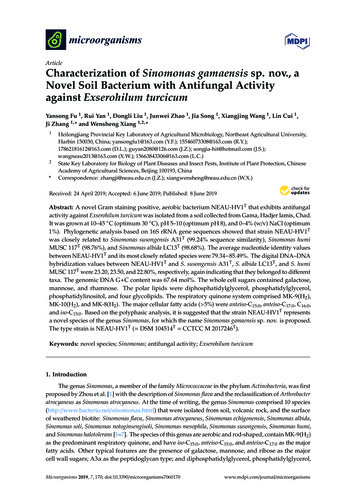
Microorganisms 2019, 7, 1704 of 122.5. In Vitro Antifungal Activity TestThe antagonistic activity of NEAU-HV1T against six phytopathogenic fungi (Fusarium oxysporum,Fusarium orthoceras, Sclerotinia sclerotiorum, Alternaria solani, Corynespora cassiicola, and Exserohilumturcicum) was evaluated using petri dish assays [39]. To further investigate the antifungal componentsproduced by NEAU-HV1T , this strain was cultured in GYM medium [40] and the inhibitory activityof the supernatant was tested. Briefly, strain NEAU-HV1T was inoculated into GYM medium andincubated at 28 C for seven days on a rotary shaker. The supernatant was obtained by centrifugationat 8000 rpm and 4 C for 10 min and subsequently filtrated with a 0.2 µm membrane filter. Thecell precipitate was extracted with an equal volume of methanol. Then, the antifungal activity wasevaluated using the well diffusion method [41], and each well contained 150 µL of the supernatantor methanol extract. In order to test the effect of pH on the antifungal stability, pH values of thesupernatant was adjusted to pH 3.0, 5.0, 7.0, and 11.0 by 1 mol/L hydrochloric acid or 1 mol/L NaOH,and the adjusted supernatant was kept at 4 C for 24 h. Afterward, the pH values were readjusted to7.0. To examine the effect of temperature on antifungal activity, the supernatant was placed in a waterbath at 40, 60, 80, and 100 C for 30 min, and then cooled to room temperature. A total of 200 µL of thetreated supernatant was added into 25 mL of sterile potato dextrose agar (PDA), and the mixture waspoured into 9 cm plates. The PDA containing 200 µL of sterilized water was used as the control. Then,5 mm plugs of E. turcicum were placed at the center of each plate, and all plates were placed at 28 Cfor seven days. The antifungal activity was evaluated when the fungal mycelium reached the edges ofthe control plates.3. Results and Discussion3.1. Polyphasic Taxonomic Characterization of NEAU-HV1TStrain NEAU-HV1T was aerobic, Gram staining positive, and non-motile. Light microscopyat 1000 magnification showed that the cells formed bent rods after cultivation for 12 h and thenfragmented into a coccoid shape at 24 h, and the changed shape was also observed by transmissionelectron microscopy (Figure 1). This metamorphic feature of rod-coccus growth cycle was also observedfor other species of Sinomonas such as S. susongensis A31T , S. halotolerans CFH S0499T , S. mesophilaMPKL 26T , and S. echigonensis CW59T [2,4–6]. Neither flagella nor spore were found. Colonies onNA were circular, convex, and pale yellow, with a smooth surface and diameter of 0.5–1.5 mm afterincubation for 48 h at 28 C. Strain NEAU-HV1T was able to grow at 10–45 C (optimum, 30 C), at pH5–10 (optimum, pH 8), and with 0–4% (w/v) NaCl (optimum, 1%). Phenotypic properties useful fordistinguishing NEAU-HV1T from its three phylogenetic neighbors are shown in Table 1 and Table S1.The detailed characteristics of strain NEAU-HV1T are given in the species description.
Microorganisms 2019, 7, 170Microorganisms 2019, 7, x FOR PEER REVIEW5 of 125 of 12T T NA agar. (A,B) represent the lightFigure 1. The isticsofofstrainstrainNEAU-HV1NEAU-HV1onon NA agar. (A) and (B) representTT after formicrographsof Gram-stainingcells of strainNEAU-HV1after incubation12 and 24h,12respectively.incubationforand 24 h,the light micrographsof Gram-stainingcellsof strain pectively. (C) and (D) represent the transmission electron micrograph of negatively staining cellsafterincubationfor 12T afterand 24h, respectively.incubationfor 12 and 24 h, respectively.of strainNEAU-HV1BLASTsequence analysisof the16S rRNAsequenceGenBank/EMBL/DDBJT and(1518Table 1. Differentialcharacteristicsbetweenstrain geneNEAU-HV1closelybp;relatedmembers of theTaccessionMF418645) indicated that strain NEAU-HV1 was related to members of the genusgenusnumberSinomonas.Sinomonas. The EzBioCloud analysis showed that strain NEAU-HV1T was most closely related to S.TTA31S. albida LC13Thumi MUSC 117TCharacteristicNEAU-HV1T withsusongensisA31T , S. humi MUSC117T , andS.S.susongensisalbida LC13a gene sequence S.similarityof 99.24,Nitrate reduction ‒ ‒T98.76, and 98.68%, respectively. In the neighbor joining tree (Figure 2), strain NEAU-HV1 clusteredProduction of urease ‒‒with members of the genus Sinomonas and formed an independent subclade with the most closelyHydrolysis of Starch‒‒ ‒TTTrelatedstrainsof S. susongensis A31of theCitrateutilization , S. albida LC13 , and S. humi MUSC‒ 117 . The topology neighborjoining treewas further confirmedusing theand minimum‒evolutionVoges–Proskauerreaction maximum likelihood methods(Figuresof:S1 and S2). De novo assembly of the genome sequencing data of strain NEAU-HV1TUtilization ‒ ‒ 2). Theresulted L-Tyrosinein 39 contigs, an N50 of 233,829bp, and an averagegenome coverageof 441.0 (TableTL-Glycine‒ bp and its DNA G C content was 67.64 draft genomesize of strain NEAU-HV1was 4,320,429mol% ‒ for strainL-ArginineNEAU-HV1T , which is withinthe range (66.6–71.8mol%) previouslyobtained for species ofL-Threonine ‒ showedthe genusSinomonas [5]. A phylogenetictree (Figure‒ 3) based on the conservedcore genomeL-AlanineT T‒Tthat NEAU-HV1 was most related to S. susongensis A31 , S. albida LC13 , and S. humi MUSC 117T ,Creatine‒ ‒ supporting the phylogenetic analysis based on 16S rRNA gene sequences (Figure 2). The dDDHRaffinose ‒ T (NCBI accession number NZ SSCL00000000) and S.values (Table2) between strain NEAU-HV1L-Rhamnose ‒TT (NZ SWDW00000000), and S. humi MUSCsusongensisA31 (NZ SSNH00000000),S. albida LC13D-Mannitol‒ ‒117T (NZ JTDL00000000)were 23.20,23.50, and 22.80%,respectively, which were belowInositol‒‒‒ the 70%L-Arabinose ‒ The values of OrthoANIu,‒DDH speciesboundary as recommendedby Meier-Kolthoffet al. [28].D-Xylose NEAU-HV1T with S. susongensis A31‒T , S. albidaANI-MUMmer(ANIm), and ANI-Blast(ANIb) of strainLactose ‒LC13T , andS. humi MUSC 117T (Table3) were below‒the 94–96% cut-off value previously proposedforD-FructoseD-GalactitolD-GalactoseG C mol% ‒ 67.64‒ 68.64 69.46‒ ‒67.16
Microorganisms 2019, 7, 170Microorganisms 2019, 7, x FOR PEER REVIEW6 of 126 of 12T represents a putative novel species of thespecies delimitation[42],wereindicatingthatstrainAll dataobtainedfromthisNEAU-HV1study. , positive;‒, negative. All strains were positive for thegenus Sinomonas.utilization of glucose, sucrose, ribose, maltose, mannose, sorbose, L-aspartic acid, L-proline, L-glutamine, L-serine, L-asparagine, L-glutamic acid, decomposition of cellulose, and the hydrolysis ofT and closely related members of thebetweenstrainNEAU-HV1Table 1. Differential2S production,liquefaction of gelatin, hydrolysis of Tweensaesculin.characteristicsAll strains werenegativefor Hgenus Sinomonas.20, 40, and 80, and the methyl red test.TNEAU-HV1TS. susongensisA31rRNAS.genealbidasequenceLC13TS.humi bp;MUSC117Tsequenceanalysisof the 16S(1518GenBank/EMBL/DDBJT was related- to members of the genusNitratereductionnumber MF418645) - that strain NEAU-HV1 accessionindicatedProductionofurease Sinomonas. The EzBioCloud analysis showed that strain NEAU-HV1T was -most closely related to S.Hydrolysis of Starch TTsusongensisMUSC 117T , and S. albida LC13similarity of 99.24,Citrateutilization A31 , S. humi - with a gene sequence Voges–Proskauer98.76, and 98.68%, respectively.In the neighbor joining tree(Figure 2), strainNEAU-HV1T clustered reactionwith members of the genus Sinomonas and formed an independent subclade with the most closelyUtilization of:T. The topology of therelatedsusongensis A31T,- S. albida LC13T, andL-Tyrosinestrains of S. S. humi MUSC 117L-Glycine neighbor joining tree was further confirmed using the maximum likelihood and minimum evolutionL-Arginine methods (Figures S1 and S2). De novoassembly of the genome sequencingdata of strain NEAUL-Threonine T resulted in 39 contigs, an N50 of 233,829 bp, and an average genome coverage of 441.0 (TableHV1L-Alanine T was 4,320,429Creatine G C content was 67.642).The draft genome -size of strain NEAU-HV1bp and its DNARaffinose T, which- is within the rangemol% for strain NEAU-HV1(66.6–71.8 mol%) previously obtained forL-Rhamnose species of the genus Sinomonas[5]. A phylogenetictree (Figure3) based on theD-Mannitol - conserved core genomeT was most relatedT, and S. humi MUSCInositol that NEAU-HV1 showedto S. susongensisA31T, S. albida LC13L-Arabinose T, supporting the phylogenetic analysis based on 16S rRNA gene sequences (Figure 2). The dDDH117D-Xylose T (NCBI accession number NZ SSCL00000000) and S.valuesstrain NEAU-HV1Lactose (Table 2) between D-Fructose - S. albida LC13T (NZ SWDW00000000), - and S. humi MUSC 117Tsusongensis A31T (NZ SSNH00000000),D-Galactitol (NZ JTDL00000000) were 23.20, 23.50,and 22.80%, respectively,which werebelow the 70% DDHD-Galactose speciesrecommended68.64by Meier-Kolthoff69.46et al. [28]. The valuesG Cmol%boundary as67.6467.16 of OrthoANIu, ANIT with S. susongensis A31T, S. HV1All data were obtained from this study. , positive; -, negative. All strains were positive for the utilization ofT, andT ,valueL-asparagine,humi MUSC3) weretheL-glutamine,94–96% cut-offpreviously proposedL-glutamic acid, decomposition of cellulose, and the hydrolysis of aesculin. All strainswere negative for H2 EAU-HV1representsaputativenovel speciesproduction, liquefaction of gelatin, hydrolysis of Tweens 20, 40, and 80, and the methyl red test.of the genus Sinomonas.CharacteristicBLASTFigure 2. Neighborjoiningtree showingtheshowingphylogeneticposition of strainNEAU-HV1(1518 bp) T (1518 bp)Figure 2.Neighborjoining treethe phylogeneticpositionof strain TNEAU-HV1and related taxabased hatwere alsoand relatedtaxa16S sequences.rRNA geneFilledsequences.circlesindicatebranchesthat were alsorecovered ds.Bootstrapvalues 50%recovered using the maximum-likelihood and minimum evolution methods. Bootstrap values 50%(based on 1000replications)shown at branchpoints. atBar,0.01 substitutionsnucleotideposition.(basedon 1000arereplications)are shownbranchpoints. Bar,per0.01substitutionsper nucleotideposition.
Microorganisms 2019, 7, 170Microorganisms 2019, 7, x FOR PEER REVIEW7 of 127 of 12TTableTable 2.2. General features of the genomegenome ofof NEAU-HV1NEAU-HV1T.FeaturesFeaturesGenomeGenomesize (bp)size (bp)DNA G C(%)DNAG C (%)NumberNumberof contigsof contigsNumberNumberof CDS of CDSRNA genesRNA genesrRNA rRNAtRNA tRNAN50 (bp)N50 (bp)N90 (bp)N90 (bp)Numbers of genomics islandsNumbers of genomics islandsNumbers of prophageNumbers of prophageNumbers of CRISPRNumbers of 33Figure 3. Neighbor joining tree showing the phylogenetic position of strain NEAU-HV1NEAU-HV1TT relative toclosely related type strains based on the core genome.genome.Table 3. The digital DNA–DNA hybridization (DDH) and average nucleotide identity (ANI) valuesTable 3. The digital DNA–DNAhybridization (DDH) and average nucleotide identity (ANI) valuesbetween strain NEAU-HV1T and the closely related members of the genus Sinomonas.between strain NEAU-HV1T and the closely related members of the genus Sinomonas.StrainsStrainsS. susongensisA31TS. susongensisA31TTS.albidaLC13TS. albida LC13S. humi MUSC 117TS. humi MUSC 117TDDH (%)DDH u80.2380.2380.0880.0879.7279.72ANImANIbANIm85.16 85.16 79.3485.49 85.49 79.4585.1778.9485.17ANIb79.3479.4578.94The whole cell sugars of strain NEAU-HV1TT were galactose, mannose, and rhamnose, whichThe whole cell sugars of strain NEAU-HV1 were galactose, mannose, and rhamnose, whichwere consistent with type strains of Sinomonas [6]. The major cellular fatty acids ( 5% of the total fattywere consistent with type strains of Sinomonas [6]. The major cellular fatty acids ( 5% of the totalacids) of strain NEAU-HV1T wereT anteiso-C15:0 (44.6%), anteiso-C17:0 (27.0%), C16:0 (20.5%), and iso-C15:0fatty acids) of strain NEAU-HV1 were anteiso-C15:0 (44.6%), anteiso-C17:0 (27.0%), C16:0 (20.5%), and(7.9%). The fatty acid profile of strain NEAU-HV1T was similarto those of closely related strains S.T wasiso-C15:0 (7.9%). The fatty acid profile of strain NEAU-HV1similar to those of closely relatedT, which support the placement of strainsusongensis A31T, S. albidaT LC13T, and S. humiMUSC117T , and S. humi MUSC 117T , which support the placementstrains S. susongensisA31,S.albidaLC13NEAU-HV1T withinT the genus Sinomonas. However, some quantitative differences clearlyof strain NEAU-HV1 within the genus Sinomonas. However, some quantitative differences clearlydistinguished strain NEAU-HV1TT from the other reference strains (Table 4). For example, C16:0 (20.5%)distinguished strain NEAU-HV1 from the other referenceTstrains (Table 4). For example, C16:0 (20.5%)was detected as a major component in strain NEAU-HV1, while it was a minor component ( 5%) inwas detected as a major component in strain NEAU-HV1T , while it was a minor component( 5%) in theT, but the threethe related strains. Additionally, iso-C16:0 was not detected in strain NEAU-HV1related strains. Additionally, iso-C16:0 was not detected in strain NEAU-HV1T , but the three referencereference strains had this component at 3.8, 6.8, and 11.4%, respectively. The polar lipids of strainTstrains had this component at 3.8, 6.8, and 11.4%, respectively. The polar lipids of strain NEAU-HV1NEAU-HV1T were diphosphatidylglycerol, phosphatidylglycerol, phosphatidylinositol, and fourwere diphosphatidylglycerol, phosphatidylglycerol, phosphatidylinositol, and four glycolipids (Figureglycolipids (Figure S3). This pattern was similar to the three related strains, which containedS3). This pattern was similar to the three related strains, which contained diphosphatidylglycerol,diphosphatidylglycerol, phosphatidylglycerol, and phosphatidylinositol as the major componentsphosphatidylglycerol, and phosphatidylinositol as the majorcomponents [2,5,7]. The respiratory[2,5,7]. The respiratory quinone systemTof strain NEAU-HV1T consisted of MK-9(H2), MK-10(H2), andquinone system of strain NEAU-HV1 consisted of MK-9(H2 ), MK-10(H2 ), and MK-8(H2 ) (approx.MK-8(H2) (approx. 89, 10, and 1%, respectively). The menaquinone composition was in agreement89, 10, and 1%, respectively). The menaquinone composition was in agreement with the previouswith the previous descriptions that MK-9(H2) was the predominant menaquinone of members ofSinomonas [2,5,7]. A comparison of the menaquinones in strain NEAU-HV1T and three other related
Microorganisms 2019, 7, 1708 of 12descriptions that MK-9(H2 ) was the predominant menaquinone of members of Sinomonas [2,5,7]. Acomparison of the menaquinones in strain NEAU-HV1T and three other related strains demonstratedthat the menaquinone composition of NEAU-HV1T was very similar to that of S. susongensis A31T [5],however, the minor component MK-8(H2 ) in NEAU-HV1T became the major component in S. albidaLC13T and no MK-8(H2 ) was detected in S. humi MUSC 117T [2,7]. The DNA G C content of strainNEAU-HV1T was 67.64 mol%, which was within the range (66.6–71.8 mol%) previously obtained forspecies of the genus Sinomonas [5].Table 4.Cellular fatty acid compositions (%) of strain NEAU-HV1T and its closestphylogenetic neighbors.Fatty AcidNEAU-HV1TS. susongensis A31TS. albidus LC13TS. humi MUSC 7:0Iso-C17:0C18:1 2–, Not detected. Fatty acids found in amounts 1.0% in the strains are not shown. All data were obtained fromthis study.On the basis of morphological, physiological, chemotaxonomic, and phylogenetic results, strainNEAU-HV1T is considered to represent a novel species within the genus Sinomonas, for which thename Sinomonas gamaensis sp. nov. is proposed.3.2. Description of Sinomonas gamaensis sp. nov.Sinomonas gamaensis sp. nov. (ga.ma.en’sis N.L. fem. adj. gamaensis of or belonging to Gama, thelocation of the soil sample from which the type strain was isolated).Cells are aerobic, Gram staining positive, non-motile, bent rods at the beginning phase of growth,then fragments into coccoid shape. Colonies are circular, convex and pale yellow, with a smoothsurface and diameter of 0.5–1.5 mm after incubation on NA for 48 h at 28 C and grows at 10–45 C(optimum, 30 C), at pH 5–10 (optimum, pH 8), and with 0–4% (w/v) NaCl (optimum, 1%). Positivefor decomposition of cellulose, nitrate reduction, urease, citrate utilization, and Voges–Proskauerreaction; but negative for H2 S production and methyl red reaction. Aesculin is hydrolyzed, butgelatin, starch, and Tween 20, 40, and 80 are not. The following compounds are utilized as the solesource of carbon and energy: L-alanine, L-arabinose, L-arginine, D-fructose, D-galactose, lactose,raffinose, L-rhamnose, L-threonine, L-tyrosine, and D-xylose. The following compounds are notutilized as the sole source of carbon and energy: creatine, D-galactitol, L-glycine, inositol, D-mannitol.Acid is produced from D-fructose, and D-turanose; but not from N-acetylglucosamine, D-arabinose,L-arabinose, arbutin, D-cellobiose, L-fucose, D-galactose, D-glucose, D-lactose, D-maltose, D-mannose,methyl α-D-mannopyranoside, L-rhamnose, D-ribose, salicin, D-sorbitol, D-tagatose, trehalose, andD-xylose. Positive for nitrate reduction, indole production, arginine dihydrolase, urease; but negativefor D-glucose fermentation. Activity is detected for esterase (C4), esterase lipase (C8), lipase (C14),valine aryla
For core-genome analysis, cores genes were extracted from the genomes of NEAU-HV1T and its related strains using the USEARCH program with BPGA (Bacterial Pan Genome Analysis tool), with 50% sequence identity cut-o [31]. The concatenated amino acid sequences of core genes were aligned to generate the phylogenetic tree using the neighbor joining .