Transcription
Geng et al. BMC Complementary Medicine and 4(2022) 22:92BMC ComplementaryMedicine and TherapiesOpen AccessRESEARCHHuaier attenuates the adverse effectsof pyroptosis by regulating the methylationof rat mesangial cells: an in vitro studyWenjia Geng1†, Can Tu2,3†, Dahao Chen2†, Zhaoyu Lu2,4,5, Wei Mao2,4,5* and Hanyu Zhu6*AbstractBackground: Pyroptosis is a highly programmed inflammatory cell death process that represents an innate immuneresponse. In this study, the occurrence of pyroptosis in rat mesangial cells (RMCs) and the effect of Huaier (Trametesrobiniophia Murr) on this process were investigated.Methods: RMCs were incubated with OX7 antibodies (0.5 μg/ml, 2.5 μg/ml, 10 μg/ml), normal rat serum (NRS) andHuaier (1 mg/ml, 5 mg/ml, 10 mg/ml). RMC morphology was observed under a light microscope and by immunofluorescence. Lactate dehydrogenase (LDH) release was assessed using the CytoTox 96 Non-Radioactive CytotoxicityAssay Kit. Western blot assays were performed, and then the RMCs were incubated with the methylase DNMT3B andthe demethylase 5-aza-2′-deoxycytidine.Results: Morphological, LDH, immunofluorescence and western blot analyses showed that RMCs were lysed whenstimulated with OX7 antibodies and NRS. RMC lysis released inflammatory cytokines (interleukin-18, interleukin-1β,monocyte chemoattractant protein-1 and intracellular adhesion molecule-1), and Huaier protected RMCs by controlling lysis and the levels of inflammatory cytokines. Lysis was mediated by pyroptosis due to the positive expression ofGSDME. The methylase DNMT3B reduced the expression of GSDME induced by OX7 together with NRS. Furthermore,Huaier significantly suppressed the expression of GSDME, which was increased by 5-aza-2’-deoxycytidine.Conclusions: Pyroptosis might occur in RMCs, and Huaier can protect RMCs by upregulating the methylation of agroup of molecules.Keywords: Rat mesangial cells, Pyroptosis, Huaier, GSDME, Methylation*Correspondence: maowei@gzucm.edu.cn; hanyuzhu301@126.com†Wenjia Geng, Can Tu and Dahao Chen contributed equally to this workand share first authorship.5Department of nephrology, Guangdong Provincial Hospital of ChineseMedicine, 111 Dade Road, Yuexiu District, Guangzhou, Guangdongprovince, China6Department of Nephrology, The First Medical Center, Chinese PLAGeneral Hospital, Chinese PLA Institute of Nephrology, State KeyLaboratory of Kidney Diseases, National Clinical Research Centerfor Kidney Diseases, No.28 Fuxing Road, Haidian District, Beijing, ChinaFull list of author information is available at the end of the articleIntroductionPyroptosis is a highly programmed inflammatory celldeath process that is an innate immune response [1, 2].GSDME (DFNA5) is one of the main executor proteins ofpyroptosis and can be cleaved by activated caspase-3. Theself-inhibition of GSDME is released, and N-terminal fragments are exposed. The N-terminal fragments perforate inthe cell membrane, thereby causing pyroptosis [3, 4].Recently, an increasing number of studies have shownthat the caspase-3/GSDME-dependent pyroptosis pathway plays a role in kidney disease [5–7]. However, inthe field of mesangial proliferative glomerulonephritis The Author(s) 2022. Open Access This article is licensed under a Creative Commons Attribution 4.0 International License, whichpermits use, sharing, adaptation, distribution and reproduction in any medium or format, as long as you give appropriate credit to theoriginal author(s) and the source, provide a link to the Creative Commons licence, and indicate if changes were made. The images orother third party material in this article are included in the article’s Creative Commons licence, unless indicated otherwise in a credit lineto the material. If material is not included in the article’s Creative Commons licence and your intended use is not permitted by statutoryregulation or exceeds the permitted use, you will need to obtain permission directly from the copyright holder. To view a copy of thislicence, visit http:// creat iveco mmons. org/ licen ses/ by/4. 0/. The Creative Commons Public Domain Dedication waiver (http:// creat iveco mmons. org/ publi cdoma in/ zero/1. 0/) applies to the data made available in this article, unless otherwise stated in a credit line to the data.
Geng et al. BMC Complementary Medicine and Therapies(2022) 22:92(MsPGN), there is almost no research on pyroptosis. MsPGN is one of the most common pathologicalsymptoms among chronic kidney diseases such as IgAnephropathy and membranous nephropathy. Acuteanti-thy1 MsPGN in rats is a classic and stable modelto mimic the pathogenesis of MsPGN [8]. This modelis established by tail-vein injection of OX7 antibody (amonoclonal anti-Thy 1.1 antibody). This model is characterized by a decrease in the number of mesangial cells(MCs) in 1–3 days, with the least number of cells on day3. Then MCs proliferate gradually, reaching the highest cell number at 7 days and then gradually decreasing.Previously, it was thought that this cellular reductionwas caused by C5b-9 attacking mesangial cells to induceapoptosis [9]. Then, it was discovered that attack may initiate signal transduction leading to apoptosis and DNArupture in the nucleus [10]. This phenomenon is similarto pyroptosis. Therefore, this study simulated the modelof acute anti-thy1 MsPGN in vitro.Urine protein excretion and the proliferation of MCsin acute anti-Thy-1 MsPGN might be inhibited byTrametes robiniophia Murr (Huaier), as we previouslyreported [11]. As a medicinal fungus in China, Huaierwas first described by Gehong in the 340 s. Another traditional Chinese medicine book stated that Huaier dispels wind, breaks blood and reinforces a healthy qi. Aseries of pharmacological studies showed that Huaiercould promote tumor cell apoptosis and inhibit tumorcell proliferation [12, 13], increase sensitivity to chemotherapy drugs [14], promote autophagy [15, 16] andregulate the immune response [13, 17]. In kidney disease, Huaier protects podocytes against adriamycininduced cytotoxicity in the context of nephropathy,possibly by reversing the mitochondrial dysfunction viaPGC-1α overexpression [18]. However, whether Huaierinterferes with pyroptosis is unknown. Therefore, weinvestigated whether pyroptosis occurred in RMCs andwhether Huaier played a renoprotective role by affectingpyroptosis and also assessed the mechanism by whichHuaier exerts its renal protective effect. We hope toprovide new ideas for a better understanding of humanMsPGN pathogenesis in the future and new therapeuticdrugs for the treatment of MsPGN from the perspectiveof traditional Chinese medicine.MethodsMaterialsThe RMC (HBZY-1) line was obtained from the American Type Culture Collection (ATCC, Manassas, VA,USA) and incubated at 37 C in an atmosphere of 5% CO2/95% O2. The monoclonal OX7 antibody waskindly provided by the People’s Liberation Army General Hospital. Normal rat serum (NRS) was preparedPage 2 of 12from the sera of 10 male Sprague-Dawley (180–220 g,6–8 weeks) rats and filtered with a 0.22 μm sterilefilter. All SD rats were provided by the LaboratoryAnimal Services Center of Guangzhou Universityof Chinese Medicine (Guangzhou, China). The ratswere housed in the specific-pathogen-free (SPF) animal breeding room of the Academy of Chinese Medicine of Guangdong Province and given free access towater. After 1 week of adaptive feeding, the rats wereanaesthetized, and blood was collected. All proceduresand assays were approved by the Ethics Committee ofAnimal Experiments, Guangdong Provincial Hospitalof Chinese Medicine (approval no. 2019041). All theprotocols were performed in accordance with relevantguidelines and regulations. The study is also in accordance with the ARRIVE guidelines.Huaier extract is the electuary ointment of Huaier(provided by Gaitianli Medicine, Jiangsu, China),which is obtained by extracting Huaier fungi by usingan aqueous solution, ultrafiltration and concentration[19]. In brief, Huaier fungi were heated and boiled inan aqueous solution for 6 h. Then, the supernatant wascollected, and the residue was boiled with the aqueoussolution again. This process was repeated 3 times. Thecollected supernatants were combined, centrifuged,and filtered with a 1–30 kDa ultrafiltration membrane.Then, the supernatants were vacuum dried underreduced pressure to obtain the Huaier extract.One gram of Huaier extract was dissolved in 10 ml ofphosphate-buffered saline (PBS). Then, the Huaier solution was filtered with a 0.22 μm sterile filter and storedat 20 C.Morphology of pyroptotic cellsIn this study, OX7 plus NRS was used for modeling.OX7 is an anti-thy 1 monoclonal antibody that can specifically bind to the thy-1 antigen on RMCs to form anantigen-antibody complex. NRS contain complementso that using OX7 plus NRS can simulate the pathogenesis of acute anti-thy1 MsPGN in vivo [20, 21].RMCs at passages 5 to 10 were grown in RPMI media(Gibco) with 10% fetal bovine serum (FBS). Then, thecells were placed in 6-well plates at 4.8 105 cells/welland allowed to adhere overnight. After being washedthree times with PBS, the cells were placed in serumfree media for an additional 12 h. The cells were sensitized by incubation with OX7 antibodies (0.5 μg/ml,2.5 μg/ml and 10 μg/ml) at 37 C for 60 min, followed byincubation with 8% NRS or serum-free RPMI. Moreover, Huaier (10 mg/ml) or an equal volume of PBS wasadded. RMC morphology was observed and recordedby inverted microscopy (Olympus) within 24 h.
Geng et al. BMC Complementary Medicine and Therapies(2022) 22:92Measuring cell lysisRMCs (4 103/ml) were cultured in 96-well plates andincubated with OX7 (2.5 μg/ml) at 37 C for 60 min.Then, the cells were then incubated with 8% NRS orserum-free RPMI containing different Huaier concentrations (1 mg/ml, 5 mg/ml, 10 mg/ml) or an equal volume of PBS. At 2 h, 4 h and 8 h, 50 μl of supernatant wasplaced in a new 96-well plate after centrifugation. Then,50 μl of CytoTox 96 Reagent was added to each well, andthe plate was covered with foil to protect it from lightaccording to the instructions of the CytoTox 96 NonRadioactive Cytotoxicity Assay Kit (Promega Corporation). The 96-well plates were incubated for 30 min atroom temperature. Finally, 50 μl of stop solution wasadded to each well, and the absorbance at 490 nm wasanalyzed.Immunofluorescence analysis of GSDME and OX7 in RMCsRMCs (1.2 105/ml) were seeded in 6-well plates andgrown to 30–40% confluence. Cells were incubatedwith OX7 (2.5 μg/ml) at 37 C for 60 min and thenincubated with 8% NRS or serum-free RPMI. Different Huaier solutions (1 mg/ml, 5 mg/ml, 10 mg/ml)were added concurrently. After being incubated for4 h, the culture medium was removed. Then, the cellswere washed with PBS three times, fixed with 4% paraformaldehyde, permeabilized with 0.5% Triton X-100at 4 C and subsequently blocked with 0.1% bovineserum albumin (BSA). Thereafter, the cells were incubated with anti-GSDME antibodies (55 kD, 35 kD, 25kD, sc393162, Santa Cruz) or OX7 antibodies at 4 Covernight. On the second day, Cy3-labeled goat antimouse antibodies were added and incubated for 1 h atroom temperature. DAPI was added to stain the nuclei.Then, the expression of GSDME or OX7 in RMCswas observed by laser scanning confocal microscopy.In addition, it should be noted that GSDME 55 kD isGSDME full length (GSDME-FL), GSDME 35 kD is theGSDME N terminus (GSDME-NT), and GSDME 25 kDis the GSDME C terminus (GSDME-CT) [4, 22]. Afteractivation of GSDME-FL, GSDME-NT and GSDMECT can be formed. The perforating effect that causespyroptosis is mainly accomplished by GSDME-NT [23].Western blot analysis of the expression of GSDMEand related proteins in RMCsRMCs (4.8 105/ml) were seeded in 6-well plates andgrown to 50% confluence. Cells were incubated withOX7 (2.5 μg/ml) at 37 C for 60 min. Then, the cells wereincubated with 8% NRS or serum-free RPMI and different Huaier concentrations (1 mg/ml, 5 mg/ml, 10 mg/ml) or an equal volume of PBS. After being incubatedfor 4 h, the culture medium was removed. The cellsPage 3 of 12were washed with PBS three times. After removing themedia, the cells were lysed with RIPA buffer (ThermoFisher Scientific, USA) and collected in 1.5 ml EP tubes.Approximately 20 μg of protein was loaded into eachlane, and after electrophoresis the proteins were transferred onto PVDF membranes. The primary antibodiesincluded anti-mouse monoclonal GSDME, anti-rabbitpolyclonal PARP (116 kD, #9542, CST), anti-polyclonalcaspase-3 (35 kD, 19 kD, 17 kD, #9662, CST), monoclonal rabbit IL-18 (44 kD, 52,914, Abcam), and polyclonalanti-MCP-1 (13–15 kD, NBP1–07035, Novusbio). Themembranes were then incubated with secondary antimouse IgG (#7076, CST) labelled with HRP or antirabbit IgG (#7074, CST) labelled with HRP. The levelsof GAPDH, detected by a rabbit monoclonal antibody(37 kD, #2118, CST), β-actin, detected by an antimonoclonal mouse antibody (43 kD, #3700, CST) andαβ-tubulin, detected by an anti-polyclonal rabbit antibody (55 kD, #2148, CST) were measured as loadingcontrols. Immunostained bands were detected using anECL kit (Bio-Rad, California, USA).GSDME expression was reduced by DNMT3BRMCs (4.8 105/ml) were seeded in 6-well plates andgrown to 50% confluence. Cells were incubated withOX7 (2.5 μg/ml) at 37 C for 60 min and then incubated with 8% NRS or serum-free RPMI. Furthermore,DNMT3B (1 nmol, 10 nmol, 20 nmol, 40 nmol, 400 nmol)was added, and serum-free RPMI was added to the control group. After 4 h, cell culture was terminated, proteinwas extracted from the after two washes with PBS, andthe expression of GSDME was determined.GSDME expression was increased by 5‑aza‑DCand decreased by HuaierRMCs (1.2 105/ml) were seeded in 6-well plates. Cellswere incubated with OX7 (2.5 μg/ml) at 37 C for 60 minand then incubated with 8% NRS or serum-free RPMI.Moreover, Huaier (1 mg/ml), 5-aza-DC (10 μM) or an equalvolume of PBS was added. After 48 h of incubation, the culture medium was removed. Then, the cells were washedwith PBS three times. Cellular proteins were extracted, andwe performed western blot analysis as described above.Statistical analysisThe data were analyzed by SPSS 23.0 (New York, USA)and GraphPad Prism 5.01 (San Diego, USA). For continuous variables, analysis of variance was used fornormally distributed data, and the Kruskal-Wallis Htest was used for nonnormally distributed data. Pairwise comparisons were performed using the leastsignificant difference t test. P 0.05 was consideredstatistically significant.
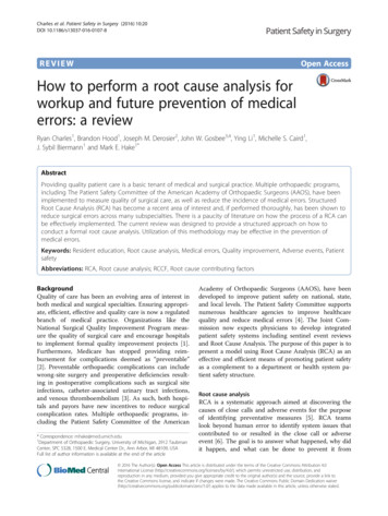
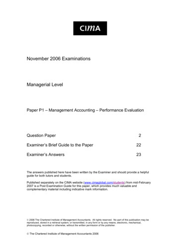
Geng et al. BMC Complementary Medicine and Therapies(2022) 22:92Page 4 of 12Fig. 1 Morphological changes in RMCs after OX7 (0.25 μg/ml,2.5 μg/ml,5 μg/ml,10 μg/ml), 8% NRS and Huaier (10 mg/ml) stimulation under a lightmicroscopeResultsOX7 antibodies plus NRS induced RMC lysis, while Huaierprotected RMCsFirst, RMCs in the control and the OX7 group grewnormally or underwent apoptosis, as shown by microscopy. In contrast, cells stimulated with OX7 (0.5 μg/ml,2.5 μg/ml, 10 μg/ml) plus NRS experienced membraneswelling and subsequent lysis, resulting in the releaseof cytosolic contents. Moreover, RMCs in the Huaier(10 mg/ml) group grew normally (Fig. 1). Second, compared with the OX7 NRS, Huaier (5 mg/ml and 10 mg/ml) significantly reduced LDH release at 2 h and 4 h. AtFig. 2 LDH release from RMCs after OX7, NRS and Huaier stimulation (2a, LDH release in each group at 2 h; 2b, LDH release in each group at 4 h; 2c,LDH release in each group at 6 h). *, compared to the control group, P 0.05. #, compared to the OX7 NRS group, P 0.05. , compared to theOX7 group, P 0.05. , compared to the NRS group, P 0.05. #, compared to the OX7 NRS group, P 0.05
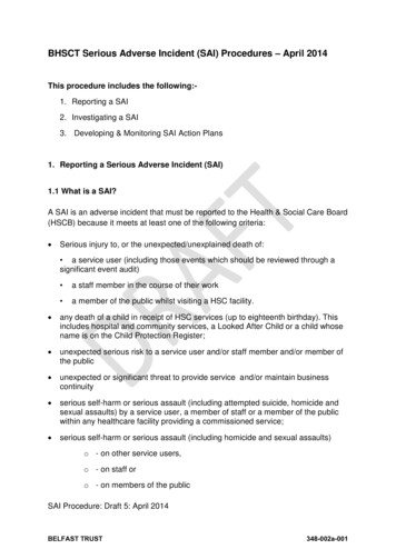
Geng et al. BMC Complementary Medicine and Therapies(2022) 22:92Page 5 of 12Fig. 3 Morphological changes in RMCs after OX7, NRS and Huaier stimulation, as shown by immunofluorescence analysis8 h, compared with the OX7 NRS group (Fig. 2a, band c). Third, RMCs in the control group appeared tohave obvious membranes, while cells in the OX7 NRSgroup showed a weakened membrane, as indicated byimmunofluorescence. Control and OX7 NRS RMCshad intact nuclei. Moreover, Huaier (5 mg/ml) partiallyprotected RMCs, and Huaier (10 mg/ml) protectedRMCs even better than the lower dose (Fig. 3).Cleaved caspase-3 expression in the OX7 NRSgroup was decreased compared to that in the control group. However, Huaier increased the expressionof cleaved caspase-3 (Fig. 4a, d and e). There was no
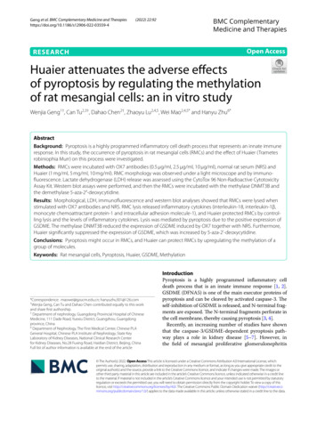
Geng et al. BMC Complementary Medicine and Therapies(2022) 22:92Page 6 of 12Fig. 4 Expression of caspase-3 and PARP in RMCs stimulated by OX7, NRS and Huaier. (a western blot results of pro-caspase-3, cleaved caspase-3and PARP; b relative intensity ratio of caspase-3 to β-actin; c relative intensity ratio of caspase-3 to α/β-tubulin; d relative intensity ratio of cleavedcaspase-3 19 kD to β-actin; e relative intensity ratio of cleaved caspase-3 17 kD to β-actin). , Huaier 1 mg/ml; , Huaier 5 mg/ml; , Huaier10 mg/ml. *, compared to the control group, P 0.05. #, compared to the OX7 NRS group, P 0.05. , compared to the OX7 group, P 0.05. ,compared to the NRS group, P 0.05. #, compared to the OX7 NRS group, P 0.05differences in pro caspase-3 (Fig. 4a and b). In addition, poly ADP-ribose polymerase (PARP) expressionin the OX7 NRS group was significantly decreasedcompared to that in the control, OX7 and NRS groups.However, Huaier increased PARP expression in a concentration-dependent manner (Fig. 4a and c). The lowexpression levels of cleaved caspase-3 and PARP in theOX7 NRS group indicated the consumption or inactivation of these factors, which needs to be furtherverified.RMC lysis released inflammatory cytokines, whichwas reduced by HuaierIL-18, MCP-1, ICAM-1 and IL-1β were significantlyincreased compared to those in the control group.However, in the Huaier (10 mg/ml) group, there weredecreased in the expression levels of ICAM-1, IL-18and MCP-1 (Fig. 5a, b, d and e). Although Huaier alsodecreased the expression of IL-1β, there was no significant difference (Fig. 5c).RMC lysis was mediated by pyroptosis, and Huaier reducedthe expression of GSDMEAccording to Shaofeng’s research [4], GSDME isa new biomarker of pyroptosis in many cell types.GSDME-NT is the fragment that forms holes in thecell membrane and induces pyroptosis. Hence, weexamined the expression of GSDME by western blotting and immunofluorescence assays. We found thatthe expression of GSDME in the control group andthe NRS group was low, while GSDME-NT levels inthe OX7 group and the OX7 NRS group increasednotable, as shown by western blotting. And theexpression of GSDME was significantly reduced in
Geng et al. BMC Complementary Medicine and Therapies(2022) 22:92Page 7 of 12Fig. 5 Expression of IL-18, MCP-1, ICAM-1 and IL-1β after RMCs were stimulated by OX7, NRS and Huaier (a relative intensity ratio of ICAM to β-actin;b expression of IL-1β in the cell culture supernatant; c relative intensity ratio of IL-18 to β-actin; d relative intensity ratio of MCP-1 to β-actin). ,Huaier 1 mg/ml; , Huaier 5 mg/ml; , Huaier 10 mg/ml. *, compared to the control group, P 0.05. #, compared to the OX7 NRS group,P 0.05. , compared to the OX7 group, P 0.05. , compared to the NRS group, P 0.05. #, compared to the OX7 NRS group, P 0.05Huaier (5 mg/ml) group than in the OX7 NRS group(Fig. 6a, b and c). Consistent with this result, immunofluorescence analysis of GSDME showed that thecontrol group was negative, while the OX7 group andthe OX7 NRS group were positive. The positive signal was attenuated by increasing concentrations ofHuaier (Fig. 7).The initiating methylase DNMT3B could reduce RMCpyroptosis induced by OX7 antibodies plus complementDNMT3B is an initiating DNA methyltransferasethat catalyzes the formation of methylation siteson unmethylated DNA double strands and downregulates gene expression. Compared with the control group, the OX7 NRS, OX7 NRS DNMT3B(1 nmol), OX7 NRS DNMT3B (10 nmol), andOX7 NRS DNMT3B (20 nmol) groups exhibited significant differences. Likewise, there was a significantreduction in the OX7 NRS DNMT3B (400 nmol)group compared with the OX7 NRS group (Fig. 8a, band c).5‑Aza‑2’‑deoxycytidine (5‑Aza‑DC) increased GSDMEexpression induced by OX7 antibodies plus NRS,while Huaier attenuated this expressionIn contrast to DNMT3B, 5-aza-DC causes the demethylation of DNA. In cancer cell lines, this compound canreverse the effect of silencing GSDME [24]. In this study,5-aza-DC increased the expression of GSDME afterthe cells were incubated with OX7 and NRS. Huaier isknown as Trametes robiniophila Murr and grows onthe stalks of old locust trees. The results of the presentstudy showed that Huaier could reduce the 5-aza-DCenhanced expression of GSDME. These results suggested that Huaier could exert an antipyroptotic effectby enhancing GSDME methylation (Fig. 9a, b and c).
Geng et al. BMC Complementary Medicine and Therapies(2022) 22:92Page 8 of 12Fig. 6 Expression of GSDME after RMCs were stimulated with OX7, NRS and Huaier, as shown by western blotting (a western blot results ofGSDME-FL and GSDME-NT; b relative intensity ratio of GSDME-FL to β-actin; c relative intensity ratio of GSDME-NT to β-actin). , Huaier 1 mg/ml; , Huaier 5 mg/ml; , Huaier 10 mg/ml. *, compared to the control group, P 0.05. #, compared to the OX7 NRS group, P 0.05. #,compared to the OX7 NRS group, P 0.05DiscussionIn this study, we found that RMC pyroptosis could beinduced by OX7 antibodies plus NRS, and Huaier couldplay a renoprotective role by affecting pyroptosis. Mesangial cells can undergo pyroptosis consistent with previousstudies. Yuxuan Zhang et al. [25] found that mesangialcells undergo pyroptosis in rats with unilateral ureteralobstruction (UUO). Jin-Feng Zhan et al. [26] found thatlncRNA-Neat1 can promote high glucose-induced ratMC pyroptosis.Huaier can play a protective role by suppressingpyroptosis and the release of inflammatory factors inMCs. Huaier is the fruiting body of Trametes robiniophila Murr, which grows on the rods of aged Sophorajaponica trees and belongs to the Basidiomycota phylum, the Polyporaceae family, and the Trametes genus[27]. Huaier was first published in Behind the Elbowand can cure colonic wind poison. Previous studieshave confirmed that Huaier can play an antitumor roleby regulating immune functions, such as increasing theindex of immune organs, regulating the number of lymphocytes and regulating the secretion of cytokines. YingChen et al [28] found that Huaier could increase theexpression of Duffy antigen receptor for chemokines(DARC) and reduce its ligands, such as CC chemokineligand 2 (CCL-2), CXC chemokine ligand 8 (CXCL-8,IL-8), matrix metalloproteinase 2 (MMP-2), and CXCchemokine ligand 1 (CXCL-1). DARC plays a negativerole in breast cancer metastasis. Yi Sun et al. [13] foundthat Huaier polysaccharide can inhibit the proliferationof cholangiocarcinoma on the one hand and promote theproliferation of spleen cells in BALB/c mice on the otherhand, inducing the production of nitric oxide synthaseand thus making macrophages produce more nitric oxideand enhancing macrophage phagocytosis. These studiesconfirmed the immunomodulatory role of Huaier.However, our findings are contrary to those ofJun Xie et al., who found that Huaier increased the
Geng et al. BMC Complementary Medicine and Therapies(2022) 22:92Page 9 of 12Fig. 7 Expression of GSDME after RMCs were stimulated by OX7, NRS and Huaier, as shown by immunofluorescence analysispyroptosis of non-small-cell lung cancer cells, therebyreducing their proliferation and playing a therapeuticrole [29]. Tumor cells were the object of Jun Xie’s study,which showed that increasing pyroptosis reduced proliferation. This is consistent with the findings of FengShao. However, MCs are a normal cell type inherentin the kidney. In addition to being a form of cell death,pyroptosis can cause a strong inflammatory response[30]. In recent years, pyroptosis has been graduallycharacterized by swelling, rupture, and the loss of intactcell morphology [31]. Pyroptotic cells release their cellular contents, causing a strong inflammatory responseand ultimately activating the innate immune response.Excessive inflammatory responses might also stimulate
Geng et al. BMC Complementary Medicine and Therapies(2022) 22:92Page 10 of 12Fig. 8 The methylase DNMT3B can attenuate GSDME expression in RMCs induced by OX7 plus NRS (a western blot results of GSDME-FL andGSDME-NT; b relative intensity ratio of GSDME-FL to GAPDH; c relative intensity ratio of GSDME-NT to GAPDH). , OX7 (0.5 μg/ml) plus Huaier (1 mg/ml). *, compared to the control group, P 0.05. #, compared to the OX7 NRS group, P 0.05. #, compared to the OX7 NRS group, P 0.05Fig. 9 The demethylating agent 5-aza-DC can upregulate the expression of GSDME in RMCs, while Huaier can attenuate the upregulated GSDMEexpression (a western blot results of GSDME-FL and GSDME-NT; b relative intensity ratio of GSDME-FL to GAPDH; c relative intensity ratio ofGSDME-NT to GAPDH). , OX7 (0.5 μg/ml) plus Huaier (1 mg/ml).*, compared to the OX7 NRS group, P 0.05. #, compared to the OX7 NRS HRgroup, P 0.05
Geng et al. BMC Complementary Medicine and Therapies(2022) 22:92the excessive proliferation of normal cells [32]. Ourresearch results suggest that Huaier can suppress thepyroptosis of RMCs and the release of inflammatoryfactors, which may attenuate the excessive proliferationof cells caused by the inflammatory response, therebyexerting a protective effect on renal cells.Further, Huaier could play a protective role by upregulating the methylation of a group of molecules in MCs.GSDME is silenced due to promoter methylation incancers [24, 33, 34]. DNA methylation is catalyzed byDNA methyltransferases (DNMTs), which transfer amethyl group to the 5th carbon atom of cytosine to form5-methylcytosine (5MC). DNMT3A and DNMT3B canestablish new methylation patterns on unmodified DNA,and these are called initiating methylation enzymes,while DNMT1 copies DNA methylation patterns fromparent DNA strands to newly synthesized daughterDNA strands during DNA replication. This study confirmed that the expression of GSDME could be attenuated by DNMT3B, which was consistent with Duan Yet al.’s study [35]. On the other hand, 5-aza-DC, a methyltransferase inhibitor, simulates cytosine and blocksmethyltransferase activity to reduce DNA methylationlevels. As an epigenetic regulatory reagent, 5-aza-DC iswidely used to examine the hypomethylation of cellularDNA. Kim used 5-Aza-DC to activate silenced genes incolon cancer and found that GSDME was activated themost frequently, in up to 40% of cases [24]. The resultsof this study also showed that 5-Aza-DC could furtherincrease the expression of GSDME in RMCs.The results of our study indicated that Huaier protectedRMCs by reducing pyroptosis and preventing the releaseof inflammatory cytokines. Pyroptosis can be activatedby innate immune-dependent pattern recognition receptors (PRRs) to disrupt extraneous antigen replication andkill bacteria through perforation, which is a part of innateimmunity [36, 37]. This finding suggests that Huaiermediated regulation may also occur through the immuneresponse. In addition, it was further shown that Huaiercould reduce the upregulated GSDME expression inRMCs after demethylation, suggesting that Huaier couldplay a protective role by regulating RMC methylation.ConclusionWe found that OX7 antibodies together with NRSinduce pyroptosis in RMCs. Huaier plays a protective role by upregulating the methylation of a group ofmolecules in RMCs.AbbreviationsRMCs: Rat mesangial cells; NRS: Normal rat serum; LDH: Lactate dehydrogenase; IL-18: Interleukin-18; IL-1β: Interleukin-1β; MCP-1: Monocytechemoattractant protein-1; ICAM-1: Intracellular adhesion molecule-1;Page 11 of 12MsPGN: Mesangial proliferative glomerulonephritis; FBS: Fetal bovine serum;PARP: Poly ADP-ribose polymerase; DNMTs: DNA methyltransferases; 5-MC:5-methylcytosine; 5-Aza-DC: 5-Aza-2’-deoxycytidine; DARC : Duffy antigenreceptor for chemokines; CCL-2: CC chemokine ligand 2; CXCL-1: CXCchemokine ligand 1; CXCL-8 (IL-8): CXC chemokine ligand 8; MMP-2: Matrixmetalloproteinase 2.Supplementary InformationThe online version contains supplementary material available at https:// doi. org/ 10. 1186/ s12906- 022- 03559-4.Additional file 1. : Expression of caspase-3 and PARP in RMCs stimulatedwith OX7, NRS and Huaier.Additional file 2. : Expression of IL-18, MCP-1, ICAM-1 and IL-1β after thestimulation of RMCs with OX7, NRS and Huaier.Additional file 3. : Expression of GSDME after the stimulation of RMCswith OX7, NRS and Huaier.Additional file 4. : shows that the methylase DNMT3B can attenuateGSDME expression in RMCs induced by OX7 plus NRS.Additional file 5. : shows that the demethylating agent 5-aza-DC canupregulate the expression of GSDME in RMCs, while Huaier can attenuatethe upregulated GSDME expression.AcknowledgmentsWe thank Dr. Wenjun Pan, Dr. Yiqi Yang and Ms. Yingchun He for their helpwith our study.Authors’ contributionsWenjia Geng, Wei Mao and Hanyu Zhu conceived the study. Can Tu andDahao Chen performed the study and analyzed the results of each assay.Zhaoyu Lu assisted with the experiment implementation and results interpretation. The author(s) read and approved the final manuscript.FundingThis study was supported by grants from the National Natural Sciences Foundation of China (grant number 81804056) and the State KeyLaboratory of Dampness Syndrome of Chinese Medicine (grant numberSZ2020ZZ07).Availability of data and materialsThe datasets used and/or analyzed during the current study are available fromthe corresponding author upon reasonable request.DeclarationsEthics approval and consent to participateAll experiments were assessed an
ditional Chinese medicine book stated that Huaier dis-pels wind, breaks blood and reinforces a healthy qi. A series of pharmacological studies showed that Huaier could promote tumor cell apoptosis and inhibit tumor cell proliferation [1312], increase sensitivity to chem, - otherapy drugs [14], promote autophagy [16] and 15,