Transcription
353Glucagon-like peptide-1 stimulates human insulin promoter activityin part through cAMP-responsive elements that lie upstream anddownstream of the transcription start siteColin W Hay*, Elaine M Sinclair*, Giovanna Bermano,Elaine Durward, Mohammad Tadayyon1 and Kevin DochertySchool of Medical Sciences, University of Aberdeen, Institute of Medical Sciences, Foresterhill, Aberdeen AB25 2ZD, UK1Department of Vascular Biology, GlaxoSmithKline Pharmaceuticals, Third Avenue, Harlow, CM19 5AW, UK(Requests for offprints should be addressed to K Docherty; Email: k.docherty@abdn.ac.uk)*(C W Hay and E M Sinclair contributed equally to this work)(M Tadayyon is now at Department of Metabolic Research, Boehringer Ingelheim, 88937 Biberach, Germany)AbstractGlucagon-like peptide-1 (GLP-1) is a peptide hormonesecreted from the enteroendocrine L-cells of the gut andwhich acts primarily to potentiate the effects of glucose oninsulin secretion from pancreatic -cells. It also stimulatesinsulin gene expression, proinsulin biosynthesis and affectsthe growth and differentiation of the islets of Langerhans.Previous studies on the mechanisms whereby GLP-1regulates insulin gene transcription have focused on the ratinsulin promoter. The aim of this study was to determinewhether the human insulin promoter was also responsiveto GLP-1, and if so to investigate the possible role ofcAMP-responsive elements (CREs) that lie upstream(CRE1 and CRE2) and downstream (CRE3 and CRE4)of the transcription start site. INS-1 pancreatic -cellswere transfected with promoter constructs containingfragments of the insulin gene promoter placed upstream ofIntroductionGlucagon-like peptide-1 (GLP-1) is a peptide hormonesecreted by the enteroendocrine L-cells of the smallintestine in response to food intake (Kieffer & Habener1999, Drucker 2001). GLP-1 plays an important role inlowering blood glucose levels primarily through its abilityto potentiate the stimulatory effects of glucose on insulinsecretion from pancreatic -cells (Holz et al. 1993). It doesthis through a G-protein-coupled receptor that acts toincrease intracellular cAMP levels (Thorens 1992). It alsoaffects blood glucose levels through its inhibitory effects ongastric emptying (Nauck et al. 1997), suppression ofappetite (Turton et al. 1996), and inhibition of glucagonsecretion from -cells (Komatsu et al. 1989).GLP-1 is a particularly important regulator of thepancreatic -cell with diverse effects on insulin secretion,the firefly luciferase reporter gene. GLP-1 was found tostimulate the human insulin promoter, albeit to a lesserdegree than the rat insulin promoter. Mutagenesis ofCRE2, CRE3 and CRE4 blocked the stimulatory effectof GLP-1 while mutagenesis of CRE1 had no effect.Analysis of nuclear protein binding to the four CREsshowed that, while they share some proteins, each CREsite is unique. Stimulation of transcription by GLP-1through CRE2, CRE3 and CRE4 resulted in alteredprotein binding that was different for each of the CREsites involved. Collectively, these data show that the fourhuman CREs are not simply multiple copies of the ratCRE site and further emphasise that the human insulinpromoter is distinct from the rodent promoter.Journal of Endocrinology (2005) 186, 353–365gene expression, proinsulin biosynthesis and islet cellgrowth and neogenesis (Drucker et al. 1987, Fehmann& Habener 1992, Edvell & Lindstrom 1999, Xu et al.1999, Stoffers et al. 2000). It also induces differentiation of pancreatic exocrine cells to an endocrinephenotype (Zhou et al. 1999). Multiple signalling pathways and second messengers are involved in the responseof the -cell to these pleiotropic actions of GLP-1(Habener 2001).All of the studies to date on the effects of GLP-1 oninsulin gene transcription have focused on the regulationof the rat insulin I promoter. However, since GLP-1 (or itsanalogues) may have a potentially important role in thetreatment of type 2 diabetes (Doyle & Egan 2001), itis important to understand how it affects expression ofthe human insulin gene. Expression of the insulin geneis regulated by promoter sequences located up toJournal of Endocrinology (2005) 186, 353–3650022–0795/05/0186–353 2005 Society for Endocrinology Printed in Great BritainDOI: 10.1677/joe.1.06205Online version via http://www.endocrinology-journals.orgDownloaded from Bioscientifica.com at 09/04/2022 07:57:03PMvia free access
354C W HAY, E M SINCLAIRand others· Glucagon-like peptide-1 effects on human insulin promoter activityapproximately 400 bp from the start site. Although thearrangement of regulatory sequences in the rat insulin Iand human insulin promoters is roughly similar (Germanet al. 1995), there are marked differences in their regulation (Clark & Docherty 1992, Melloul et al. 2002). Giventhe importance of cAMP in mediating the effects ofGLP-1, we were interested in determining whether thehuman insulin promoter was responsive to GLP-1 and, ifso, also determining the possible role of cAMP-responsiveelements (CREs).Materials and MethodsCell cultureINS-1 cells that were directly descended from the originalrat insulinoma cell line developed by Asfari et al. (1992)were cultured in RPMI 1640 containing 11·1 mM -glucose and 2 mM -glutamine, supplemented with10% foetal bovine serum (FBS), 1 mM sodium pyruvate,10 mM Hepes balanced salt solution and 50 µM -mercaptoethanol. Cells were maintained at 37 C without antibiotics in a humidified atmosphere containing 95%air and 5% CO2. Cells were passaged by trypsinisation andsubcultured on a weekly basis.RadioimmunoassaysTotal insulin-like immunoreactivity (ILI) in INS-1 cellculture medium was detected with an in-house radioimmunoassay employing guinea pig polyclonal antibodiesraised against human (pro)insulin (1011 from LincoResearch, Biogenesis, Poole, Dorset, UK). Radioimmunoassays were calibrated using rat insulin (LincoResearch) and performed in duplicate. ILI results werecalculated as ng/ml per 24 hours.respectively. These were generated by PCR amplificationof phins300 using the following primers: 5 -CGAGCTCCTGTGAGCAGGGACAG-3 and 5 -TCCCCCGGGGGAATGCTTCACGAG-3 for phINS260LUC, and 5 CGAGCTCAGATCTTCCCACAGAC-3 and 5 -TCCCCCGGGGGAATGCTTCACGAG-3 for phINS171LUC.Mutagenesis of CRE3 at position 18 to generateplasmid phINS171m1LUC was achieved by using theQuik Change Site-Directed Mutagenesis Kit (Stratagene,Amsterdam, The Netherlands) and the following primers:5 -CCTCCAGGACAGGCTAAATCAGAAGAGGCCATC-3 and its complementary reverse. Mutagenesisof CRE4 at position 61 to generate plasmidphINS171m2LUC was achieved using the primer 5 -GTTCCAAGGGCCTTTAAATCAGGTGGGCTCAGG-3and its complementary reverse. The DNA constructphINS171m3LUC contained mutations at CRE3 andCRE4 using the above primers.A further construct (phINS356LUC), containing a–356 to 14 fragment of the human insulin promoter inthe pGL3 plasmid, was obtained from Dr J. Xu, Universityof Hong Kong, Hong Kong. Sequencing of this constructshowed the presence of an A to G substitution at position–189 that was immediately upstream of the CRE2 consensus sequence. This putative error was corrected bymutagenesis as described above using the primer 5 -GGTCCTGAGGAAGAGGTGCTGACGACC-3 and itsreverse complement. Similarly, the CRE1 and CRE2 siteswere mutated using the following primers and theirreverse complements: CRE1 5 -CTGGTTAAGACTCTAATAAACCGCTGGTCCTGAGGAAG-3 and CRE25 -CTGAGGAAGAGGTGCTAAAGACCAAGGAGATCTTCC-3 . Constructs were created that lacked CRE1(phINS356m1LUC), CRE2 (phINS356m2LUC) andboth CRE1 and CRE2 (phINS356m3LUC).The mutations and integrity of final products wereconfirmed by DNA sequencing.Plasmid DNA constructsThe rat insulin I promoter ( 410 to 1bp) construct,pFOXLUC410, and the control vector pFOXLUC wereprovided by Dr M. German (University of California atSan Francisco Medical School, San Francisco, CA, USA).The human insulin promoter construct was generated byPCR amplification of a –364 to 148 bp fragment ofDNA from the plasmid phins300 that contains a XhoIfragment ( 5 kb) of hINS-1 (Bell et al. 1980) clonedinto the SalI site of pBR322 plasmid. The primers usedhad the sequence 5 -CGAGCTCGACAGCAGCGCAAAGA-3 and 5 -TCCCCCGGGGGAATGCTTCACGAG-3 . The amplified fragment was inserted upstream ofthe firefly luciferase sequences within plasmid pGL3(Promega) to generate phINS364LUC. The plasmidsphINS260LUC and phINS171LUC contained fragmentsspanning the regions –260 to 148 and –171 to 148Journal of Endocrinology (2005) 186, 353–365TransfectionsCells were grown to 80–90% confluency, trypsinised andtransferred to six-well plates at a density of 4 105 cellsper well; 72 h later, the cells were transfected usingLipofectamine Plus reagent in a 1 ml transfection cocktailcontaining 1 µg plasmid DNA. Cells were incubated for3 h in the transfection mixture and then overnight in2 ml of normal growth medium. The medium was thenreplaced with 2 ml RPMI containing 1 mM sodiumpyruvate, 10 mM Hepes balanced salt solution, 50 µM -mercaptoethanol, 3 mM glucose and 10% foetal bovineserum, and the cells incubated for a further 24 h. Cellswere washed once with minimal medium before testsubstances or solvent alone were added to a final volume of2 ml per well. Unless specified, cells were incubated inminimal medium containing 11·1 mM glucose withwww.endocrinology-journals.orgDownloaded from Bioscientifica.com at 09/04/2022 07:57:03PMvia free access
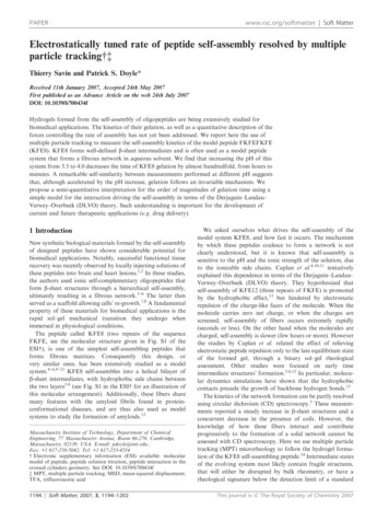
Glucagon-like peptide-1 effects on human insulin promoter activity ·C W HAY, E M SINCLAIRand others 355the test substances for 4 h at 37 C in a humidifiedatmosphere.Luciferase assaysCells were washed once with PBS and lysed with 400 µlPassive Lysis Buffer (Promega). Cell lysis was carried outfor 15 min with shaking and cell debris was removed bycentrifugation at 13 000 g for 5 min. Then, 10 µl cellextract were added to 350 µl of A buffer (15 mMMgSO4.7H2O, 30 mM glycylglycine, 2 mM Na2ATP,pH 7·8). To this, 150 µl of G buffer (30 mM glycylglycine, pH 7·8) containing 0·5 mM luciferin were injectedand the luminescence read at 560 nm using a BertholdLumat LB 9501/16. Protein content was measured usingthe Bio-Rad DC Protein Assay (Bio-Rad) with BSA as astandard.Nuclear extractsCells were grown to approximately 80% confluence in9 cm Petri dishes and treated exactly as for luciferaseassays. They were grown in complete RPMI 1640medium containing 10% foetal bovine serum and 3 mMglucose for 24 h, then washed once with minimal mediumbefore test substances or solvent alone were added. Cellswere incubated in minimal medium containing 11·1 mMglucose with the test substances for 4 h at 37 C. Nuclearextracts were prepared by the method of Dignam et al.(1983) in the presence of protein phosphatase inhibitors(5 mM -glycerophosphate and 100 µM Na3VO4). Theprotein content was determined using the Bio-Rad DCProtein Assay with BSA as a standard.Electrophoretic mobility shift assaysThe coding sequences of the 30 bp oligonucleotides of thehuman insulin gene CRE sites were: CRE1, 5 -TAAGACTCTAATGACCCGCTGGTCCTGAGG-3 ; CRE2,5 -GGAAGAGGTGCTGACGACCAAGGAGATCTT-3 ;CRE3, 5 -CCAGGACAGGCTGCATCAGAAGAGGCCATC-3 ; CRE4, 5 -CCAAGGGCCTTTGCGTCAGGTGGGCTCAGG-3 . The double-stranded CRE oligonucleotides were labelled with T4 polynucleotide kinase(New England Biolabs, Hitchin, UK) and [ -32P]ATP(Amersham), followed by purification using Quick SpinColumns (Roche). Nuclear binding assays were carriedout in a total volume of 20 µl of 20 mM Hepes, pH 7·9,70 mM KCl, 0·2 mM EDTA, 0·5 mM dithiothreitol and10% glycerol. Nuclear extracts (10 µg) were incubated onice for 15 min with 1 µg poly(dI-dC).poly(dI-dC) asnon-specific competitor along with other competitors orantibodies when indicated. Oligonucleotide competitionbinding assays were carried out with 300 molar excessof specific competing unlabelled CRE oligonucleotide.Antibody competition assays had the addition of eitherwww.endocrinology-journals.orgFigure 1 Response of INS-1 -cells to glucose and GLP-1. INS-1cells were incubated for 24 h in medium containing 3 mM glucoseand foetal bovine serum. The cells were then pre-incubated inHepes-balanced buffer containing 3 mM glucose with 0·2% BSAfor 1 h followed by stimulation for 45 min in Hepes-balancedbuffer containing either 3 or 11·1 mM glucose in the presence orabsence of 10 nM GLP-1. The levels of insulin secreted weremeasured by radioimmunoassay. The data represent themeans S.E.M. of three independent experiments carried out intriplicate and are expressed as fold increases in insulin secretioncompared with 3 mM glucose. Statistical analyses of increases ininsulin secretion, which were relevant to specific assays, werecalculated. Cells without GLP-1: 11·1 mM glucose compared with3 mM glucose, P 0·001. GLP-1 treated cells: comparison withthe same concentration of glucose without GLP-1, **P 0·01,***P 0·001.1 µl rabbit polyclonal anti-human pancreatic duodenalhomeobox-1 (PDX-1) serum (a gift from Dr C. Wright,Vanderbilt University Medical Center, Nashville, TN,USA) or 1 µl rabbit serum as a control. Labelled oligonucleotide corresponding to 60 fmol was added and incubation was carried out at room temperature for 30 min.The reaction products were resolved on 6% nondenaturingpolyacrylamide gels run in TGE buffer, pH 8·5 (50 mMTris, 380 mM glycine, 2 mM EDTA). The gels weredried and subjected to autoradiography at 70 C.Statistical analysisStatistical analysis was performed using a BlackwellScience statistical package (Blackwell Scientific Publications, Oxford, UK) and statistical significance wasmeasured by analysis of variance.ResultsEffect of GLP-1 on human insulin promoter activityGLP-1 is known to potentiate the effects of glucose oninsulin secretion and gene expression. In preliminaryexperiments the responsiveness of INS-1 -cells toglucose and GLP-1 was investigated (Fig. 1). INS-1 cellsresponded to glucose with a significant 2·5-fold increase ininsulin secretion when cultured in the presence of11·1 mM compared with 3 mM glucose. Treatment withJournal of Endocrinology (2005) 186, 353–365Downloaded from Bioscientifica.com at 09/04/2022 07:57:03PMvia free access
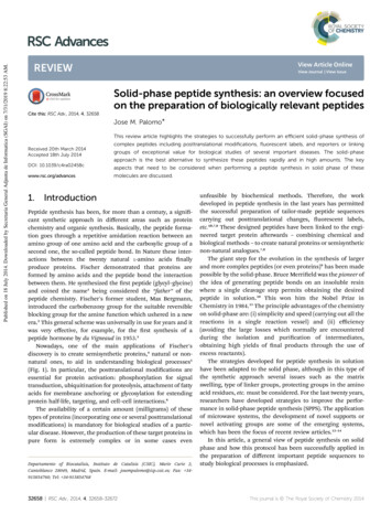
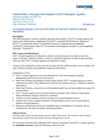
356C W HAY, E M SINCLAIRand others· Glucagon-like peptide-1 effects on human insulin promoter activityFigure 2 Schematic representation of the human insulin promoter constructs. The scale represents nucleotides relative tothe transcription start site 1and forward arrow. The positions of the four CREs and major cis-acting elements are indicated.10 nM GLP-1 resulted in significant increases in insulinsecretion in both 3 and 11·1 mM glucose. Insulin secretionincreased 1·4-fold in the presence of GLP-1 in cellscultured in 3 mM glucose compared with 3 mM glucosealone and by 2·7-fold in cells cultured in GLP-1 and11 mM glucose compared with 11 mM glucose alone. Inaddition, Northern blot analysis showed that the GLP-1stimulated increase in insulin secretion was accompaniedby 1·7-fold (P 0·001) and 1·5-fold (P 0·05) increases inthe amount of preproinsulin mRNA in low and highglucose respectively (data not shown). Thus, INS-1 -cellsrespond to GLP-1 by increasing insulin secretion in aglucose-dependent manner and were used for all furtherstudies.More detailed studies on the mechanism of GLP-1stimulation of the insulin promoter were carried out usingluciferase reporter constructs containing the rat insulin Ipromoter (pFOXLUC410) and a series of human insulinJournal of Endocrinology (2005) 186, 353–365promoter constructs (Fig. 2). Initial experiments usingpFOXLUC410, which contained the rat insulin I promoter ( 410 to 1bp) in the pFOXLUC vector,showed that GLP-1 stimulated the activity of the ratinsulin I promoter both in 3 mM glucose (2-fold increase)and in 11·1 mM glucose (2·4-fold increase) (Fig. 3). Themagnitude of this effect on the rat insulin promoter was inkeeping with previous studies using a similar rat insulinpromoter construct in INS-1 cells (Skoglund et al. 2000,Kemp & Habener 2001, Chepurny et al. 2002). In detaileddose–response experiments (data not shown), a maximaleffect of GLP-1 was observed at 10 nM and thisconcentration was used in all further experiments. Thehuman insulin promoter contains an inhibitory elementbetween –279 and –258 (Boam et al. 1990) and regulatorysequences that lie downstream of the transcription start site(Inada et al. 1999). When the human insulin promoterconstruct phINS364LUC (Fig. 2), which contains thewww.endocrinology-journals.orgDownloaded from Bioscientifica.com at 09/04/2022 07:57:03PMvia free access
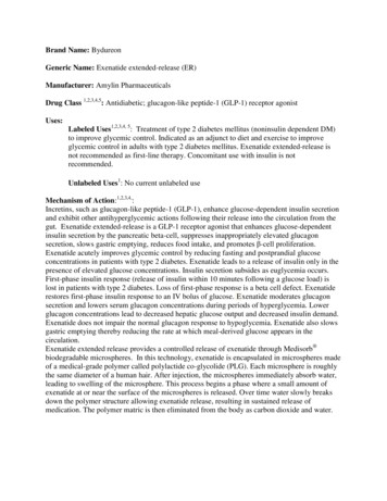
Glucagon-like peptide-1 effects on human insulin promoter activity ·C W HAY, E M SINCLAIRand others 357Figure 3 Effect of GLP-1 on rat 1 and human insulin promoter constructs in INS-1 cells.INS-1 cells were transfected with 1 g of the indicated plasmids. Construct pFOXLUC410contained the rat insulin I promoter ( 410 to 1bp) in the pFOXLUC vector and thevarious human insulin promoter constructs are outlined in Fig. 2. Cells were cultured for24 h in medium containing low glucose (3 mM) and foetal bovine serum, after which thecells were washed once with serum-free medium and cultured in serum-free mediumcontaining 3 or 11·1 mM glucose in the presence or absence of 10 nM GLP-1 for 4 h.Whole-cell extracts were then prepared and firefly luciferase activity measured. The resultsare presented as luciferase activity in cells incubated in GLP-1 relative to those incubatedin 3 mM (open bars) or 11·1 mM glucose (shaded bars) in the absence of GLP-1. Thedata represent the means S.E.M. of four independent experiments carried out in triplicateand the significance of GLP-1 stimulation was determined. *P 0·05, **P 0·01,***P 0·001.inhibitory and downstream elements, was used, there wasa significant stimulation by GLP-1 of promoter activity in3 mM glucose (1·12-fold increase) and 11·1 mM glucose(1·25-fold increase) (Fig. 3). GLP-1 also stimulated theactivity of the human insulin promoter constructphINS260LUC (Fig. 2), which lacked the inhibitoryelement (1·23- and 1·27-fold in 3 and 11 mM glucoserespectively) (Fig. 3). The inhibitory element, which alsofunctions as a glucose response element in transfected islets(Sander et al. 1998), does not therefore appear to affect theresponsiveness of the insulin promoter to GLP-1. Ingeneral the human insulin promoter constructs weremuch less responsive to GLP-1 than the rat insulinpromoter (Fig. 3). There was no effect of glucose orGLP-1 on the control vectors pFOXLUC or pGL3 (datanot shown).Role of CREs in regulating the human insulin promoterFurther experiments were designed to determine whetherCREs within the human insulin promoter might beinvolved in mediating the effects of GLP-1. There are fourCREs within the human insulin promoter located at 210 (CRE1, TGACCCGC), 183 (CRE2, TGACGACC), 18 (CRE3, TGCATCAG) and in the first intronat 61 (CRE4, TGCGTCAG) (Fig. 2). CRE2 is the onlyone conserved between humans and rodents (Philippewww.endocrinology-journals.org& Missotten 1990, Inagaki et al. 1992). The absenceof sequences that included CRE1 and CRE2 inphINS171LUC, and CRE3 and CRE4 inphINS356LUC did not diminish the effect of GLP-1 onpromoter activity (Fig. 3). This suggests that CREs that lieupstream and downstream of the transcription start sitemight be involved.The individual roles of CRE1 and CRE2 were studiedby mutating these responsive elements in the construct phINS356LUC as outlined in Fig. 2. Similarly, theroles of CRE3 and CRE4 were investigated by creatinga series of mutations in the construct phINS171LUC(Fig. 2). In order to determine if all four CRE sites weretranscriptionally active, initial experiments looked at theeffect of the mutations on the rates of basal transcription.Mutagenesis of CRE1 (phINS356m1LUC) and ofCRE2 (phINS356m2LUC) resulted in a reduction ofbasal transcription to 0·63 and 0·65 relative to theunmutated construct respectively (Fig. 4A). ConstructphINS356m3LUC, in which both CRE1 and CRE2were mutated, displayed an even greater reduction in basaltranscription to 0·4 relative to the unmutated construct(Fig. 4A). Mutagenesis of CRE3 (phINS171m1LUC), ofCRE4 (phINS171m2LUC) and of both CRE3 andCRE4 (phINS171m3LUC) also led to reductions in basaltranscription to 0·70, 0·58 and 0·66 relative to theunmutated construct respectively (Fig. 4B). These dataJournal of Endocrinology (2005) 186, 353–365Downloaded from Bioscientifica.com at 09/04/2022 07:57:03PMvia free access
358C W HAY, E M SINCLAIRand others· Glucagon-like peptide-1 effects on human insulin promoter activityFigure 4 Effect of CRE mutations on basal transcription rates.INS-1 cells that had been transfected with appropriate plasmidswere cultured for 24 h in medium containing low glucose (3 mM)and foetal bovine serum. Following this, the cells were washedonce with serum-free medium and incubated for 4 h in serum-freemedium containing 11·1 mM glucose. Whole-cell extracts werethen prepared and firefly luciferase activity measured. The resultsare presented as luciferase activity relative to that in cellstransfected with plasmids containing wild-type CRE sites.(A) phINS356LUC series of constructs. (B) phINS171LUC series ofconstructs. The data represent the means S.E.M. of threeindependent experiments carried out in triplicate and thesignificance of reductions in transcriptional activity wasdetermined. ***P 0·001.show that all four CRE sites in the human insulinpromoter are transcriptionally active and that there couldbe constitutive cAMP/PKA activity in INS-1 cells.Role of individual CREs in the human insulin promoterresponse to GLP-1Both constructs phINS356LUC and phINS171LUCshowed significant stimulation by GLP-1 (Fig. 5). Mutagenesis of CRE1 (phINS356m1LUC) had no effecton GLP-1 stimulation while mutagenesis of CRE2(phINS356m2LUC) completely abolished the stimulatoryeffect of GLP-1 (Fig. 5A). In keeping with these results,Journal of Endocrinology (2005) 186, 353–365Figure 5 Response of CRE sites to GLP-1. INS-1 cells weretransfected with plasmids containing wild-type or mutated CREsites as indicated. Cells were cultured for 24 h in mediumcontaining low glucose (3 mM) and foetal bovine serum, afterwhich the cells were washed once with serum-free medium andcultured in serum-free medium containing 11·1 mM glucose inthe presence or absence of 10 nM GLP-1 for 4 h. Whole-cellextracts were then prepared and firefly luciferase activitymeasured. The results are presented as luciferase activity in cellsincubated in GLP-1 relative to those transfected with the sameplasmid incubated in the absence of GLP-1. (A) Effect of GLP-1on phINS356LUC series of constructs. (B) Effect of GLP-1 onphINS171LUC series of constructs. The data represent themeans S.E.M. of three independent experiments carried out intriplicate. Statistical analyses, which were relevant to specificassays, were calculated. Wild type: the significance of GLP-1stimulation, ***P 0·001. CRE1: the significance of no reduction inGLP-1 stimulation in the construct with a mutated CRE1 sitecompared with wild type, ‡P 0·05. All other constructs: thesignificance of reduction in GLP-1 stimulation in constructs withmutated CREs compared with wild type, P 0·05, P 0·001.the construct, in which both CRE1 and CRE2were mutated (phINS356m3LUC), was also unresponsive to GLP-1 (Fig. 5A). Mutagenesis of CRE3(phINS171m1LUC) and of CRE4 (phINS171m2LUC)diminished the stimulatory effect of GLP-1 from a 1·30fold increase for the unmutated construct to 1·18- and1·19-fold increases respectively (Fig. 5B), while thewww.endocrinology-journals.orgDownloaded from Bioscientifica.com at 09/04/2022 07:57:03PMvia free access
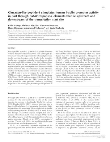
Glucagon-like peptide-1 effects on human insulin promoter activity ·C W HAY, E M SINCLAIRand others 359construct phINS171m3LUC, in which both CRE3 andCRE4 were mutated, completely blocked the effects ofGLP-1 (Fig. 5B). This would suggest that CRE3 andCRE4 act in a cumulative manner. In summary, thestimulatory effect of GLP-1 on the human insulin promoter is mediated through CRE2, CRE3 and CRE4,while CRE1 is not involved.The response of individual CREs in the human insulinpromoter to forskolinGLP-1 is thought to act primarily through a G-proteincoupled receptor leading to increased intracellular cAMPlevels (Thorens 1992). The responsiveness of the differentCRE sites to cAMP was investigated using forskolin, astimulator of adenylate cyclase. Although forskolin raisescAMP levels in an unphysiological manner and cannotbe used in a qualitative manner, it is widely recognisedthat increased intracellular levels of cAMP can mimicsignalling pathways. Both constructs phINS356LUC andphINS171LUC showed stimulation upon treatment withforskolin (Fig. 6). The contribution of CRE1 and CRE2sites to forskolin responsiveness was investigated using thephINS356LUC mutant constructs. Loss of CRE2 resultedin a concomitant loss of responsiveness to forskolin, whilemutation of CRE1 had only a minor effect (Fig. 6A).Mutagenesis of CRE3 and CRE4 had minimal effect onforskolin stimulation on phINS171LUC, however thedouble mutation of CRE3 and CRE4 did result in asignificant reduction in stimulation (Fig. 6B). The inabilityof the CRE3 and CRE4 mutations to significantlydiminish forskolin effects may reflect the promiscuity ofbinding specificities for the eight or so members of thebZIP family of transcription factors that are known torecognise CRE-related sequences. It is highly significant,however, that whereas the CRE3 and CRE4 mutationsonly weakly inhibited forskolin stimulation, they producedsignificant reductions in the stimulatory effect of GLP-1as described above. This would suggest that GLP-1 isacting on CRE3 and CRE4 mainly by a mechanism otherthan through an increase in active cAMP-dependenttranscription factors.The role of cAMP in GLP-1 stimulation of the humaninsulin promoter was further examined by using H-89, theinhibitor of protein kinase A (PKA). While H-89 (10 µM)completely abolished the stimulatory effect of forskolinon the phINS260LUC construct (P 0·01), it onlypartially inhibited the stimulatory effect of GLP-1 onphINS260LUC construct by 48% (P 0·01) and 39%(P 0·001) in 3 and 11·1 mM glucose respectively (datanot shown), lending further support to the view thatGLP-1 can act through cAMP-dependent and noncAMP-dependent intracellular pathways. These findingsare in keeping with the observations that H-89 is ineffective in blocking GLP-1 stimulation of the rat insulin Ipromoter (Skoglund et al. 2000).www.endocrinology-journals.orgFigure 6 Response of CRE sites to forskolin. INS-1 cells weretransfected with plasmids containing wild-type or mutated CREsites as indicated. Cells were cultured for 24 h in mediumcontaining low glucose (3 mM) and foetal bovine serum, afterwhich the cells were washed once with serum-free medium andcultured in serum-free medium containing 11·1 mM glucose in thepresence or absence of forskolin (10 M) for 4 h. Whole-cellextracts were then prepared and firefly luciferase activitymeasured. The results are presented as luciferase activity incells incubated in forskolin relative to cells transfected with thesame plasmid incubated in the absence of forskolin. (A) Effectof forskolin on phINS356LUC series of constructs. (B) Effect offorskolin on phINS171LUC series of constructs. The data representthe means S.E.M. of three independent experiments carried out intriplicate. Statistical analyses, which were relevant to specificassays, were calculated. Wild type: the significance of forskolinstimulation, ***P 0·001. All other constructs: the significance ofreduction in forskolin stimulation in constructs with mutated CREscompared with wild type, P 0·001.Nuclear protein binding to individual CRE sitesNuclear proteins were purified from INS-1 cells that hadbeen grown in 3 mM glucose for 24 h followed by 4 h inminimal medium containing 11·1 mM glucose. When theproteins were incubated with labelled oligonucleotidescontaining each of the four human insulin promoter CREsites and analysed by electrophoretic mobility shift assay,multiple retarded bands were observed. Unique patterns ofJournal of Endocrinology (2005) 186, 353–365Downloaded from Bioscientifica.com at 09/04/2022 07:57:03PMvia free access
360C W HAY, E M SINCLAIRand others· Glucagon-like peptide-1 effects on human insulin promoter activityFigure 7 Nuclear protein binding to CRE sites. Nuclear extracts from INS-1 cells that hadbeen incubated for 24 h in 3 mM glucose followed by 4 h in medium containing11·1 mM glucose were incubated with labelled oligonucleotides containing the humaninsulin promoter CRE sites and resolved by electrophoretic mobility shift analysis. SpecificDNA–protein complexes are indicated with arbitrary labels. (A) Nuclear extract wasincubated with the labelled CRE oligonucleotide indicated above the lane. The differentsamples were run on adjacent lanes in the same gel. (B) Labelled CRE1 oligonucleotidewas incubated with nuclear extract that had either received no additional treatment (lane1), been pre-incubated for 15 min on ice with rabbit serum (lane 2) or pre-incubated for15 min on ice with rabbit polyclonal anti-human PDX-1 serum (lane 3).protein binding were formed with each CRE site andthe various DNA–protein complexes have been assignedarbitrary names (Fig. 7A). Each CRE displayed one or twomajor bands including (1A and 1D, 2E, 3B and 3D, and4B and 4D) and a number of minor bands. Antibodycompetition studies showed the major complex 1D contained PDX-1 (Fig. 7B) and removal of PDX-1 permittedanother protein to bind and form the complex 1H.Clearly, the presence of the overlapping PDX-1 bindingsite affects the binding of proteins and possibly the activityof the CRE1 site. Many complexes were common toseveral CRE sites. For example, bands corresponding tothe CRE1 complex 1A were also observed with CRE3Journal of Endocrinology (2005) 186, 353–365and CRE4 (3C and 4B respectively). The major band inthe CRE2 lane (2E) was present in all the other lanes as1C, 3D and 4D, while one of the two major complexescreated with CRE3 (3B) had analogou
were washed once with minimal medium before test substances or solvent alone were added to a final volume of 2 ml per well. Unless specified, cells were incubated in minimal medium containing 11·1 mM glucose with 354 C W HAY, E M SINCLAIR and others · Glucagon-like peptide-1 effects on human insulin promoter activity