Transcription
Chaignat et al. BMC Infectious Diseases 2014, 4RESEARCH ARTICLEOpen AccessPerformance of seven serological assays fordiagnosing tularemiaValérie Chaignat1, Marina Djordjevic-Spasic2, Anke Ruettger3, Peter Otto1, Diana Klimpel1, Wolfgang Müller1,Konrad Sachse3, George Araj4, Roland Diller1 and Herbert Tomaso1*AbstractBackground: Tularemia is a rare zoonotic disease caused by the Gram-negative bacterium Francisella tularensis.Serology is frequently the preferred diagnostic approach, because the pathogen is highly infectious and difficult tocultivate. The aim of this retrospective study was to determine the diagnostic accuracy of tularemia specific tests.Methods: The Serazym Anti-Francisella tularensis ELISA, Serion ELISA classic Francisella tularensis IgG/IgM, an in-houseELISA, the VIRapid Tularemia immunochromatographic test, an in-house antigen microarray, and a Western Blot (WB)assay were evaluated. The diagnosis tularemia was established using a standard micro-agglutination assay. In total,135 sera from a series of 110 consecutive tularemia patients were tested.Results: The diagnostic sensitivity and diagnostic specificity of the tests were VIRapid (97.0% and 84.0%), Serion IgG(96.3% and 96.8%), Serion IgM (94.8% and 96.8%), Serazym (97.0% and 91.5%), in-house ELISA (95.6% and 76.6%),WB (93.3% and 83.0%), microarray (91.1% and 97.9%).Conclusions: The diagnostic value of the commercial assays was proven, because the diagnostic accuracy was 90%.The diagnostic sensitivity of the in-house ELISA and the WB were acceptable, but the diagnostic accuracy was 90%.Interestingly, the antigen microarray test was very specific and had a very good positive predictive value.Keywords: Serology, Tularemia, Diagnostic sensitivity, Diagnostic specificityBackgroundTularemia is a zoonotic disease caused by Francisellatularensis, a highly infectious, facultative intracellular,Gram-negative bacterium. The clinical presentation inhumans depends on the route of infection and variesfrom relatively mild skin lesions and lymphadenopathyto life-threatening pneumonia and/or septicemia [1].Due to the low infectious dose and the ease of dissemination by aerosols, this bacterium was categorized as ahighly dangerous biological agent by the Centers forDisease Control in Atlanta (Georgia, USA) [2-4].The diagnostic repertoire comprises cultivation methods,PCR assays, and serological tests [5-8]. Because of the highrisk of laboratory infections associated with cultivation ofF. tularensis, serology is frequently preferred. Serologicalassays are usually easy to perform, allow high throughput,* Correspondence: herbert.tomaso@fli.bund.de1Institute of Bacterial Infections and Zoonoses, Friedrich-Loeffler-Institut(Federal Research Institute for Animal Health), Naumburger Str. 96a, Jena07743, GermanyFull list of author information is available at the end of the articleand cause little risk of infection for laboratory workers[9-12]. However, some patients never seroconvert, whileothers stay seropositive for years after an infection, and/ormay have cross- reacting antibodies due to infections withother bacteria (e.g. Yersinia spp., Brucella spp.) [13,14].The aim of this study was to compare novel assays withwell-established diagnostic tools using a comprehensivecollection of human sera.MethodsSerum samplesThe present retrospective study was conducted in accordance with the STARD guidelines to assess the diagnostic accuracy and the clinical value of the respectiveassays [15,16]. The study population included a consecutive series of 110 patients in an endemic area of tularemia in Serbia that had a history of potential risk ofexposure and/or had clinical symptoms compatible withtularemia. In accordance with WHO guidelines, patientswith typical symptoms were regarded as tularemia cases, 2014 Chaignat et al.; licensee BioMed Central Ltd. This is an Open Access article distributed under the terms of the CreativeCommons Attribution License (http://creativecommons.org/licenses/by/2.0), which permits unrestricted use, distribution, andreproduction in any medium, provided the original work is properly credited.
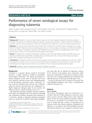
Chaignat et al. BMC Infectious Diseases 2014, 4if at least one serum sample was positive in the microagglutination assay (MAT) for tularemia [9]. If the firstserum sample was negative, paired serum samples weretested after two weeks or later. In total 135 sera were collected between 1999 and 2009. Patients were excluded, ifdocumentation was not complete or if the sample volumewas not sufficient to perform all diagnostic tests assessedin this study. All patients signed an informed consent andthe use of the samples for this study was granted by theEthics Committee of the Medical Faculty, Niš University(Number 01-4608-3, 9th July 2009).In order to assess potential cross-reactivity, 94 sera ofhumans without known history of tularemia were testedincluding 20 seropositive samples from patients withculture-proven brucellosis from Lebanon. The company“Mikrogen” (Martinsried, Germany) kindly provided 74sera collected from blood donors in Germany, whichhad been purchased from the Bavarian Red Cross (Munich,Germany). All control sera were anonymized in relation topatient data.AssaysMAT assayThis test was performed in Serbia to establish the diagnosisand was regarded as the reference method in this study.Briefly, serial 2-fold dilutions of sera (25 μl) were mixedwith an equal volume of formalin-inactivated F. tularensissubsp. holarctica (LVS) whole cell suspension (OD560 1.0).The reactions were performed in round-bottom microtiterplates (96-well; NUNC, Roskilde, Denmark). The plateswere read out after incubation at 37 C for 18 h. Agglutinations at dilutions of 1:20 or higher were considered MATpositive.All commercial assays were performed and interpretedaccording to the manufacturer’s instructions:The Serazym Anti-Francisella tularensis ELISA (Serazym ELISA) (Seramun Diagnostica GmbH, Heidesee OTWolzig, Germany) detects all classes of antibodies. ThePage 2 of 6Serion ELISA classic Francisella tularensis IgG/IgM (IgGSerion ELISA/IgM Serion ELISA) (Institut Virion/SerionGmbH, Würzburg, Germany) allows separate detection ofIgG and IgM. The VIRapid (VIRapid) (Vircell S.L., SantaFé, Spain) is an immunochromatographic lateral flow test(ICT) (Figure 1).The Yersinia recomline kit (Yersinia IgG 2.0 kit,Mikrogen) was used to determine, which sera had antiYersinia antibodies that could potentially cause crossreactions.In-house developed assaysThe in-house ELISA was designed to detect anti-FrancisellaIgG antibodies. The assay published by Porsch-Ozcürümezet al. [11] was performed with some modifications. Briefly,the coating antigen was a purified lipopolysaccharide (LPS)obtained from F. tularensis subsp. holarctica (ATCC 29684)(Micromun, Greifswald, Germany). The secondary antibodywas a horseradish peroxidase-conjugated goat anti-humanIgG (Millipore, Schwalbach, Germany). Receiver operatingcharacteristic (ROC) curves were used to determine thecut-off value (MedCalc Software Version 13.0.4, Oostend,Belgium).The Western Blot assay (WB) was developed as a modification of the test published by Schmitt et al. [10]. TheF. tularensis LPS was purchased from Micromun. Thesecondary antibody was a purified recombinant proteinA/G, which was alkaline phosphatase labeled (PierceBiotechnology, Rockford, Illinois, USA) diluted 1:5,000.The microarray detecting anti-Francisella antibodiesused two different preparations of antigen: Whole-cellbacterial antigen of Francisella tularensis subsp. holarcticastrain LVS and commercially available purified LPS werecompared. Whole-cell bacterial antigen was obtained aftergrowth for 48 h on Cysteine Heart Agar enriched withchocolatized red blood cells at 37 C in ambient atmosphere. Bacterial pellets were harvested and diluted inphosphate-buffered saline containing 5–20 mM sucroseABFigure 1 Hand-held immunochromatographic test for the serological diagnosis of tularemia (VIRapid ; Vircell S.L., Santa Fé, Spain).A test is interpreted as positive, if the specific line and the control line are positive (A). The test is valid, but negative, if only the control line isvisible (B).
Chaignat et al. BMC Infectious Diseases 2014, 4Page 3 of 6to final antigen concentrations between 0.5 and 1 mg protein/ml. Subsequently, the antigen preparations were spotted onto microarray chips as previously described [17] andassembled in the ArrayStrip 8-well microtiter strip format(Alere Technologies GmbH, Jena, Germany). To evaluatethe impact of the secondary antibody (Protein A/G-HRP,Thermo Fisher Scientific GmbH, Schwerte, Germany) onthe background, it was added to the tularemia antigenswithout the patient sera. Results from this analysis showedno direct reactivity due to the secondary antibody.Antigen-antibody reaction in array strip vesselsExamination of sera was conducted using the ProteinBinding Kit (Alere Technologies GmbH) with the following modifications: The incubation buffer P1 was supplemented with 1% (w/v) milk powder (Marvel; PremierInternational Foods UK Ltd, Dublin, Ireland), and blocking buffer consisted of P1 containing 3% (w/v) milk powder. All incubation and washing steps were performed at37 C using a heated horizontal tube shaker (BioShakeiQ; Quantifoil Instruments GmbH, Jena, Germany). TheArrayStrips were initially conditioned with 100 μl of incubation buffer at 400 strokes per minute (spm) for5 min. The supernatant was discarded and the microarrays were blocked by incubation with 100 μl blockingbuffer at 300 spm for 5 min. Prior to incubation, serumsamples were diluted 1:100 in incubation buffer in a separate tube. After transfer of 100 μl of diluted serum into thearray vessel, the antigen-antibody reaction was allowed toproceed at 300 spm for 30 min. The supernatant was discarded, and the arrays were washed using 150 μl incubation buffer at 400 rpm for 5 min. The supernatant wasagain discarded and the arrays were incubated with 100 μlof a 1:10,000 dilution of HRP-conjugated Protein A/G(Thermo Fisher Scientific GmbH) in incubation buffer at300 spm for 30 min. Subsequently, the ArrayStrips werewashed in two steps as above. Finally, 100 μl of the readyto-use HRP substrate D1 (part of the Protein Binding Kit,Alere Technologies GmbH) were added for staining (25 C,10 min, no shaking). Before the final reading, the stainingsolution was completely removed. The outcome of theantigen-antibody reaction was measured using an ArrayMate transmission reader (Alere Technologies GmbH).Signal intensity data were processed using the Iconoclustsoftware, version 3.3 (Alere Technologies GmbH). Pilottests with positive and negative control sera were used toempirically determine a cut-off value of 0.3 intensity units(data not shown).The diagnostic value was determined based on thefollowing parameters: Diagnostic sensitivity [TP/(TP FN)] 100; Diagnostic specificity [TN/(TN FP)] 100;Positive Predictive Value (PPV) [TP/(TP FP)] 100;Negative Predictive Value (NPV) [TN/(TN FN)] 100;Diagnostic accuracy (TP TN)/(TP TN FP FN) 100. (TP true positive, TN true negative, FN falsenegative, FP false positive).ResultsThe parameters used to assess the diagnostic accuracyfor all serological assays are presented in Table 1. Thediagnostic sensitivity and diagnostic specificity of the testswere: VIRapid (97.0% and 84.0%), Serion IgG (96.3% and96.8%), Serion IgM (94.8% and 96.8%), Serazym (97.0%and 91.5%), in-house ELISA (cut-off 0.18; 95.6% and76.6%), WB (93.3% and 83.0%), microarray (91.1% and97.9%). The cut-off for the in-house ELISA was set to 0.18based on a ROC analysis in order to achieve a relativelyhigh diagnostic sensitivity (Figure 2, Additional file 1).The microarray test with spotted whole antigen of F.tularensis was used for the final evaluation, because pilotTable 1 Performance of serological assays for diagnosing tularemiaTrue positiveFalse negativeIn-house ELISACut-off 0.18In-house ELISACut-off 0.33VIRapidWBSerazymELISASerion ELISAIgGSerion se positive221215168332True negative7282797886919192Diagnostic sensitivity [%]95.687.497.093.397.096.394.891.1Diagnostic specificity [%]76.687.284.083.091.596.896.897.9PPV [%]85.490.889.788.794.297.797.798.4NPV [%]92.382.895.289.795.694.892.988.5Diagnostic accuracy [%]87.887.391.789.194.896.595.693.9The following tests were evaluated: Western blot (WB); Serion ELISA classic Francisella tularensis IgG/IgM (Institut Virion\Serion GmbH, Würzburg, Germany) (IgGand IgM Serion ELISA); Serazym Anti-Francisella tularensis (Seramun Diagnostica GmbH, Heidesee OT Wolzig, Germany) (Serazym ELISA); VIRapid Tularemia (Vircell S.L.,Santa Fé, Spain) (VIRapid); antigen microarray (array) and an in-house ELISA. (TP true positive, TN true negative, FN false negative, FP false positive).The diagnostic value was determined based on the following parameters: Diagnostic sensitivity [TP/(TP FN)] 100; Diagnostic specificity [TN/(TN FP)] 100;Positive Predictive Value (PPV) [TP/(TP FP)] 100; Negative Predictive Value (NPV) [TN/(TN FN)] 100;Diagnostic accuracy (TP TN)/(TP TN FP FN) 100.
Chaignat et al. BMC Infectious Diseases 2014, 4Figure 2 Receiver operating characteristic (ROC) curves wereused to determine the cut-off value for the in-house ELISA(MedCalc software version 13.0.4, Oostend, Belgium).experiments showed that spotted LPS lead to many falsenegative results. The results of the Yersinia recomline kitshowed that 48% of the sera of tularemia cases (65/135)and 39% of non-tularemia sera (37/94) had antibodiesagainst Yersinia spp., but their relevance regarding crossreactions could not be determined. In the microarray nocross-reactions with anti-Brucella antibodies in 20 serologically positive sera of culture-proven brucellosis patients were observed in the microarray. Cross-reactionswith anti-Brucella antibodies were observed in the following assays: VIRapid [7], Serion IgG [2], Serion IgM [3],Serazym [2], in-house ELISA [10], WB [10].DiscussionTularemia is a zoonotic disease that has broken out naturally in many countries in recent years. The disease requires special attention because F. tularensis can be usedas a bioterrorist agent [3,4,18]. Infections may remainundiagnosed, because the symptoms of tularemia are frequently non-specific [17,18]. This has serious consequences,because F. tularensis is resistant to cephalosporins and delayed adequate antibiotic therapy can lead to complicationsor even death [19-21]. In the past, several serological assayshave been established and evaluated [6]. However, laboratories still face a capacity gap regarding reliable bedsidetests and simple tools to assess cross-reactions. Althoughtularemia is endemic in many European countries, casesoccur only sporadically [18]. Therefore, laboratories arerequested to perform diagnostic tests for tularemia onlyrarely, which may make it less attractive to set up andvalidate ELISAs and WB assays. For field-use, a noncommercial in-house ICT was developed to support aPage 4 of 6presumptive diagnosis of tularemia [5]. However, routinelaboratories and other first responders need ready-to-usecommercial products that are certified for in vitro diagnostic use.In this study, seven different serological assays for tularemia were evaluated including a commercially availableICT, and an antigen microarray. The assays were testedwith a comprehensive number of sera obtained from tularemia cases and a collection of potentially cross-reactingsera with anti-Brucella and anti-Yersinia antibodies. Thecommercial ELISA assays showed excellent performanceregarding diagnostic specificity and diagnostic sensitivityand proved their diagnostic value.Schmitt et al. [10] showed that the combination of anin-house ELISA and a WB assay was suitable for screening and found no cross-reactions with antibodies againstBrucella spp., E. coli, Salmonella spp., or Yersinia enterocolitica. Although the in-house ELISA in our study hadan acceptable diagnostic sensitivity of 95.6% (cut-off 0.18), the diagnostic accuracy of this ELISA and also ofthe WB assays established in this study were not as expected, because the correspondence with the referencetest was 90%. This might be explained by the use of different staining protocols to detect the antigen-antibodyreaction. In other studies, anti-human anti-IgG antibodieswere used as secondary antibodies, whereas we used alkaline phosphatase-labeled protein A/G for the WB assayand protein G labeled with HRP for the in-house ELISA.This combination was chosen, because it allows the use ofthe assays for samples of various mammal species, whichis important for screening the animal reservoir and indicator animals [6,10,11]. Interestingly, a recently publishedcompetitive ELISA using biotin-labelled anti-LPs Mabs,streptavidin-peroxidase and tetramethylbenzidine as enzyme substrate allowed screening of humans (and animals,i.e. rabbits and mice) with a diagnostic sensitivity anddiagnostic specificity of 93.9 and 96.1%, respectively [22].The VIRapid assay has a very good diagnostic sensitivity (97.0%) and is a useful hand-held test kit for field orbedside use, when only small numbers of samples need tobe tested. This is in line with the good results obtained ina study in Turkey that included 236 sera of 109 tularemiacases where the test showed a diagnostic sensitivity of99.3% and a diagnostic specificity of 94.6%. A study inSpain with 321 patients also found a diagnostic sensitivityof 95.5% and a diagnostic specificity of 100% [23]. InTurkey, 4 out of 50 (8%) sera of brucellosis patients crossreacted, while in the present study, this was observed in 7out of 20 serologically and culture-proven Brucella positive samples [24].The microarray showed a good diagnostic specificity(97.9%) and may be a promising tool for simultaneousdetection of antibodies to different antigens and for assessing cross-reactions. Therefore, studies are ongoing to
Chaignat et al. BMC Infectious Diseases 2014, 4combine Francisella antigens with those of other zoonoticpathogens on a microarray. Anti-Yersinia antibodies canbe expected in approximately one third of the populationand cross-reactions with tularemia might play a role [25].However, our data do not allow reliable conclusions.A limitation of this study is the relatively low numberof non-tularemia sera (n 94). Therefore, the value ofthese tests could not be assessed for use in large-scaleseroprevalence studies.ConclusionIn our hands, the commercial ELISA assays have provento be the method of choice for testing large numbers ofsamples. The VIRapid is of practical use as a bedside diagnostic assay to corroborate a clinical diagnosis, but it isneither designed nor useful for high throughput screening.The microarray is a promising tool for assessing crossreactions and studies are ongoing to combine these targetswith other zoonotic pathogens.Additional fileAdditional file 1: Receiver operating characteristic (ROC) curvesdata used to determine the cut-off value for the in-house ELISA(MedCalc Software Version 13.0.4, Oostend, Belgium).Competing interestsThe VIRapid Tularemia assay was kindly provided by Vircell (Vircell S.L., SantaFé, Spain).Authors’ contributionsVC supervised the ELISA, WB and hand-held ICT and drafted the manuscript.MD investigated and treated the patients in Serbia. AR conducted allexperiments using the protein microarray, processed the numerical data andparticipated in the design of the serology microarray. PO supervised ELISAsand WB and drafted the manuscript. DK and WM established the in-houseELISA and the WB. KS conceived the design of the serology microarray,participated in data processing and wrote part of the manuscript.GA diagnosed the brucellosis cases in Lebanon and revised the manuscript.RD performed the ROC analysis and drafted parts of the manuscript.HT designed the study, coordinated the experiments, and finalized themanuscript. All authors read and approved the final manuscript.AcknowledgementsWe are grateful to Renate Danner, Karola Zmuda, Elke Mueller, AnikaRuppert, Sabine Scharf, and Susann Bahrmann for their excellent technicalassistance. Vircell kindly provided test strips. The research leading to theseresults has received funding from the European Union Seventh FrameworkProgramme (FP7/2007-2013) under grant agreement n 222633 and grantagreement n 261810.Author details1Institute of Bacterial Infections and Zoonoses, Friedrich-Loeffler-Institut(Federal Research Institute for Animal Health), Naumburger Str. 96a, Jena07743, Germany. 2Clinical Centre Niš, Clinic for Infectious Diseases, BulevarZorana Djindjica 48, Niš 18000, Serbia. 3Institute of Molecular Pathogenesis,Friedrich-Loeffler-Institut (Federal Research Institute for Animal Health),Naumburger Str. 96a, Jena 07743, Germany. 4Clinical Microbiology,Department of Pathology & Laboratory Medicine, American University ofBeirut Medical Center, Beirut 1107-2020, Lebanon.Received: 2 August 2013 Accepted: 15 April 2014Published: 5 May 2014Page 5 of 6References1. Ellis J, Oyston PC, Green M, Titball RW: Tularemia. Clin Microbiol Rev 2002,15(4):631–646.2. Sjostedt A: Tularemia: history, epidemiology, pathogen physiology, andclinical manifestations. Ann N Y Acad Sci 2007, 1105:1–29.3. Rotz LD, Khan AS, Lillibridge SR, Ostroff SM, Hughes JM: Public healthassessment of potential biological terrorism agents. Emerg Infect Dis 2002,8(2):225–230.4. Dennis DT, Inglesby TV, Henderson DA, Bartlett JG, Ascher MS, Eitzen E,Fine AD, Friedlander AM, Hauer J, Layton M, Lillibridge SR, McDade JE,Osterholm MT, O’Toole T, Parker G, Perl TM, Russell PK, Tonat K, WorkingGroup on Civilian Biodefense: Tularemia as a biological weapon: medicaland public health management. JAMA 2001, 285(21):2763–2773.5. Splettstoesser W, Guglielmo-Viret V, Seibold E, Thullier P: Evaluation of animmunochromatographic test for rapid and reliable serodiagnosis ofhuman tularemia and detection of Francisella tularensis-specificantibodies in sera from different mammalian species. J Clin Microbiol2010, 48(5):1629–1634.6. Splettstoesser WD, Tomaso H, Al Dahouk S, Neubauer H, Schuff-Werner P:Diagnostic procedures in tularaemia with special focus on molecularand immunological techniques. J Vet Med B Infect Dis Vet Public Health2005, 52(6):249–261.7. Grunow R, Splettstoesser W, McDonald S, Otterbein C, O’Brien T, Morgan C,Aldrich J, Hofer E, Finke EJ, Meyer H: Detection of Francisella tularensis inbiological specimens using a capture enzyme-linked immunosorbentassay, an immunochromatographic handheld assay, and a PCR.Clin Diagn Lab Immunol 2000, 7(1):86–90.8. Johansson A, Berglund L, Eriksson U, Goransson I, Wollin R, Forsman M,Tarnvik A, Sjostedt A: Comparative analysis of PCR versus culture fordiagnosis of ulceroglandular tularemia. J Clin Microbiol 2000, 38(1):22–26.9. WHO: World health organization guidelines on Tularaemia. In Epidemicand Pandemic Alert and Response. Geneva: World Health Organization; 2007.10. Schmitt P, Splettstoesser W, Porsch-Ozcürümez M, Finke EJ, Grunow R:A novel screening ELISA and a confirmatory Western blot useful fordiagnosis and epidemiological studies of tularemia. Epidemiol Infect 2005,133(4):759–766.11. Porsch-Ozcürümez M, Kischel N, Priebe H, Splettstoesser W, Finke EJ,Grunow R: Comparison of enzyme-linked immunosorbent assay, Westernblotting, microagglutination, indirect immunofluorescence assay, andflow cytometry for serological diagnosis of tularemia. Clin Diagn LabImmunol 2004, 11(6):1008–1015.12. Rusnak JM, Kortepeter MG, Hawley RJ, Anderson AO, Boudreau E, Eitzen E:Risk of occupationally acquired illnesses from biological threat agents inunvaccinated laboratory workers. Biosecur Bioterror 2004, 2(4):281–293.13. Ericsson M, Sandstrom G, Sjostedt A, Tarnvik A: Persistence of cell-mediatedimmunity and decline of humoral immunity to the intracellular bacteriumFrancisella tularensis 25 years after natural infection. J Infect Dis 1994,170(1):110–114.14. Bockemühl J, Roth J: Brucella titers in subclinical infections due toYersinia enterocolitica serotype O:9 in a pig-breeding farm (author’stransl). Zentralbl Bakteriol Orig A 1978, 240(1):86–93.15. Bossuyt PM, Reitsma JB, Bruns DE, Gatsonis CA, Glasziou PP, Irwig LM,Moher D, Rennie D, de Vet HC, Lijmer JG: Standards for reporting ofdiagnostic accuracy group: the STARD statement for reporting studiesof diagnostic accuracy: explanation and elaboration: the standards forreporting of diagnostic accuracy group. Croat Med J 2003, 44(5):639–50.16. Bossuyt PM, Reitsma JB, Bruns DE, Gatsonis CA, Glasziou PP, Irwig LM,Lijmer JG, Moher D, Rennie D, de Vet HC: Standards for reporting ofdiagnostic accuracy group: towards complete and accurate reportingof studies of diagnostic accuracy: the STARD initiative: the standardsfor reporting of diagnostic accuracy group. Croat Med J 2003,44(5):635–8.17. Anjum MF, Tucker JD, Sprigings KA, Woodward MJ, Ehricht R: Use ofminiaturized protein arrays for Escherichia coli O serotyping. Clin VaccineImmunol 2006, 13(5):561–567.18. Tärnvik A, Priebe HS, Grunow R: Tularaemia in Europe: an epidemiologicaloverview. Scand J Infect Dis 2004, 36(5):350–5.19. Eliasson H, Back E: Tularaemia in an emergent area in Sweden: ananalysis of 234 cases in five years. Scand J Infect Dis 2007, 39(10):880–889.20. Evans ME, Gregory DW, Schaffner W, McGee ZA: Tularemia: a 30-yearexperience with 88 cases. Medicine (Baltimore) 1985, 64(4):251–269.
Chaignat et al. BMC Infectious Diseases 2014, 4Page 6 of 621. Meric M, Willke A, Finke EJ, Grunow R, Sayan M, Erdogan S, Gedikoglu S:Evaluation of clinical, laboratory, and therapeutic features of 145tularemia cases: the role of quinolones in oropharyngeal tularemia.APMIS 2008, 116(1):66–73.22. Sharma N, Hotta A, Yamamoto Y, Fujita O, Uda A, Morikawa S, Yamada A,Tanabayashi K: Detection of Francisella tularensis-specific antibodies inpatients with tularemia by a novel competitive enzyme-linked immunosorbent assay. Clin Vaccine Immunol 2013, 20(1):9–16.23. Cubero Ribas A, Menegotto F, González Cabrero S, Gutiérrez P,Bratos Pérez MA, Delgado JM, Orduña A: Evaluación de un nuevo testinmunocromatográfico (Virapid Tularemia) para el diagnóstico serológico detularemia humana. Malaga, Spain: 2011.24. Kiliç S, Celebi B, Yeşilyurt M: Evaluation of a commercialimmunochromatographic assay for the serologic diagnosis of tularemia.Diagn Microbiol Infect Dis 2012, 74(1):1–5.25. Tomaso H, Mooseder G, Dahouk SA, Bartling C, Scholz HC, Strauss R, Treu TM,Neubauer H: Seroprevalence of anti-Yersinia antibodies in healthy Austrians.Eur J Epidemiol 2006, 21(1):77–81.doi:10.1186/1471-2334-14-234Cite this article as: Chaignat et al.: Performance of seven serologicalassays for diagnosing tularemia. BMC Infectious Diseases 2014 14:234.Submit your next manuscript to BioMed Centraland take full advantage of: Convenient online submission Thorough peer review No space constraints or color figure charges Immediate publication on acceptance Inclusion in PubMed, CAS, Scopus and Google Scholar Research which is freely available for redistributionSubmit your manuscript atwww.biomedcentral.com/submit
Mate transmission reader (Alere Technologies GmbH). Signal intensity data were processed using the Iconoclust software, version 3.3 (Alere Technologies GmbH). Pilot tests with positive and negative control sera were used to empirically determine a cut-off value of 0.3 intensity units (data not shown). The diagnostic value was determined based .